Medicines & Treatments

Tesamorelin (5mg / 10mg)
Tesamorelin is a synthetic polypeptide composed of 44 amino acids analogous to growth hormone-releasing hormone. The N-terminus of the compound has been modified compared to growth hormone-releasing hormone, the modification of which researchers suggest may lead to improved stability.(1) Tesamorelin has been studied for its potential mechanism of action, posited to be similar to growth hormone-releasing hormones (GHRH) receptors located at the anterior pituitary gland, possibly leading to increased production and secretion of growth hormones. Growth hormones may act on several cells, including hepatocytes, stimulating the systemic synthesis of insulin-like growth factor-1 (IGF-1). In addition, growth hormone may also stimulate IGF-1 production locally, inside various tissues.(1) Overview IGF-1 has been posited to be the main anabolic mediator of growth hormone, potentially working to stimulate growth and inhibit programmed cell death.(1) On the other hand, growth hormone itself is suggested to be lipolytic, inducing fat breakdown at specific adipose depots, such as abdominal and visceral fat depositions. Tesamorelin appears to stimulate the release of growth hormone, and consequently IGF-1, by potentially interacting with the GHRH receptors in the anterior pituitary gland cells. When Tesamorelin interacts with the GHRH receptor, it is hypothesized that this interaction might alter the receptor's structure, potentially initiating communication pathways within the cell. It is also theorized that Tesamorelin might enhance the production of cyclic adenosine monophosphate (cAMP) in certain cells. This process may occur through the stimulation of adenylate cyclase, an enzyme that converts adenosine triphosphate (ATP) into cAMP. Increased cAMP levels may lead to the activation of protein kinase A (PKA), an enzyme deemed critical for transmitting signals within cells. Activated PKA may phosphorylate various target proteins, triggering a cascade of cellular responses. The conjectural stimulation of the GHRH receptor by Tesamorelin and the cAMP-PKA signaling pathway might promote the secretion and distribution of growth hormone (hGH) from somatotroph cells in the pituitary gland. Research indicates that this peptide may lead to an estimated 69% increase in overall growth hormone levels, measured by the area under the curve (AUC), and a reported 55% increase in the mean pulse area of the growth hormone. However, it does not seem to influence the frequency or peak levels of growth hormone pulses. Additionally, IGF-1 levels apparently surged by 122%.(3) The N-terminus and C-terminus of the GHRH molecule are altered in Tesamorelin, potentially lending stability to the peptide and possibly increasing the compound's resistance to enzyme deactivation compared to natural GHRH.(4) Focusing on the specific alterations, the C-terminus of Tesamorelin is modified by the addition of a trans-3-hexenoic acid group. This particular change, often referred to as an omega-amino acid modification, is believed to potentially reinforce the peptide's defense against enzymatic breakdown. On the other end, the N-terminus is modified by the attachment of an acetyl group, represented by the chemical notation CH₃CO-. This acetylation might enhance not only the molecule's stability but also its biological activity. As a result of these specific modifications, Tesamorelin is designated chemically as N-(trans-3-hexenoyl)-[Tyr1]hGRF(1–44)NH2 acetate, highlighting the specific alterations made to the peptide. Chemical Makeup Molecular Formula: C221H366N72O67S Molecular Weight: 5136 g/mol Other Known Titles: (3E)-hex-3-enoylsomatoliberin Research and Clinical Studies Tesamorelin Peptide and Lipodystrophy Lipodystrophy models refer to abnormal or pathological fat distribution and metabolism. The primary feature of lipodystrophy is the irregular distribution of fat into depots, leading to loss of fat (lipoatrophy) from specefic areas, and accumulation of excess fat (lipohypertrophy) in other regions. This abnormal fat distribution is often associated with serious negative metabolic changes, including insulin resistance, elevated cholesterol and triglyceride levels. Test models exhibiting lipodystrophy report low levels of GH and IGF-1. Researchers studying Tesamorelin's action and potential impact, suggest that the peptide may positively influence lipid metabolism, especially in lipodystrophy models. For example, two phase III studies(6) were conducted with 806 test subjects over 26 weeks, followed by another 26-week extension. Each of the 806 test subjects had immunodeficiencies and lipodystrophy. The subjects were divided into two groups; one group with 543 subjects was presented with Tesamorelin, and the remaining 263 subjects were presented with a placebo for 26 weeks. After this duration, the Tesamorelin subjects were again randomly divided into 2 groups, in which one group continued Tesamorelin influence, and the other half was presented with a placebo for another 26 weeks. At week 26, the researchers observed a significant decrease in visceral adipose tissue level amongst the Tesamorelin subjects, at least 15.4%. Additionally, the levels of triglyceride and cholesterol were reported significantly decreased compared to the placebo group. Tesamorelin Peptide and Immunodeficient Fat Fractions Researchers posit that serious immunodeficiencies may induce non-alcoholic fatty liver disease (NAFLD), which in clinical cases is reported in nearly 40% of HIV-positive test models.(7) In this study,(5) 61 test subjects with HIV and a high hepatic fat fraction (HFF) were selected as test models. These subjects were influenced with Tesamorelin or a placebo for 12 months. The rate of HFF was monitored at the end of the study. After 12 months, it was reported by the researchers that 35% of subjects presented with Tesamorelin exhibited an apparent reduction in HFF rate by less than 5% vs. only 4% of subjects receiving placebo exhibited any HFF reduction. There was no reported alteration in the glucose levels. Tesamorelin Peptide and Cognition In this clinical study,(8) immunodeficient models with mild cognitive impairment were observed. The main intent of this study was to determine Tesamorelin's potential effect on neurological functioning. 100 subjects, aged more than 40 years, participated in this trial and underwent Tesamorelin presentation daily for 6 months, followed by all absence of Tesamorelin influence for the next 6 months. Then Tesamorelin was re-introduced once a day for another 6 months. The primary outcome of this study was reported in changes in neurocognitive performance measured by the Global Deficit Score (GDS) after 6 and 12 months. This study is underway, and the final results have not been published. Tesamorelin Peptide and Insulin The main aim of this study(9) was to determine any potential Tesamorelin might exhibit in altering insulin sensitivity. In this clinical trial setting, 53 test subjects with Type II diabetes were observed in this 12-week randomized trial. The subjects were divided into three groups, each receiving a lower or higher concentration of Tesamorelin or a placebo. Following the study period of 12 weeks, the concentration of fasting glucose, glycosylated hemoglobin, and diabetes control was measured. There was no reported significant reduction in either of these parameters. The results of all three groups appeared to be indifferent. Tesamorelin Peptide and Muscle Tissue In a research investigation, the possible impacts of Tesamorelin on the structural quality of muscle tissues were evaluated using computed tomography (CT) scans.(10) Computed tomography (CT) is an imaging tool that combines X-rays and computer technology to produce detailed pictures of internal structures. The findings from this study tentatively suggested a potential association between Tesamorelin and improvements in the density and overall volume of muscle tissues. It was observed that specific muscle groups, particularly the rectus abdominis, psoas major, and paraspinal muscles, exhibited more noticeable variations. These variations consisted of either increased muscle density and volume or decreased fat within the muscle tissue. From a statistical perspective, the alterations in muscle density and size or the reduction in fat content in these specific muscles were significantly different when compared to results from a control group receiving a placebo. Tesamorelin Peptide and Visceral Fat Visceral obesity involves the accumulation of excess fat around and within internal organs, a condition often observed in models of lipodystrophy—a disorder characterized by abnormal distribution of fat cells. This form of excessive fat accumulation is potentially linked to several metabolic issues. These issues include insulin resistance, a diminished ability to respond to insulin leading to elevated blood glucose levels. Additionally, visceral obesity is associated with the development of atherosclerosis, a condition where plaque builds up in the arteries, elevated levels of low-density lipoprotein (LDL) cholesterol, and hyperuricemia, an excess of uric acid. The significance of these models extends beyond aesthetic concerns, indicating that lipodystrophy may precipitate profound metabolic disturbances. In addressing these challenges, Tesamorelin, a synthetic form of the growth-hormone-releasing factor, has been proposed as a possibly positive avenue for further development. Research into Tesamorelin has suggested it may lead to a reduction of up to 25% in visceral fat among lipodystrophy models.(11) Tesamorelin peptide is available for research and laboratory purposes only. Please review and adhere to our Terms and Conditions before ordering. References: Clinical and Research Information on Drug-Induced Liver Injury [Internet]. Bethesda (MD): National Institute of Diabetes and Digestive and Kidney Diseases; 2012-. Tesamorelin. [Updated 2018 Oct 20]. https://www.ncbi.nlm.nih.gov/books/NBK548730/ Spooner, L. M., & Olin, J. L. (2012). Tesamorelin: a growth hormone-releasing factor analogue for HIV-associated lipodystrophy. The Annals of pharmacotherapy, 46(2), 240–247. https://doi.org/10.1345/aph.1Q629 Stanley TL, Chen CY, Branch KL, Makimura H, Grinspoon SK. Effects of a growth hormone-releasing hormone analog on endogenous GH pulsatility and insulin sensitivity in healthy men. J Clin Endocrinol Metab. 2011 Jan;96(1):150-8. doi: 10.1210/jc.2010-1587. Epub 2010 Oct 13. PMID: 20943777; PMCID: PMC3038486. https://www.ncbi.nlm.nih.gov/pmc/articles/PMC3038486/ Ferdinandi ES, Brazeau P, High K, Procter B, Fennell S, Dubreuil P. Non-clinical pharmacology and safety evaluation of TH9507, a human growth hormone-releasing factor analogue. Basic Clin Pharmacol Toxicol. 2007 Jan;100(1):49-58. doi: 10.1111/j.1742-7843.2007.00008.x. PMID: 17214611. https://pubmed.ncbi.nlm.nih.gov/17214611/ Stanley, T. L., Fourman, L. T., Feldpausch, M. N., Purdy, J., Zheng, I., Pan, C. S., Aepfelbacher, J., Buckless, C., Tsao, A., Kellogg, A., Branch, K., Lee, H., Liu, C. Y., Corey, K. E., Chung, R. T., Torriani, M., Kleiner, D. E., Hadigan, C. M., & Grinspoon, S. K. (2019). Effects of tesamorelin on non-alcoholic fatty liver disease in HIV: a randomised, double-blind, multicentre trial. The lancet. HIV, 6(12), e821–e830. https://www.ncbi.nlm.nih.gov/pmc/articles/PMC6981288/ Falutz J, Mamputu JC, Potvin D, Moyle G, Soulban G, Loughrey H, Marsolais C, Turner R, Grinspoon S. Effects of tesamorelin (TH9507), a growth hormone-releasing factor analog, in human immunodeficiency virus-infected patients with excess abdominal fat: a pooled analysis of two multicenter, double-blind placebo-controlled phase 3 trials with safety extension data. J Clin Endocrinol Metab. 2010 Sep;95(9):4291-304. doi: 10.1210/jc.2010-0490. Epub 2010 Jun 16. PMID: 20554713. https://pubmed.ncbi.nlm.nih.gov/20554713/ Tesamorelin Effects on Liver Fat and Histology in HIV. https://clinicaltrials.gov/ct2/show/NCT02196831 Phase II Trial of Tesamorelin for Cognition in Aging HIV-Infected Persons. https://clinicaltrials.gov/ct2/show/record/NCT02572323 Clemmons, D. R., Miller, S., & Mamputu, J. C. (2017). Safety and metabolic effects of tesamorelin, a growth hormone-releasing factor analogue, in patients with type 2 diabetes: A randomized, placebo-controlled trial. PloS one, 12(6), e0179538. https://www.ncbi.nlm.nih.gov/pmc/articles/PMC5472315/ Adrian S, Scherzinger A, Sanyal A, Lake JE, Falutz J, Dubé MP, Stanley T, Grinspoon S, Mamputu JC, Marsolais C, Brown TT, Erlandson KM. The Growth Hormone Releasing Hormone Analogue, Tesamorelin, Decreases Muscle Fat and Increases Muscle Area in Adults with HIV. J Frailty Aging. 2019;8(3):154-159. doi: 10.14283/jfa.2018.45. PMID: 31237318; PMCID: PMC6766405. https://www.ncbi.nlm.nih.gov/pmc/articles/PMC6766405/ Sivakumar T, Mechanic O, Fehmie DA, Paul B. Growth hormone axis treatments for HIV-associated lipodystrophy: a systematic review of placebo-controlled trials. HIV Med. 2011 Sep;12(8):453-62. doi: 10.1111/j.1468-1293.2010.00906.x. Epub 2011 Jan 25. PMID: 21265979. Dr. MarinovDr. Marinov (MD, Ph.D.) is a researcher and chief assistant professor in Preventative Medicine & Public Health. Prior to his professorship, Dr. Marinov practiced preventative, evidence-based medicine with an emphasis on Nutrition and Dietetics. He is widely published in international peer-reviewed scientific journals and specializes in peptide therapy research.

KPV (4mg)
Alpha melanocyte-stimulating hormone (a-MSH) is also classified as a melanotropin. a-MSH is an endogenous peptide hormone, composed of 13 amino acids, and considered to play a role in metabolic function, as well as other biological processes. Scientists isolated a fragment of this protein hormone and identified its potential biological action, naming it KPV peptide. KPV comprises three amino acids: Lysine, Proline, and Valine.(1) This peptide is a C-terminal fragment of the a-MSH protein hormone, which is considered the primary amino acid sequence in the hormone responsible for its properties.(2) A study(2) was published in 1989 explaining how the tripeptide was isolated and its biological potential determined. Upon discovering that the COOH terminal peptide in the a-MSH hormone is the primary amino acid messenger sequence, scientists conducted preliminary research to determine if KPV might prevent an excessive increase in vasopermeability and excessive swelling of blood vessels. As a part of the study, scientists isolated the KPV peptide and presented it to experimental mice to determine its potential to mitigate swelling in their ears. After the completion of the study, the researchers reported that the isolated fragment appeared to have inhibited the swelling. KPV's potential anti-inflammatory action may be induced by inactivating the inflammatory pathways.(3) It may also possibly inhibit the synthesis and release of the pro-inflammatory cytokine cells in intestinal and immune cells. Chemical Makeup Molecular Formula: C16H30N4O4 Molecular Weight: 342.43 g/mol Other Known Titles: MSH (11-13), ACTH(11-13), alpha-MSH(11-13) Research and Clinical Studies KPV Peptide and Intestinal Protection A study(4) was conducted on murine models to determine the peptide's potential on intestinal inflammation. The experiment was conducted on mice induced with bowel dysfunction. These mice were divided into two groups: one group was given the peptide, and the other was given the placebo. After the study, researchers reported that the peptide mice exhibited reduced inflammatory cells and anti-enzymatic symptoms. Another study(4) was conducted on a murine model of inflamed intestines, which involved the exposure of a chemical-induced compound of KPV and a chemical called hyaluronic acid. This chemical-induced KPV compound was given to the mice, with the added hyaluronic acid supplementation, intended to aid targeted delivery of the peptide to specific locations in the intestine. The results observed mitigated swelling in the intestine. KPV Peptide and Intestinal Cells One study(5) was conducted on a cell culture of inflamed intestinal cells. The main purpose of this study was to determine the peptide’s potential against inflammation. Inflamed intestinal cells were isolated and exposed to either the KPV peptide or a placebo. Upon exposure to the peptide, these cells were examined, and results indicated that even nanomolar concentrations of the peptide appeared to have led to anti-inflammatory results. The researchers suggested that the KPV peptide appeared to mainly act via PepT1 expression in these intestinal cells, suggesting that PepT1 may play a role in transporting the peptide to the site of inflammation. Another study delved into the potential of KPV in addressing ulcerative inflammation of the colonic mucosa cells.(6) The researchers hypothesized that KPV might mitigate inflammatory responses within colonic cells, by accelerating mucosal healing and alleviating inflammation of the colonic mucosa. The action mechanism was suggested to involve the targeted delivery of KPV to inflamed colonic tissues, where it may exert anti-inflammatory potential. The researchers found that KPV may have exhibited a capacity to protect mucosal surfaces and downregulate TNF-α, a key marker of inflammation. Further trials in two murine models of intestinal inflammation suggest that KPV may have led to significant improvements, including earlier recovery, significant regain of body weight, and a reduction in inflammatory infiltrates, in the colonic tissue.(7) These outcomes were further supported by a notable decrease in myeloperoxidase (MPO) activity, indicating reduced neutrophil accumulation and inflammation in colonic tissue following KPV exposure. Moreover, the study explored whether KPV's anti-inflammatory actions may have been linked to the melanocortin-1 receptor (MC1R), suggesting that the action of KPV might at least be partially independent of MC1R signaling. One 1984 study aimed to evaluate the potential antipyretic action of the peptide,(8) wherein rabbits were given KPV peptide to examine its potential action on the nervous system. Following the study, researchers suggested that the peptide exhibited antipyretic potential, reducing the rabbits’ body temperature to optimal levels. KPV Peptide and Inflammation Studies A comparative study analysis(9) was conducted to examine the potential of a-MSH and KPV on the swelling (inflammation) of organs. An experiment was conducted on the mice with swollen ears due to skin rashes and dermatitis. The mice were divided into two groups - one was given an irritant (to induce ear swelling) and then exposed to the peptide, and the other with the irritant and the a-MSH molecule. After 24 hours, both groups exhibited apparently equal improvement in reducing ear swelling. After 2 weeks, researchers ceased exposure to both compounds, and only the irritant was given, with the results observing that the a-MSH mice appeared to continue to show reduced swelling compared to the other group. KPV Peptide and Wound Healing Wound healing is a complex biological process comprised of three general phases: inflammation, proliferation, and remodeling of the skin, tissue, or cells. This process is characterized by different types of cells and concentrations of cytokines in the wounded area. Though every wound and associated cells affected by the wound may differ, most cells possess a melanocortin 1 receptor (MC1R) receptor. This receptor is where the a-MSH hormone binds, and researchers suggest that a-MSH hormone analogs, such as KPV peptide, may also bind to these receptors.(10) Another study explored the potential of KPV in enhancing corneal epithelial wound healing with a particular focus on the possible involvement of nitric oxide (NO) in these actions. Following mechanical abrasion to damage the corneal epithelium, the tissue was exposed to variable concentrations of the KPV peptide. The progress of epithelial wound healing was meticulously tracked and analyzed through computerized software, comparing the mean area of the epithelial defect among experimental groups at multiple time intervals. The findings suggested a potential acceleration in the healing process in corneal tissues compared to placebo. Specifically, within 60 hours, all corneas exposed to KPV appeared to have complete re-epithelialization, a stark contrast to the placebo group, where none of the corneas appeared to have achieved full healing. This accelerated healing effect was apparently hindered by pre-treatment with the nitric oxide synthase inhibitor, Nω-nitro-l-arginine methyl ester (l-NAME), suggesting that the facilitating impact of KPV on corneal epithelial wound healing might be linked to NO activity within the corneal tissue. Further in vitro experiments with corneal epithelial cells (RCE) exposed to different concentrations of KPV reportedly exhibited a stimulation of cell viability at 1 and 10 μM concentrations. These results suggest that KPV may not only accelerate corneal epithelial wound healing but also might stimulate cell viability, hinting at a broader reparative role that may involve NO dynamics.(11) KPV Peptide and Scar Formation A study(12) was conducted to further understand the potential of KPV peptide in scar recovery. The experiment was conducted in murine models, half of the group of young mice were exposed to KPV, and the other served as a control group. Half an hour after exposure, these mice underwent two surgical 6.5 mm wide incisions in their dorsal skin under anesthesia. The wound healing and scar formation were analyzed on days 3, 7, 40, and 60. On days 3 and 7, researchers observed that the peptide mice appeared to show improved healing on the skin, possibly due to reduced levels of inflammatory cells such as leukocytes and mast cells. On days 40 and 60, it was observed that the peptide mice exhibited a lesser scar area than the control group. KPV peptide is available for research and laboratory purposes only. Please review and adhere to our Terms and Conditions before ordering. References: Dalmasso, G., Charrier-Hisamuddin, L., Nguyen, H. T., Yan, Y., Sitaraman, S., & Merlin, D. (2008). PepT1-mediated tripeptide KPV uptake reduces intestinal inflammation. Gastroenterology, 134(1), 166–178. https://doi.org/10.1053/j.gastro.2007.10.026 Hiltz ME, Lipton JM. Antiinflammatory activity of a COOH-terminal fragment of the neuropeptide alpha-MSH. FASEB J. 1989 Sep;3(11):2282-4. https://pubmed.ncbi.nlm.nih.gov/2550304/ Brzoska T, Luger TA, Maaser C, Abels C, Böhm M. Alpha-melanocyte-stimulating hormone and related tripeptides: biochemistry, antiinflammatory and protective effects in vitro and in vivo, and future perspectives for the treatment of immune-mediated inflammatory diseases. Endocr Rev. 2008 Aug;29(5):581-602. doi: 10.1210/er.2007-0027. Epub 2008 Jul 8. https://pubmed.ncbi.nlm.nih.gov/18612139/ Klaus Kannengiesser, MD, Christian Maaser, MD, Jan Heidemann, MD, Andreas Luegering, MD, Matthias Ross, MD, Thomas Brzoska, PhD, Markus Bohm, MD, Thomas A. Luger, MD, Wolfram Domschke, MD, Torsten Kucharzik, MD, Melanocortin-derived tripeptide KPV has anti-inflammatory potential in murine models of inflammatory bowel disease, Inflammatory Bowel Diseases, Volume 14, Issue 3, 1 March 2008, Pages 324–331, https://doi.org/10.1002/ibd.20334 Dalmasso G, Charrier-Hisamuddin L, Nguyen HT, Yan Y, Sitaraman S, Merlin D. PepT1-mediated tripeptide KPV uptake reduces intestinal inflammation. Gastroenterology. 2008 Jan;134(1):166-78. https://pubmed.ncbi.nlm.nih.gov/18061177/ Xiao, B., Xu, Z., Viennois, E., Zhang, Y., Zhang, Z., Zhang, M., Han, M. K., Kang, Y., & Merlin, D. (2017). Orally Targeted Delivery of Tripeptide KPV via Hyaluronic Acid-Functionalized Nanoparticles Efficiently Alleviates Ulcerative Colitis. Molecular therapy : the journal of the American Society of Gene Therapy, 25(7), 1628–1640. https://doi.org/10.1016/j.ymthe.2016.11.020 Kannengiesser K, Maaser C, Heidemann J, Luegering A, Ross M, Brzoska T, Bohm M, Luger TA, Domschke W, Kucharzik T. Melanocortin-derived tripeptide KPV has anti-inflammatory potential in murine models of inflammatory bowel disease. Inflamm Bowel Dis. 2008 Mar;14(3):324-31. doi: 10.1002/ibd.20334. PMID: 18092346. D.B. Richards, J.M. Lipton, Effect of α-MSH 11–13 (lysine-proline-valine) on fever in the rabbit, Peptides, Volume 5, Issue 4, 1984, Pages 815-817, ISSN 0196-9781, https://doi.org/10.1016/0196-9781(84)90027-5 Luger, T. A., & Brzoska, T. (2007). alpha-MSH related peptides: a new class of anti-inflammatory and immunomodulating drugs. Annals of the rheumatic diseases, 66 Suppl 3(Suppl 3), iii52–iii55. https://www.ncbi.nlm.nih.gov/pmc/articles/PMC2095288/#!po=3.33333 Brzoska T, Luger TA, Maaser C, Abels C, Böhm M. Alpha-melanocyte-stimulating hormone and related tripeptides: biochemistry, antiinflammatory and protective effects in vitro and in vivo, and future perspectives for the treatment of immune-mediated inflammatory diseases. Endocr Rev. 2008 Aug;29(5):581-602. https://pubmed.ncbi.nlm.nih.gov/18612139/ Bonfiglio V, Camillieri G, Avitabile T, Leggio GM, Drago F. Effects of the COOH-terminal tripeptide alpha-MSH(11-13) on corneal epithelial wound healing: role of nitric oxide. Exp Eye Res. 2006 Dec;83(6):1366-72. doi: 10.1016/j.exer.2006.07.014. Epub 2006 Sep 11. PMID: 16965771. de Souza KS, Cantaruti TA, Azevedo GM Jr, Galdino DA, Rodrigues CM, Costa RA, Vaz NM, Carvalho CR. Improved cutaneous wound healing after intraperitoneal injection of alpha-melanocyte-stimulating hormone. Exp Dermatol. 2015 Mar;24(3):198-203. https://pubmed.ncbi.nlm.nih.gov/25431356/ Dr. MarinovDr. Marinov (MD, Ph.D.) is a researcher and chief assistant professor in Preventative Medicine & Public Health. Prior to his professorship, Dr. Marinov practiced preventative, evidence-based medicine with an emphasis on Nutrition and Dietetics. He is widely published in international peer-reviewed scientific journals and specializes in peptide therapy research.

Mod GRF 1-29 & GHRP-2 Blend (10mg)
GHRP-2 and Modified GRF 1-29 peptides are synthetic peptides developed with the intention to aid in synthesis, secretion, and regulation of growth hormone synthesis. These peptides appear to exert synergistic action when taken together as a blend. GHRP-2 is a synthetic peptide that apparently activates the receptors for ghrelin.(1) Ghrelin is a naturally occurring peptide containing 28 amino acids that is considered by scientists to regulate growth hormones, and increase appetite.(2) This is why it is also referred to as ‘the hunger hormone.’ GHRP-2 appears to activate the ghrelin receptors in the pituitary gland, which are also called growth hormone secretagogue receptors, and thus may potentially stimulate growth hormone release. Thus, GHRP-2 has been characterized by researchers as a growth hormone secretagogue (GHS). Modified GRF 1-29 (or Mod GRF 1-29) is a man-made peptide analog to the naturally occurring growth hormone-releasing hormones (GHRH). It is made of the first 29 amino acids of the native hormone, which appears sufficient for activating the GHRH receptors in the somatotroph cells in the anterior pituitary gland and triggering growth hormone release. Structurally similar to GRF 1-29, the peptide contains a chain of 29 amino acids and is slightly modified for proposed durability by substituting four of the amino acids in the original amino acid-chain.(3) These modifications appear to enhance the pharmacokinetics of the peptide. GHRP-2 and Mod GRF 1-29 have been studied for their individual potential to stimulate the somatotroph cells in the pituitary gland, possibly triggering the secretion of growth hormone.(4) Chemical Makeup (3)(5) Molecular formula: Modified GRF 1-29: C152H252N44O42 GHRP-2: C45H55N9O6 Molecular weight: Modified GRF 1-29: 3367.9 g/mol GHRP-2: 817.9 g/mol Other known titles: Modified GRF 1-29: Mod GRF 1-29, CJC-1295 without DAC GHRP-2: Pralmorelin, Growth hormone-releasing peptide-2 Research and Clinical Studies Modified GRF 1-29 & GHRP-2 Peptide Blend and Tolerability Research studies in animal models(5) have conducted to understand the various potential actions of GHSs such as GHRP-2. According to researchers, the presentation of the peptides in both guinea pigs and rabbits did not result in any serious or profound action on the tested experimental models. The only reported change was seen was an increase in the motility rate of the isolated ileum of rabbits and increased muscle contractions in the isolated ileum of the guinea pigs. No other impacts were observed on the kidneys, respiratory, gastric, and blood systems. As per Furuta S et al., these results suggest that the peptide “has no serious general pharmacological effects at dose levels showing GH-releasing activity in the experimental animals. Therefore, it is concluded that the peptide [may be useful in] diagnosing serious GH deficiency and treating short stature.” (5) Modified GRF 1-29 & GHRP-2 Peptide Blend and Appetite Due to its potential to activate the ghrelin receptors not just in the pituitary gland but also in other areas, GHRP-2 is considered the likely compound for inducing hunger and appetite increase in this peptide blend. A clinical trial(6) was carried out in which seven test subjects were observed. The subjects in the trial were divided into two groups – one experimental peptide group and a placebo (saline) group receiving each of the compounds for approximately 5 hours. After peptide presentation, all subjects were taken to buffet-style meals to measure their food intake. The peptide group were reported to have consumed approximately 35% more food measured in kilocalories than the saline group, with each subject exhibiting increased appetite when measured against their body weight. Moreover, the concentration of growth hormone also appeared to increase significantly in the peptide test subjects. These results indicate a possibility that one of the additional actions of the peptide blend may be increased food intake and appetite, particularly due to the presence of GHRP-2. General Research in Growth Hormone Secretagogues (GSHs), Growth Hormone Releasing Peptides (GHRPs) A literature review(7) aimed to evaluate the potential impacts of growth hormone secretagogues (GSHs) such as GHRP-2. As part of some of the studies included in the review, subjects were monitored for physiological changes after being presented with the peptide. The results suggested that these peptides appeared to yield an increased growth rate in younger subjects, with increased appetite, and increased lean mass in mature subjects. In obese test subjects, these peptides appeared to stimulate a reduction in bone turnover, increased lean mass, and improved sleep cycle. The researchers stated that these peptides appear to “increase lean body mass, reduce fat mass, increase exercise tolerance and maximum oxygen uptake, enhance muscle strength, and improve linear growth.”(7) Modified GRF 1-29 & GHRP-2 Peptide Blend and Growth Hormone Deficiency Since 2000, several clinical studies have been conducted on test models of growth hormone deficiency. Models presented with the GHRP-2 peptide stimulating growth hormone release indicated that this peptide might produce action in two ways, including (i) possibly stimulating the pituitary gland to release growth hormones and (ii) acting on the arcuate nucleus of the hypothalamus. While GHRP-2 appears to yield high concentrations of growth hormone, it remains to be seen how its action is exerted. In addition, these studies suggested that these peptides may impact food intake and sleep cycle via receptor-specific agonist actions.(8) In another GHRP-2 study focusing on young test subjects, six growth hormone-deficient subjects facing growth failure were presented with different concentrations of the peptide for eight months.(9) All subjects were monitored for any significant rise in growth hormone levels and toxicity levels during this period. Throughout the study, there appeared to be a steep rise in the levels of growth hormones in all subjects for the duration of the study and a little time after. As per V Mericq et al., the study suggested that the peptide was “well tolerated and [may] stimulate GH secretion.” Modified GRF 1-29 & GHRP-2 Peptide Blend and Hormones In one clinical study,(10) the primary objective was to understand the impact of GHRP-2 presentation on growth hormones, cortisol, prolactin, and adrenocorticotropic hormone (ACTH) levels in male test subjects. All subjects were divided into two groups based on their age – the first group had six mature subjects aged between 22 and 27 years, and the second group had 6 mature subjects aged between 66 and 73 years. Both groups were presented with GHRP-2. While the growth hormone levels increased in both groups, the younger group was observed to have a significant increase compared to the elderly subjects. It also appeared to stimulate increased levels of ACTH and cortisol hormones and a mild increase in prolactin levels. The results supported the hypothesis that the peptide may have profound hormonal secretion capabilities in both mature and young male test subjects. Modified GRF 1-29 & GHRP-2 Peptide Blend and the GHRH receptors While GHRP-2 appears to function by binding to the GHS-R1a receptor, often referred to as the growth hormone secretagogue receptor type 1a, Mod GRF 1-29 appears to promote growth hormone release by potentially engaging with the GHRH receptors located on the somatrophs of the anterior pituitary gland. Upon binding to these receptors, Mod 1-29 appears to potentially trigger a series of intracellular signals. A notable pathway that gets activated is the adenylyl cyclase pathway, which might lead to the transformation of ATP (adenosine triphosphate) into cAMP (cyclic adenosine monophosphate). The subsequent increase in cAMP levels seems to stimulate protein kinase A (PKA), possibly resulting in the phosphorylation of specific proteins, such as the voltage-dependent calcium channels on the cellular membrane. The opening of these channels might allow calcium ions to flow into the somatotropic cells. This surge in intracellular calcium seems to encourage the secretory vesicles within these cells to discharge growth hormone into the bloodstream. Researchers have suggested that Mod GRF 1-29, through these cellular processes, may aid in the release and production of growth hormone upon interaction with GHRH receptors. Modified GRF 1-29 & GHRP-2 Peptide Blend and the Insulin-like Growth Factor-1 (IGF-1) A comprehensive review of the literature suggests that both the GHRH-mimetics like Mod GRF-1 and the GHS GHRP-2 may upregulate growth hormone levels and consecutively its main anabolic mediator IGF-1. The researchers comment that the unmodified version of Mod GRF-1 may apparently boost mean growth hormone levels by 82% in research conditions, as indicated by area under the curve (AUC) measurements. The growth hormone concentrations appeared elevated for about two hours after the intervention.(11) Other trials have also suggested a potential 64% increase in mean growth hormone levels as measured by AUC, and the increase appeared greater in studies conducted in the evening, compared to those conducted in the morning. This apparent increase in growth hormone levels has also been suggested to occur in modified versions of GRF 1-29 which closely resemble Mod GRF-1 and have been posited to reach 70-107% increase in growth hormone levels measured by AUC. This potentially translates to a significant increase in IGF-1 levels of 27-28%.(11) Another trial covered in the aforementioned review also suggested that the combination of unmodified Mod GRF 1-29 and GHSs like GHRP-2 might result in whopping 65% increase in IGF-1 levels.(11) Modified GRF 1-29 & GHRP-2 Peptide Blend and Synergistic Potential As mentioned, both Mod GRF 1-29 and GHRP-2 appear to stimulate the release of growth hormone. The available research suggests that GHRP-2 may lead to a 47-fold increase in pulsatile growth hormone secretion. Unfortunately, there is a lack of experiments revealing how much exactly Mod GRF 1-29 may upregulate growth hormone synthesis. Yet trials on another GHRH-mimetic, more specifically the non-truncated and unmodified version of Mod GRF 1-29 which has shorter half life, appear to lead to a 20-fold increase in pulsatile growth hormone secretion. It has been hypothesized that the combination of such GHRH-mimetics and the secretagogue GHRP-2 may potentially exhibit synergistic potential in regards to their apparent stimulation of growth hormone secretion. Notably, when combining GHRH and GHRP-2, the blend was observed to induce a 54-fold increase in pulsatile GH secretion compared to controls, suggesting a synergistic potential.(11) Mod GRF 1-29 & GHRP-2 blend is available for research and laboratory purposes only. Please review and adhere to our Terms and Conditions before ordering. References Garcia JM, Merriam GR, Kargi AY. Growth Hormone in Aging. [Updated 2019 Oct 7]. In: Feingold KR, Anawalt B, Boyce A, et al., editors. Endotext [Internet]. South Dartmouth (MA): MDText.com https://www.ncbi.nlm.nih.gov/books/NBK279163/ Phung LT, Sasaki A, Lee HG, Vega RA, Matsunaga N, Hidaka S, Kuwayama H, Hidari H. Effects of the administration of growth hormone-releasing peptide-2 (GHRP-2) orally by gavage and in feed on growth hormone release in swine. Domest Anim Endocrinol. 2001 Jan;20(1):9-19. https://pubmed.ncbi.nlm.nih.gov/11164330/ National Center for Biotechnology Information (2023). PubChem Compound Summary for CID 91976842, CJC1295 Without DAC. https://pubchem.ncbi.nlm.nih.gov/compound/CJC1295-Without-DAC. Roh SG, He ML, Matsunaga N, Hidaka S, Hidari H. Mechanisms of action of growth hormone-releasing peptide-2 in bovine pituitary cells. J Anim Sci. 1997 Oct;75(10):2744-8. doi: 10.2527/1997.75102744x. PMID: 9331879. https://pubmed.ncbi.nlm.nih.gov/9331879/ Furuta S, Shimada O, Doi N, Ukai K, Nakagawa T, Watanabe J, Imaizumi M. General pharmacology of KP-102 (GHRP-2), a potent growth hormone-releasing peptide. Arzneimittelforschung. 2004;54(12):868-80. doi: 10.1055/s-0031-1297042. PMID: 15646371. https://pubmed.ncbi.nlm.nih.gov/15646371/ Laferrère, Blandine et al. “Growth hormone releasing peptide-2 (GHRP-2), like ghrelin, increases food intake in healthy men.” The Journal of clinical endocrinology and metabolism vol. 90,2 (2005): 611-4. https://www.ncbi.nlm.nih.gov/pmc/articles/PMC2824650/ Sigalos, John T, and Alexander W Pastuszak. “The Safety and Efficacy of Growth Hormone Secretagogues.” Sexual medicine reviews vol. 6,1 (2018): 45-53. doi:10.1016/j.sxmr.2017.02.004 https://www.ncbi.nlm.nih.gov/pmc/articles/PMC5632578/ Rogério G. Gondo et al, Growth Hormone-Releasing Peptide-2 Stimulates GH Secretion in GH-Deficient Patients with Mutated GH-Releasing Hormone Receptor, The Journal of Clinical Endocrinology & Metabolism, Volume 86, Issue 7, 1 July 2001, Pages 3279–3283, https://doi.org/10.1210/jcem.86.7.7694. Mericq V, Cassorla F, Salazar T, Avila A, Iñiguez G, Bowers CY, Merriam GR. Effects of eight months treatment with graded doses of a growth hormone (GH)-releasing peptide in GH-deficient children. J Clin Endocrinol Metab. 1998 Jul;83(7):2355-60. https://pubmed.ncbi.nlm.nih.gov/9661608/ Emanuela Arvat, Lidia Di Vito, Barbara Maccagno, Fabio Broglio, Muni F Boghen, Romano Deghenghi, Franco Camanni, Ezio Ghigo, Effects of GHRP-2 and Hexarelin, Two Synthetic GH-Releasing Peptides, on GH, Prolactin, ACTH and Cortisol Levels in Man. Comparison with the Effects of GHRH, TRH and hCRH, Peptides, Volume 18, Issue 6, 1997, Pages 885-891, ISSN 0196-9781, https://doi.org/10.1016/S0196-9781(97)00016-8 Sinha, D. K., Balasubramanian, A., Tatem, A. J., Rivera-Mirabal, J., Yu, J., Kovac, J., Pastuszak, A. W., & Lipshultz, L. I. (2020). Beyond the androgen receptor: the role of growth hormone secretagogues in the modern management of body composition in hypogonadal males. Translational andrology and urology, 9(Suppl 2), S149–S159. https://doi.org/10.21037/tau.2019.11.30 Dr. MarinovDr. Marinov (MD, Ph.D.) is a researcher and chief assistant professor in Preventative Medicine & Public Health. Prior to his professorship, Dr. Marinov practiced preventative, evidence-based medicine with an emphasis on Nutrition and Dietetics. He is widely published in international peer-reviewed scientific journals and specializes in peptide therapy research.
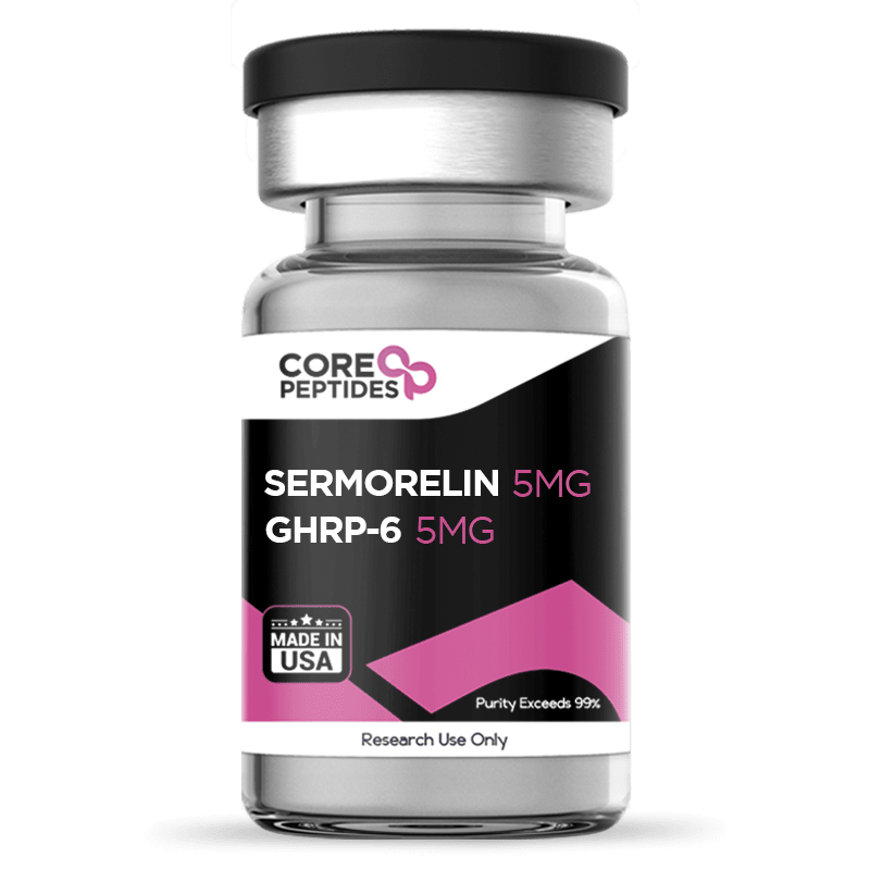
Sermorelin & GHRP-6 Blend (10mg)
Sermorelin and GHRP-6 are synthetic research peptides that both appear to interact with pituitary cells, albeit via different receptors. Sermorelin is an analog of the endogenous Growth Hormone-Releasing Hormone and is thought to act mainly at the classical GHRH receptor on anterior pituitary cells. Activating them is thought to lead to growth hormone release from pituitary cells, potentially. GHRP-6, by contrast, is classified as a growth hormone secretagogue (GHS) and appears to interact with ghrelin-sensitive receptors such as GHS-R1a, which are distinct from the GHRH receptor and may prefer partially separate intracellular signaling pathways. Activating them may also lead to growth hormone release from pituitary cells. Because these two receptor families seem to converge on the same GH-producing cells while using different upstream inputs, investigators have posited that combining Sermorelin and GHRP-6 in experimental models might provide a stronger or more nuanced stimulus to somatotroph function than either peptide alone. Chemical Makeup Other Known Titles Sermorelin: GRF 1-29 NH2 GHRP-6: SKF-110679, growth hormone-releasing hexapeptide Molecular Weight: Sermorelin:93 g/mol GHRP-6:03 g/mol Molecular Formula: Sermorelin: C149H246N44O42S GHRP-6: C46H56N12O6 Research and Clinical Studies Sermorelin & GHRP-6 Structure Sermorelin is posited to be a 29–amino acid fragment corresponding to the N-terminal portion of endogenous GHRH, with a C-terminal amidation that may help stabilize the molecule. Experimental work such as that of Clark et al. suggests that this fragment apparently retains full affinity and activation potential for pituitary GHRH receptors.(1) Consequently, this receptor activation is thought to drive cAMP-linked pathways that support growth hormone synthesis and release from pituitary cells. Thus, they commented that Sermorelin potentially “accelerates growth and increases pituitary GH content.” Research suggests that rather than being a GHRH analog, GHRP-6 appears to be a hexapeptide designed to interact with a different set of receptors on pituitary cells, referred to as ghrelin receptors. Yet, researchers such as Bowers et al. reveal that the peptide is not an analog to ghrelin, but appears derived from an opioid receptor agonist referred to as met-enkephalin.(2) The difference is that GHRP-6 is modified so that it loses opioid receptor affinity and gains ghrelin receptor affinity. Before the discovery of ghrelin receptors, these were actually termed growth hormone secretagogue 1a receptors (GHS-R1a), and the molecules that might interact with them, like GHRP-6, are suggested to be growth hormone secretagogues (GHSs). Sermorelin & GHRP-6 Affinity Towards Pituitary Cell Receptors As mentioned, Sermorelin and GHRP-6 may both interact with pituitary cells to stimulate growth hormone release, but appear to achieve that via different receptors and cellular pathways. Research by Culhane et al. suggests that Sermorelin may activate the GHRH receptors similarly to endogenous GHRH.(3) This may involve the activation of an intracellular messenger called cyclic AMP (cAMP) and the kinase PKA (protein kinase A), which together may switch on the cellular machinery that moves growth-hormone–containing vesicles to the membrane for release. On the other hand, GHRP-6 appears to target pituitary cells through the GHS-R1a receptor. Studies such as those by Yin et al. indicate that ligands of this receptor may favor activation of another messenger called phospholipase C.(4) PLC then is thought to cleave membrane PIP₂ into second messengers such as IP₃ and DAG. IP₃ may move toward intracellular calcium stores and trigger calcium release into the cytosol, while DAG remains in the membrane and potentially activates PKC (protein kinase C). Similar to PKA, PKC, in combination with the increased calcium release, may mobilize growth hormone-containing granules toward the plasma membrane of pituitary cells and promote their discharge. Sermorelin & GHRP-6 Potential for Growth Hormone Release Research on pituitary cells suggests that exposure to Sermorelin may induce an upregulation of growth hormone synthesis and release capacity. In experiments by Vittone et al., 12-hour mean growth hormone concentrations apparently rose from about 1.1 ± 0.9 µg/L to roughly 2.2 ± 1.9 µg/L, while the integrated 12-hour growth hormone output increased from around 1,114 ± 931 to about 2,032 ± 1,728 µg·min/L.(5) Further work by Khorram et al. with a slightly modified Sermorelin molecule indicates that the most pronounced potential of the peptide on growth hormone output may be exerted within the first 2 hours.(6) The researchers suggest that the 2-hour growth hormone levels appeared to rise from roughly 200–300 to about 1,100–1,600 µg·L⁻¹·min, which they described as an approximate sixfold increase. Moreover, the researchers commented that this potential may have been accompanied by an upregulation in the “levels of IGF-I (P < 0.05) and IGFBP-3 (P < 0.001), but not IGFBP-1, which remained elevated for 12 weeks.” IGF-1 stands for insulin-like growth factor-1. Peptides like these have been posited to be the main mediators of growth hormone's anabolic actions towards different cells. Growth hormone is thought to interact with growth hormone receptors on different cells and stimulate IGF-1 synthesis, which in this experiment appeared to be increased by 27–28% following the Sermorelin experimentation. Similar experiments with the ghrelin-receptor agonist GHRP-6 suggest that it may also provoke growth hormone peaks from basal values near 1–2 mU/L to around 60 mU/L, which corresponds to roughly a 30- to 50-fold increase over baseline and more than a threefold rise over usual physiological peaks of up to 20 mU/L. Researchers such as Micle et al. have interpreted these findings as a potential of GHRP-6 for strong engagement of GHS-R1a-linked signaling that drives high-amplitude secretory bursts.(7) Sermorelin & GHRP-6 Synergistic Actions The aforementioned experiment by Micle et al. also investigated the potential of GHRP-6 on growth hormone synthesis when combined with GHRH analogs. Specifically, the researchers experimented with the full-length GHRH rather than Sermorelin.(7) Nevertheless, their research suggests that the blend may lead to a peak growth hormone reaching up to 140 mU/L, roughly doubling the GHRP-6–only response of 60 mU/L and representing an approximate 6-fold increase over physiological peaks. Further research by Cordido et al. also compared the potential of GHRP-6 alone vs the synergistic potential of GHRP-6 with a GHRH analog.(8) These researchers were working with pituitary cells with much lower baseline growth hormone synthesis that was nearly undetectable. Exposure to GHRP-6 alone apparently caused an average growth hormone peak of about 6 mU/L. In contrast, exposure to a GHRH analogue alone appeared to yield a smaller growth hormone peak of roughly 2.6 mU/L, but the combination of GHRP-6 and GHRH analog led to a total peak of 16.3 mU/L, which was approximately 2.7 times the GHRP-6–only peak and 6.2 times the GHRH analog-only peak. The 12-hour growth hormone levels with GHRP-6 alone were suggested to be around 260 mU·min/L, versus 159 mU·min/L for GHRH analog, but reaching up to 729 mU·min/L when the two peptides were blended. Even though the GHRH-analog was not Sermorelin, the research suggests that a combination of GHRP-6 with a peptide like Sermorelin may induce synergistic actions on pituitary cells. The only one of these experiments to combine GHRP-6 specifically with Sermorelin was conducted by Sigalos et al.(9) The researchers combined the two peptides alongside another GHRP and their findings suggest that the blend upregulated growth hormone levels from baseline values of 160 ng/mL to roughly 250–265 ng/mL, which corresponds to an apparent 1.6-fold increase. This data further suggests that GHRP-6 and Sermorelin may exert synergistic actions, although more data is needed to evaluate their potential as a blend in laboratory research. Sermorelin & GHRP-6 blend is available for research and laboratory purposes only. Please review our Terms and Conditions before ordering. References: Clark RG, Robinson IC. Growth induced by pulsatile infusion of an amidated fragment of hGH-releasing factor in normal and GHRF-deficient rats. Nature. 1985 Mar 21-27;314(6008):281-3. doi: 10.1038/314281a0. PMID: 2858818. Bowers CY. History to the discovery of ghrelin. Methods Enzymol. 2012;514:3-32. doi: 10.1016/B978-0-12-381272-8.00001-5. PMID: 22975043. Culhane KJ, Liu Y, Cai Y, Yan EC. Transmembrane signal transduction by peptide hormones via family B G protein-coupled receptors. Front Pharmacol. 2015 Nov 5;6:264. doi: 10.3389/fphar.2015.00264. PMID: 26594176; PMCID: PMC4633518. Yin Y, Li Y, Zhang W. The growth hormone secretagogue receptor: its intracellular signaling and regulation. Int J Mol Sci. 2014 Mar 19;15(3):4837-55. doi: 10.3390/ijms15034837. PMID: 24651458; PMCID: PMC3975427. Vittone J, Blackman MR, Busby-Whitehead J, Tsiao C, Stewart KJ, Tobin J, Stevens T, Bellantoni MF, Rogers MA, Baumann G, Roth J, Harman SM, Spencer RG. Effects of single nightly injections of growth hormone-releasing hormone (GHRH 1-29) in healthy elderly men. Metabolism. 1997 Jan;46(1):89-96. doi: 10.1016/s0026-0495(97)90174-8. PMID: 9005976. Khorram O, Laughlin GA, Yen SS. Endocrine and metabolic effects of long-term administration of [Nle27]growth hormone-releasing hormone-(1-29)-NH2 in age-advanced men and women. J Clin Endocrinol Metab. 1997 May;82(5):1472-9. doi: 10.1210/jcem.82.5.3943. PMID: 9141536. Micic D, Popovic V, Kendereski A, Macut D, Casanueva FF, Dieguez C. Growth hormone secretion after the administration of GHRP-6 or GHRH combined with GHRP-6 does not decline in late adulthood. Clin Endocrinol (Oxf). 1995 Feb;42(2):191-4. doi: 10.1111/j.1365-2265.1995.tb01861.x. PMID: 7734029. Cordido F, Peñalva A, Dieguez C, Casanueva FF. Massive growth hormone (GH) discharge in obese subjects after the combined administration of GH-releasing hormone and GHRP-6: evidence for a marked somatotroph secretory capability in obesity. J Clin Endocrinol Metab. 1993 Apr;76(4):819-23. doi: 10.1210/jcem.76.4.8473389. PMID: 8473389. Sigalos JT, Pastuszak AW, Allison A, Ohlander SJ, Herati A, Lindgren MC, Lipshultz LI. Growth Hormone Secretagogue Treatment in Hypogonadal Men Raises Serum Insulin-Like Growth Factor-1 Levels. Am J Mens Health. 2017 Nov;11(6):1752-1757. doi: 10.1177/1557988317718662. Epub 2017 Aug 22. PMID: 28830317; PMCID: PMC5675260. Dr. MarinovDr. Marinov (MD, Ph.D.) is a researcher and chief assistant professor in Preventative Medicine & Public Health. Prior to his professorship, Dr. Marinov practiced preventative, evidence-based medicine with an emphasis on Nutrition and Dietetics. He is widely published in international peer-reviewed scientific journals and specializes in peptide therapy research.
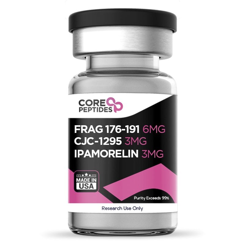
Fragment 176-191 & CJC-1295 & Ipamorelin Blend (12mg)
Researchers have suggested that growth hormone-releasing peptides (GHRPs), growth hormone secretagogues (GSHs), and analogs of growth hormones (hGH) may exhibit potential to promote growth hormone secretion. When combined, they have been suggested by researchers to exert significant action. These include increased fat metabolism, increased lean mass, regulated sleep cycle, and enhanced intestinal and cardiac functioning. CJC-1295 is a synthetic peptide composed of 29 amino acids. The peptide appears to work by activating the Growth Hormone-Releasing Hormone (GHRH) receptors in the pituitary gland. These receptors are typically activated by the native GHRH. CJC-1295 appears to be a synthetic analog made of the first 29 amino acids from the GHRH sequence, which has additional modifications, including the replacement of four amino acids in its structure and the attachment of a Drug Affinity Complex (DAC) to potentially extend the pharmacokinetics of the peptide. Ipamorelin is a synthetic pentapeptide and has been categorized by scientists as a growth hormone secretagogue (GHS). It has been posited to function similarly to Growth Hormone Releasing Peptides (GHRPs) and to potentially emulate the natural hunger hormone, ghrelin, particularly its potential to stimulate the release of hGH from the cells in the anterior pituitary gland. More specifically, Ipamorelin may mimic the function of ghrelin by activating the ghrelin receptors there, which are also known as the Growth Hormone Secretagogue receptors 1 Alpha (GHS-R1a) Fragment 176-191 peptide, as the name suggests, is a small ‘fragment’ of the growth hormone hGH composed of 15 amino acids. This peptide might target the beta-3 adrenergic receptors (ADRB3), which may potentially induce weight loss downstream. Through these receptors, it is suggested that the peptide may boost fat burning in adipose tissue cells and possibly promote 'thermogenesis' in skeletal muscle cells. Presented together, this blend may trigger a small portion of the pituitary gland, possibly stimulating the release of growth hormones. This blend has been posited by scientists to potentially synergistically enhance and regulate growth hormone concentration and maintain equilibrium levels. Chemical Makeup(1)(2)(3) Molecular formula Fragment 176-191: C78H125N23O23S2 CJC-1295 (Mod GRF 1-29): C152H252N44O42 Ipamorelin: C38H49N9O5 Molecular weight Fragment 176-191: 1817.12 g/mol CJC-1295 (Mod GRF 1-29): 3367.9 g/mol Ipamorelin: 711.8 g/mol Other known titles Fragment 176-191: AOD 9604, GH (hGH) lipolytic fragment, Somatostatin (177-191), tyrosyl CJC-1295: Mod GRF 1-29, CJC-1295 without DAC Ipamorelin Ipamorelin Acetate, IPA Research and Clinical Studies Fragment 176-191 & CJC-1295 & Ipamorelin Blend, and Growth Hormones While the fragment 176-191 appears to directly mimic the function of hGH, the peptides CJC-1295 and Ipamorelin appear to exert potential by upregulating the synthesis of hGH itself. For example, CJC-1295 has been suggested to have significant hGH-boosting potential in several phase-1 clinical trials. In the first, test subjects aged between 20 and 40 were enrolled.(4) All subjects were divided into two groups – one was presented with a saline placebo and the rest with the CJC-1295 peptide. A blood sample was collected from all subjects before and after the peptide presentation. After completion of the study, it was reported by the researchers that the peptide group appeared to exhibit a 7.5-fold increase in their growth hormone levels compared to the standard group. The trend suggested that these hormones increased gradually throughout the study and remained unchanged for at least 7 days after discontinuing presentation. Another study(5) was conducted on male test subjects aged between 20 and 60. Similar to the previous study, these subjects were divided into two groups – one was presented with a placebo while the rest were given the CJC-1295 peptide. The peptide group was given gradually increasing concentrations of the compound during the study. Upon analyzing the blood samples collected from these subjects, it was observed that there appeared to be a concentration-dependent increase, of up to 10 times, in the concentration of growth hormones among the peptide subjects. As per Madalina Ionescu et al., “The marked enhancement of trough GH levels by continuous GHRH stimulation implicates the importance of this effect on increasing IGF-I. Long-acting GHRH preparations may benefit patients with intact pituitary GH secretory capability.” (4) Similarly, Ipamorelin has also been suggested to potentially increase hGH synthesis. In one clinical study it was posited that a single presentation of the peptide may boost hGH levels by over 60-fold compared to placebo.(6) Fragment 176-191 & CJC-1295 & Ipamorelin Blend, and Lipolytic Action A clinical trial(7) was conducted in 2004 to study the peptide’s potential lipolytic (fat-burning) action. In the trial, 300 subjects were enrolled and presented with the Fragment 176-191 peptide for 12 weeks. These subjects were divided into 6 groups – one given a saline placebo, and the rest presented with different peptide concentrations. The group presented with minimal peptide presence appeared to exhibit a notable reduction in their body weight (up to 2.8 kilograms). These peptides may have also helped improve these subjects’ cholesterol profiles and glucose tolerance levels. The research team stated, “The evidence from the trial is that over 12 weeks, [the peptide] induces competitive weight loss with accompanying health benefits…” Fragment 176-191 & CJC-1295 & Ipamorelin Blend, and Regeneration Thirty-two experimental rabbits were examined as part of this 2015 study,(8) all of whom were divided into four groups of eight. All four groups were presented with placebo, Fragment 176-191 peptide, hyaluronic acid, or a combination of the peptide and hyaluronic acid for about 7 weeks. After the completion of the study, all rabbits were checked for any signs of cartilage damage. Among all groups, the rabbits presented with peptide and hyaluronic acid combination appeared to show minimal cartilage degeneration. These results suggest that the peptide may have the capability to enhance cartilage regeneration and repair. The report concluded that “Intra-articular AOD9604 using ultrasound guidance enhanced cartilage regeneration, and combined AOD9604 and HA were more effective than HA or AOD9604 alone in the collagenase-induced knee OA rabbit model.” (8) Fragment 176-191 & CJC-1295 & Ipamorelin Blend, and Body Composition As mentioned, Fragment 176-191 may potentiate a favorable shift in body composition by reducing adiposity. Yet, CJC-1295 & Ipamorelin also appear to have a significant potential via different impacts, which may include increasing lean mass for CJC-1295, and increasing overall body weight in the case of Ipamorelin. Research in the lab suggests that Ipamorelin might potentially play a role in weight gain including muscle mass gain but also adiposity, possibly due to both an elevation in hGH levels and its potential to stimulate appetite through ghrelin receptor activation. An investigation into Ipamorelin's potential impact on body fat was conducted in both hGH-deficient and hGH-intact murine models. The results indicated that Ipamorelin may have led to a modest 15% rise in body weight within a span of 2 weeks. Additionally, in both murine models, there was a relative increase in fat pad weights when compared to overall body weight.(9) On the other hand, CJC-1295 may potentially improve body composition while apparently increasing muscle mass without affecting or even possibly decreasing adiposity. Research involving murine models with a GHRH gene deletion (known as GHRHKO) noted that CJC-1295 might upregulate the synthesis of GH and potentially induce such a favorable shift in body composition. When these GHRHKO murine models were exposed to CJC-1295, they seemed to maintain standard lean mass, compared to unexposed models which failed to achieve normal lean mass. Moreover, the subcutaneous fat mass remained at control levels across all peptide-associated groups, while the GHRHKO murine models not exposed to CJC-1295 appeared to have increased adiposity.(10) Considering the potential lipolytic action of Fragment 176-191 and CJC-1295, researchers may want to consider the hypothesis that the blend of these peptides may counteract the fat-increasing potential of Ipamorelin, while further increasing its muscle building potential. Fragment 176-191 & CJC-1295 & Ipamorelin Blend, and Bone Mineral Content By increasing lean and muscle mass, the peptides and especially Ipamorelin may provide potential benefits in bone mass. In an experiment, murine subjects were either introduced to Ipamorelin or a control substance. The impact of Ipamorelin on bone mineral content was observed in real-time using dual X-ray absorptiometry (DXA) at select areas, notably the femur and L6 vertebrae. Following the study period, the femurs of the murine subjects were examined using mid-diaphyseal peripheral quantitative computed tomography (pQCT) scans. Preliminary results suggest that the peptide might have played a role in a possible weight increase and a potential rise in the tibial and vertebral bone mineral content as detected by DXA, compared to the control group. The pQCT findings hint that the increase in cortical BMC might be due to an expansion in the cross-sectional bone area.(11) Fragment 176-191 & CJC-1295 & Ipamorelin Peptide Blend is available for research and laboratory purposes only. Please review and adhere to our Terms and Conditions before ordering. References National Center for Biotechnology Information (2023). PubChem Compound Summary for CID 91976842, CJC1295 Without DAC. https://pubchem.ncbi.nlm.nih.gov/compound/CJC1295-Without-DAC National Center for Biotechnology Information (2023). PubChem Compound Summary for CID 9831659, Ipamorelin. https://pubchem.ncbi.nlm.nih.gov/compound/Ipamorelin National Center for Biotechnology Information (2023). PubChem Substance Record for SID 319360420, 386264-39-7, Source: ToxPlanet. https://pubchem.ncbi.nlm.nih.gov/substance/319360420 Ionescu M, Frohman LA. Pulsatile growth hormone secretion (GH) persists during continuous stimulation by CJC-1295, a long-acting GH-releasing hormone analog. J Clin Endocrinol Metab. 2006 Dec;91(12):4792-7. doi: 10.1210/jc.2006-1702. Epub 2006 Oct 3. PMID: 17018654. https://pubmed.ncbi.nlm.nih.gov/17018654/ Teichman SL, Neale A, Lawrence B, Gagnon C, Castaigne JP, Frohman LA. Prolonged stimulation of growth hormone (GH) and insulin-like growth factor I secretion by CJC-1295, a long-acting analog of GH-releasing hormone, in healthy adults. J Clin Endocrinol Metab. 2006 Mar;91(3):799-805. doi: 10.1210/jc.2005-1536. Epub 2005 Dec 13. PMID: 16352683. https://pubmed.ncbi.nlm.nih.gov/16352683/ Gobburu, J. V., Agersø, H., Jusko, W. J., & Ynddal, L. (1999). Pharmacokinetic-pharmacodynamic modeling of ipamorelin, a growth hormone releasing peptide, in human volunteers. Pharmaceutical research, 16(9), 1412–1416. https://doi.org/10.1023/a:1018955126402 News, Medical and Life Sciences, Obesity drug codenamed AOD 9604 highly successful in trials, 16 December 2004, https://www.news-medical.net/news/2004/12/16/6878.aspx Kwon DR, Park GY. Effect of Intra-articular Injection of AOD9604 with or without Hyaluronic Acid in Rabbit Osteoarthritis Model. Ann Clin Lab Sci. 2015 Summer;45(4):426-32. PMID: 26275694. https://pubmed.ncbi.nlm.nih.gov/26275694/ Lall, S., Tung, L. Y., Ohlsson, C., Jansson, J. O., & Dickson, S. L. (2001). Growth hormone (GH)-independent stimulation of adiposity by GH secretagogues. Biochemical and biophysical research communications, 280(1), 132–138. https://doi.org/10.1006/bbrc.2000.4065 Alba M, Fintini D, Sagazio A, Lawrence B, Castaigne JP, Frohman LA, Salvatori R. Once-daily administration of CJC-1295, a long-acting growth hormone-releasing hormone (GHRH) analog, normalizes growth in the GHRH knockout mouse. Am J Physiol Endocrinol Metab. 2006 Dec;291(6):E1290-4. doi: 10.1152/ajpendo.00201.2006. Epub 2006 Jul 5. PMID: 16822960. Svensson, J., Lall, S., Dickson, S. L., Bengtsson, B. A., Rømer, J., Ahnfelt-Rønne, I., Ohlsson, C., & Jansson, J. O. (2000). The GH secretagogues ipamorelin and GH-releasing peptide-6 increase bone mineral content in adult female rats. The Journal of endocrinology, 165(3), 569–577. https://doi.org/10.1677/joe.0.1650569 Dr. MarinovDr. Marinov (MD, Ph.D.) is a researcher and chief assistant professor in Preventative Medicine & Public Health. Prior to his professorship, Dr. Marinov practiced preventative, evidence-based medicine with an emphasis on Nutrition and Dietetics. He is widely published in international peer-reviewed scientific journals and specializes in peptide therapy research.

Receptor Grade IGF-1 LR3 (100mcg)
Insulin-like Growth factor-1, or IGF-1, is a naturally produced protein with 70 amino acids. Receptor Grade IGF-1 LR3 is a synthetic variant of the naturally occurring IGF-1, which contains an extended N-terminal structure of 13 amino acids and a replacement of the glutamic acid at residue 3 with arginine. Hence, it is named IGF-1 Long R3.(1)(2) Owing to the altered structure, Receptor Grade IGF-1 LR3 has been suggested to have increased affinity and increased anabolic potential while also binding less to IGF-1 binding proteins (IGF-1BPs). Structurally similar to insulin, this IGF-1 LR3 has the potential primarily to regulate cell tissue growth and development. This potential has been evaluated in cell growth studies, throughout which researchers first prompted the need for developing this high-potency variant. Moreover, the classification of Receptor Grade refers to the purity of the material, which is considered higher than Media Grade IGF-1 LR3. Chemical Makeup(2,3) Molecular Formula: C400H625N111O115S9 Molecular Weight: 9117.5 g/mol Other Known Titles: Long-(arg3) insulin-like growth factor-I, Insulin-like growth factor long chain R3 Research and Clinical Studies Receptor Grade IGF-1 LR3 and Anabolic Potential Unfortunately, research on the anabolic potential of IGF-1 LR3 is lacking, as the peptide is aimed towards cell culture studies. Yet, of the few experiments in murine models, researchers have suggested the significant potential of the peptide.(1) In one study, experiments were carried out on normal and dexamethasone-induced catabolic murine models. It was noted that IGF-1 LR3 might potentially be 1.5 to 2 times as anabolic as IGF-I in inducing weight gain, increasing visceral organ weights, and possibly enhancing feed use efficiency under continuous delivery conditions. Moreover, IGF-1 LR3 appeared to have retained the potential for greater potency than IGF-I in several metrics, even in studies of only intermittent exposure. Additionally, in murine models exposed to dexamethasone, it was observed that the excretion of Nτ-methylhistidine—a marker indicative of muscle protein breakdown—appeared reduced to a greater extent by IGF-1 LR3, potentially threefold more than by IGF-I. This suggests that IGF-1 LR3 may host the potential, though not consistently equivalent across all parameters, to exhibit enhanced anabolic actions under certain laboratory conditions. Therefore, Receptor Grade IGF-1 LR3 may be posited to exert even greater anabolic potential than IGF-1. To provide a comparison, several studies have researched the anabolic potential of IGF-1. For example, a study(4) was conducted in 2005 to study the potential of the peptide in models of IGF-1 deficiency. Following peptide exposure, the length and growth of the models were assessed and analyzed against control thresholds. Based on the study findings, it was observed that total length increased in all peptide-exposed models by a significant margin against the control standards. This study suggests that IGF-1, and possibly also more potent analogs like Receptor Grade IGF-1 LR3 peptide, may have some potential in mitigating growth deficiency. Receptor Grade IGF-1 LR3 and Insulin Receptor Sensitivity Research(5) has suggested that IGF proteins typically bind to IGF-1 receptors and may stimulate glucose uptake, potentially through a signaling mechanism involving PI3K and AMPK pathways. However, when studied, peptides like Receptor Grade IGF-1 LR3 appeared to induce glucose uptake not just through IGF-1 receptor interactions but also independently, possibly via other pathways or receptors. This suggests that the mechanism of glucose uptake might involve additional cellular processes beyond the traditional receptor binding. Assefa B Mahmoud et al. stated, "Multiple [...] studies reported the role of IGF-1 in enhancing insulin sensitivity and glucose metabolism. A low-serum level of IGF-1 has been associated with insulin resistance, and [...] recombinant IGF-1 has been [hypothesized] to improve insulin sensitivity and glucose metabolism.” Receptor Grade IGF-1 LR3 and Cell Lifespan Research(6) on murine models observed that common markers of physiological decline, such as muscle tears and neurological deficiencies, appeared to be mitigated for an extended period following routine exposure to the peptide. While more detailed studies and clinical trials are pending, the above preliminary study suggests that the peptide may indirectly help to increase lifespan of functional cells. As per William E. Sonntag et al., “Based on this review, we conclude that the perceived contradictory roles of growth hormone and insulin-like growth factor-1 in the genesis of the aging phenotype should not be interpreted as a controversy on whether growth hormone or insulin-like growth factor-1 increases or decreases life span but rather as an opportunity to explore the complex roles of these hormones during specific stages of the life span.” Receptor Grade IGF-1 LR3 and Muscle Cells A study(7) was conducted on female murine models to identify the IGF-1 LR3 peptide’s potential in decreasing the action of myostatin. Myostatin is considered to prevent cellular differentiation; mitigating the actions of this protein may increase lean muscle and reduce fat cell storage and fatty mass. The study's results suggested that the various IGF-1 analogs, including Receptor Grade IGF-1 LR3, appear to potentially reverse adverse myostatin and prevent apoptosis. Receptor Grade IGF-1 LR3 and Shorter Action An experimental mouse model was created for a study(8) where the IGF-1 LR3 peptide was compared to the endogenous IGF-1. Throughout the study, it was observed that when the peptide was exposed to the murine model, it appeared to quickly clear from the serum and evenly distribute into tissue. More specifically, the researchers posited that IGF-1 LR3 cleared faster as it appeared to bind to a lower degree to binding proteins than endogenous IGF-1. This reduced binding affinity means IGF-1 LR3 might circulate more freely than IGF-I. The analysis of tissue distribution patterns of IGF-1 LR3 also suggested a potentially unique localization compared to IGF-I. Elevated levels of the IGF-1 LR3 tracer were observed in tissues such as kidneys, ovaries, and adrenal glands in murine models. This distinct distribution suggests that the organs primarily involved in metabolic and reproductive functions may exhibit varying capacities for the uptake or retention of IGF-1 LR3 in contrast to IGF-I. It is hypothesized that these differences might stem from IGF-1 LR3's diminished propensity to form complexes with IGFBPs, which might influence its bioavailability and interaction with target tissues in experimental models. Nevertheless, further research suggested that a peptide with similar modifications (namely R3) to IGF-1 LR3 may exert increased anabolic potential compared to regular IGF-1 despite the shorter action.(9) Receptor Grade IGF-1 LR3 peptide is available for research and laboratory purposes only. Please review and adhere to our Terms and Conditions before ordering. References Tomas, F. M., Knowles, S. E., Owens, P. C., Chandler, C. S., Francis, G. L., Read, L. C., & Ballard, F. J. (1992). Insulin-like growth factor-I (IGF-I) and especially IGF-I variants are anabolic in dexamethasone-treated rats. The Biochemical journal, 282 ( Pt 1)(Pt 1), 91–97. https://www.ncbi.nlm.nih.gov/pmc/articles/PMC1130894/ Human Insulin-like growth factor. Protein Data Bank in Europe, https://www.ebi.ac.uk/pdbe/entry/pdb/1gzr National Center for Biotechnology Information (2023). PubChem Substance Record for SID 381123731, M9L22Y19H9, Source: ChemIDplus. Retrieved January 24, 2023 from https://pubchem.ncbi.nlm.nih.gov/substance/381123731. Anderson, L. J., Tamayose, J. M., & Garcia, J. M. (2018). Use of growth hormone, IGF-I, and insulin for anabolic purpose: Pharmacological basis, methods of detection, and adverse effects. Molecular and cellular endocrinology, 464, 65–74. https://www.ncbi.nlm.nih.gov/pmc/articles/PMC5723243/ Assefa, B., Mahmoud, A. M., Pfeiffer, A., Birkenfeld, A. L., Spranger, J., & Arafat, A. M. (2017). Insulin-Like Growth Factor (IGF) Binding Protein-2, Independently of IGF-1, Induces GLUT-4 Translocation and Glucose Uptake in 3T3-L1 Adipocytes. Oxidative medicine and cellular longevity, 2017 https://www.ncbi.nlm.nih.gov/pmc/articles/PMC5750484/ William E. Sonntag, Anna Csiszar, Raphael de Cabo, Luigi Ferrucci, Zoltan Ungvari, Diverse Roles of Growth Hormone and Insulin-Like Growth Factor-1 in Mammalian Aging: Progress and Controversies, The Journals of Gerontology: Series A, Volume 67A, Issue 6, June 2012, Pages 587–598, https://doi.org/10.1093/gerona/gls115 Naisi Li, Qiyuan Yang, Ryan G. Walker, Thomas B. Thompson, Min Du, Buel D. Rodgers, Myostatin Attenuation In Vivo Reduces Adiposity, but Activates Adipogenesis, Endocrinology, Volume 157, Issue 1, 1 January 2016, Pages 282–291. https://doi.org/10.1210/en.2015-1546 Bastian SE, Walton PE, Wallace JC, Ballard FJ. Plasma clearance and tissue distribution of labelled insulin-like growth factor-I (IGF-I) and an analogue LR3IGF-I in pregnant rats. J Endocrinol. 1993 Aug;138(2):327-36. doi: 10.1677/joe.0.1380327. PMID: 7693845. Elis S, Wu Y, Courtland HW, Cannata D, Sun H, Beth-On M, Liu C, Jasper H, Domené H, Karabatas L, Guida C, Basta-Pljakic J, Cardoso L, Rosen CJ, Frystyk J, Yakar S. Unbound (bioavailable) IGF1 enhances somatic growth. Dis Model Mech. 2011 Sep;4(5):649-58. doi: 10.1242/dmm.006775. Epub 2011 May 31. PMID: 21628395; PMCID: PMC3180229. Dr. MarinovDr. Marinov (MD, Ph.D.) is a researcher and chief assistant professor in Preventative Medicine & Public Health. Prior to his professorship, Dr. Marinov practiced preventative, evidence-based medicine with an emphasis on Nutrition and Dietetics. He is widely published in international peer-reviewed scientific journals and specializes in peptide therapy research.

Cortagen (20mg)
Cortagen is posited to be a synthetic tetrapeptide with the sequence Ala–Glu–Asp–Pro, originally designed based on amino acid analysis of the polypeptide complex cortexin. It is classified among the so-called Khavinson peptides – a family of short peptide bioregulators that are proposed to modulate endogenous regulatory systems rather than acting as classical receptor agonists or enzyme inhibitors.(1) These are low-molecular-weight peptides that are posited to be able to penetrate cells and reach the nucleus, where they may interact directly with DNA and chromatin structures in mammalian research models. In vitro studies suggest that AACC may be a preferred DNA-binding sequence for Cortagen, raising the hypothesis that it may selectively support transcription at gene sites containing this motif and thereby exert epigenetic-like actions on gene expression. Currently, it appears to be under investigation in laboratory settings for its potential to support stress signaling in different cell cultures, including nerve cell circuits. Chemical Makeup Other Known Titles: AEDP, Ala-Glu-Asp-Pro Molecular Weight: 430.4 g/mol Molecular Formula: C17H26N4O9 Research and Clinical Studies Epigenetic Potential of Cortagen Research by Lezhava et al. suggests that Cortagen may have epigenetic support for aged cells, potentially restoring the expression of genes suppressed by the aging process of the cell via condensation of the chromatin.(2) According to the researchers, Cortagen was applied to lymphoid cell cultures exhibiting an aged chromatin profile to assess its potential. Differential scanning calorimetry suggested that chromatin was thermodynamically less stable and therefore more uncondensed following Cortagen experimentation. These study authors proposed that Cortagen may partially unfold higher-order chromatin structures, such as loops of 30-nm fibers and even 10-nm nucleosomal filaments into more relaxed 5-nm fibers, implying that the “Peptidebioregulator Cortagen induces unrolling deheterochromatinization (decondensation) of total heterochromatin” in both structural and facultative chromatin domains. Consequently, ribosomal gene clusters may become more transcriptionally active, potentially supporting protein synthesis capacity in these aged cells. Yet, Cortagen does not appear to remodel all constitutive blocks as pericentromeric C-heterochromatin on chromosomes analogous to 1, 9, and 16 remained structurally stable. Cortagen also appeared to increase sister chromatid exchange (SCE) frequency in some chromosome groups, which the authors viewed as a potential cytogenetic marker of facultative heterochromatin decondensation and possible re-release of previously repressed genes. Cortagen Research into Oxidative Stress According to the research of Kozina et al., Cortagne may support lipid peroxidation and oxidative modification of proteins in mammalian research models.(3) According to the team of scientists, Cortagen exposure was associated with “decreased the content of LPO products and reduced oxidative modification of proteins” in both neural cells and the surrounding protein-rich medium. In the cells, the peptide also tended to lower later-stage products. In parallel, Cortagen appeared to reduce the accumulation of protein carbonyl groups, which act as markers of oxidative protein modification, and appeared to achieve a significant reduction in neural cells, and by about 15% in the extracellular fraction. These authors suggested that Cortagen may interfere with the chain of reactions whereby reactive lipid species attack amino groups in proteins, possibly limiting downstream carbonyl formation. Yet, Cortagen’s potential was apparent only in integrated biological models, suggesting that it may not act as a simple radical scavenger, but instead possibly modulates the generation of reactive species upstream, or alters how cells handle oxidative stress. Thus, the researchers suggest that Cortagen may exert an indirect antioxidant-like action in neural systems by dampening lipid and protein oxidation. Cortagen Research into Cellular Stress Response and Differentiation Studies involving a transcriptome-wide analysis of Cortagen’s potential on cardiac muscle cell genes suggest that the peptide may support the cellular stress response of such cultures. Specifically, Anisimov et al. suggested that Cortagen may modulate genes related to carrier proteins and membrane transport, as well as DNA synthesis and replication involved in that response.(4) This pattern of changes is interpreted as potentially supporting intracellular transport processes and proliferative or reparative programs at the transcriptional level. Cortagen also apparently up-regulated several mitochondrial genes (16S rRNA, COX3, ND5), which may theoretically interact with bioenergetics. In parallel, transcripts linked to ionic homeostasis and Ca²⁺ handling were altered, hinting at a possible support for excitation–contraction coupling pathways. Among particularly notable targets, Cortagen increased expression of stress-response genes (Pass1, Hsc70), developmental and survival-related signals (Bmp2, Wnt4), and components of mitogenic or survival signaling (Eps15, Eps15-rs). The authors posit that these coordinated transcriptional shifts may underlie some of Cortagen’s broader biological actions and suggest that the peptide may serve as a helpful laboratory tool for exploring peptide-mediated regulation of stress-response networks in vitro. Further research by Khavinson et al. suggested that by modifying gene expression, the peptide may also support cellular differentiation.(5) Experimentally, the authors examined Cortagen in an in vitro model using pluripotent embryonic ectodermal tissue. Ectodermal explants were incubated for 1 hour in solutions of Cortagen and then cultured. In the reference medium without peptides, the pluripotent ectoderm apparently gave rise only to atypical epidermis. By contrast, exposure to Cortagen induced the same pluripotent cells to differentiate into epidermal and mesenchymal cells, suggesting that these peptides may broaden the available differentiation pathways. Cortagen Research into Neurological Stress Signaling and Nerve Regeneration Cortagen has been examined in laboratory studies by Adriani et al., who have been investigating the activation of arousal- and stress-linked circuits through global output patterns. The scientists observed that the peptide apparently may support output from neural networks associated with arousal and exploratory drive, without concurrently amplifying stress-linked appraisal signals. This pattern may reflect a state in which outward-oriented processing dominates over internal “risk-checking” loops, suggesting a shift in the balance of stress-signaling within the network. The potential implications of Cortagen on nerve regeneration may better support these observations. GA group of researchers, led by Turchaninova et. al., investigated the neuroregenerative potential of Cortagen. The scientists transected sciatic nerve trunks, which were then micro-sutured and studied in vitro on a multielectrode platform. Compound action potentials (CAPs) were recorded at defined distances distal to the suture line. Cortagen apparently increased the length of the nerve segment able to conduct impulses by 27% and better-supported conduction velocity by 40% compared to placebo. The authors also suggested that the peptide may preferentially support regeneration where baseline growth is lower, rather than extending growth beyond the usual maximal range. This faster conduction is interpreted as a potential marker of more advanced functional maturation, possibly involving better-supported myelination and fiber calibre. Cortagen peptide is available for research and laboratory purposes only. Please review our Terms and Conditions before ordering. References: Khavinson, V. Kh, N. S. Lin’kova, and S. I. Tarnovskaya. "Short peptides regulate gene expression." Bulletin of experimental biology and medicine 162.2 (2016): 288-292. Lezhava, Teimuraz, et al. "Epigenetic Regulation of “Aged” Heterochromatin by Peptide Bioregulator Cortagen." International Journal of Peptide Research and Therapeutics 21.1 (2015): 157-163. Kozina, L. S. "Effects of bioactive tetrapeptides on free-radical processes." Bulletin of experimental biology and medicine 143.6 (2007): 744-746. Anisimov SV, Khavinson VKh, Anisimov VN. Elucidation of the effect of brain cortex tetrapeptide Cortagen on gene expression in mouse heart by microarray. Neuro Endocrinol Lett. 2004 Feb-Apr;25(1-2):87-93. PMID: 15159690. Khavinson V, Linkova N, Diatlova A, Trofimova S. Peptide Regulation of Cell Differentiation. Stem Cell Rev Rep. 2020 Feb;16(1):118-125. doi: 10.1007/s12015-019-09938-8. PMID: 31808038. Adriani, Walter, et al. "Modulatory effects of cortexin and cortagen on locomotor activity and anxiety-related behavior in mice." The Open Neuropsychopharmacology Journal 2.1 (2009): 22-29. Turchaninova LN, Kolosova LI, Malinin VV, Moiseeva AB, Nozdrachev AD, Khavinson VK. Effect of tetrapeptide cortagen on regeneration of sciatic nerve. Bull Exp Biol Med. 2000 Dec;130(12):1172-4. PMID: 11276314. Dr. MarinovDr. Marinov (MD, Ph.D.) is a researcher and chief assistant professor in Preventative Medicine & Public Health. Prior to his professorship, Dr. Marinov practiced preventative, evidence-based medicine with an emphasis on Nutrition and Dietetics. He is widely published in international peer-reviewed scientific journals and specializes in peptide therapy research.

Epithalon (25mg)
Research has suggested that Epithalon, (also known as AEDG peptide, tetrapeptide Epitalon, Epithalon, or Epithalone) may regulate the function of the brain, the pineal gland, and the eye retina. Studies in the peptide have spurred numerous research hypotheses, which include possible sleep regulation via pineal gland stimulation, releasing more melatonin. Studies also speculate that the peptide may stimulate the generation of telomerase, may exhibit strong antioxidant characteristics, and extend the retina's workable integrity. With recent technology and ongoing advances in the scientific field, methods have been developed to synthesize complex peptide preparations from the extracts of several different tissues. One such peptide in the tissues is ‘Epithalamin,’ naturally produced in the pineal gland. Epithalamin has been suggested to be functional in increasing melatonin production, improving the immunological and anti-carcinogenic functions in rats and mice, and restoring reproductive function in aged rodents. Utilizing the recent advancements in science, a peptide similar to Epithalamin was synthesized and titled ‘Epithalon.’ Epithalon is derived from a naturally occurring peptide belonging to both the pineal gland and eye retina.(1) Overview Epithalon is a synthetic tetrapeptide, also known as AEDG peptide, composed of amino acids Ala-Glu-Asp-Gly.(2) The peptide has been suggested to exhibit action similar to Epithalamin via various modes. Epithalamin is a related pineal peptide preparation containing Epithalon, that has been purported to potentially increase the average lifespan of various experimental models by 11–31% and may reduce mortality in murine models by a suggested 52%.(3) This potential mechanism and further studies are described below. Chemical Makeup Molecular Formula: C14H22N4O9 Molecular Weight: 390.34 g/mol Other Known Titles: Epitalon, Epithalone Research and Clinical Studies Epithalon Peptide and Longevity A study investigated the potential mechanisms of Epithalon in influencing gene expression and protein synthesis in stem cells such as gingival mesenchymal stem cells (hGMSCs). The study(2) has suggested that the peptide binds with the histones - HI/6 and HI/3 – located at different sites in tissue, which then interact with the DNA. More specifically, the study posits that Epithalon may alter chromatin structure by specifically interacting with histones, thereby modulating gene expression. This interaction might involve the peptide acting as a histone mimic, facilitating changes in chromatin dynamics. Epithalon's epigenetic regulation might involve competitive binding with histones at DNA interaction sites, increasing transcription of genes involved in neuronal differentiation. Such binding may displace other regulatory proteins, making DNA more accessible for transcriptional machinery, potentially inducing neuronal cell differentiation in retinal and periodontal ligament stem cells (hPDLSCs). This experimentation led to an upregulation of neurogenic differentiation markers, including Nestin, GAP43, β Tubulin III, and Doublecortin, in hGMSCs. Specifically, mRNA expression of these markers increased by 1.6 to 1.8 times. Considering the changes in these markers, the potential upregulation of neuronal differentiation and protein synthesis in retinal and ligament stem cells may lead to enhanced functionality. Epithalon Peptide and Oxidative Stress A recent study suggested that Epithalon might reduce intracellular reactive oxygen species (ROS) levels in aged oocyte cells, potentially exhibiting antioxidative actions.(4) The peptide was suggested to significantly lower ROS levels, unlike higher concentrations that did not demonstrate similar protective actions. Such a reduction might be critical, as oxidative stress is considered a significant factor in cellular aging. Additionally, Epithalon may help preserve the structural integrity of oocytes. It was observed to possibly decrease fragmentation in post-ovulatory aged oocytes and during parthenogenetic activation—a process where an egg develops into an embryo without fertilization. The peptide also potentially maintains spindle integrity and correct distribution of cortical granules (CG). Spindles, which are crucial structures in cell division that distribute chromosomes to daughter cells, and cortical granules, secretory vesicles important for preventing polyspermy (the fertilization of an egg by multiple sperm), are essential for normal cell division and fertilization. Epithalon might correct spindle abnormalities and prevent the misplacement of CGs, which are common issues in aged oocytes. Moreover, Epithalon exposure was associated with potential improvements in mitochondrial function, which is vital for oocyte viability. Mitochondria, known as the cell's powerhouses, are deemed crucial for ATP production and maintaining cellular metabolism. There were indications of enhanced mitochondrial membrane potential and increased mtDNA copy numbers, suggesting that Epithalon might support mitochondrial integrity and functionality as oocytes age. Finally, the peptide was suggested to reduce DNA damage and apoptosis (cell death) in aged oocytes. It appeared to lower the intensity of γH2AX signals—a marker of DNA damage—and seemed to decrease apoptosis rates, as indicated by reduced Annexin-V staining. These observations imply that Epithalon might enhance oocyte survival by potentially mitigating oxidative damage and preserving genomic stability, which are considered crucial for maintaining the overall functionality of the cells. Epithalon Peptide and Anti-Aging In order to understand the anti-aging action of various synthetic peptides, studies have been widely conducted to study their potential in cell proliferation, cell regeneration and aging, cellular apoptosis, and matrix modeling.(5) It was suggested that Epithalon may inhibit the synthesis of MM-9, which usually increases with time, and increase the proliferation and cellular regeneration process, which usually decreases with time. More specifically, the researchers posited that the peptide may have “enhanced the expression of Ki-67 and CD98hc that are less intensively synthesized during cell aging.” Epithalon also seemed to inhibit the activity of Caspase-3, a crucial enzyme believed to facilitate apoptosis (programmed cell death). The researchers commented that the peptide “suppressed caspase-dependent apoptosis that increases during aging of cell cultures.” By potentially restraining Caspase-3 activity, Epithalon might support cellular longevity and decrease apoptosis, enhancing its regenerative capabilities. Epithalon Peptide and Fetal Studies This study(6) was conducted to understand the proliferative potential of the peptide on fetal fibroblastic cells. Pulmonary fibroblasts were isolated from the 24-week-old fetus, and it was observed that these fibroblasts appeared to lose their proliferative function at the 34th passage. These cells possessed extremely small telomeres sizes – smaller than what they originally were during the 10th passage. When Epithalon was presented in these otherwise aging cells, it appeared to stimulate the development of telomeres, causing them to increase and restore their normal size. As a result of this size elongation, the telomeres appeared to cause 10 extra cell divisions than usual seen in the control cells. Thus, this study suggested that Epithalon overcame the Hayflick limit and extended the normal cell cycle in the cells.(6) Epithalon Peptide and Lymphocytic Cells In this clinical study,(7) lymphocytic cells were isolated and cultured from subjects aged between 76 and 80 years. The purpose of this study was to determine the action of Epithalon on ribosomal cell activity and its impact on denaturation and polymorphism of heterochromatin. The outcome of this study, following the delivery of the cell culture with Epithalon, was that the peptide appeared to induce activation of the ribosomal genes and decondensation of the heterochromatin. Consequently, it appeared to induce the release of genes that were otherwise suppressed due to the aging of the chromosomal regions. This study suggested that Epithalon might have the potential to modify the chromosome regions in the aging cells, activate chromatin, and restore cellular activities that were otherwise suppressed or delayed in geriatric subjects. Epithalon Peptide and Anti-Mutagenic Action In this 2011 study,(8) three different mice models were used to determine the action of the peptide on chromosomal aberrations. The three mice models were – SAMP-1 female mice with accelerated aging and wild rats SAMR-1 and SHR (both female mice). Upon delivery of Epithalon, it was observed that the incidence of the chromosomal aberrations in the bone marrow of SAMP-1 mice with accelerated aging appeared to be almost 2 times higher than the other two models. When the peptide was presented at 2 months in the mice, it appeared to decrease the chromosomal aberrations in all three models, the highest being in the SAMP-1 mice with accelerated cell aging. In combination with melatonin at night, given with water, there was no reported impact on the action of the peptide. This study suggested that Epithalon may possess anti-mutagenic potential. Epithalon Peptide and Cancer Cells In this study,(9) one-year-old female (C3H/He) mice with tumors on the reproductive organs (mammary glands and ovaries) were observed. The tumors on the mammary glands included several variants of the invasive ductal carcinogenic cells, whereas, in the ovaries, the tumors found were granulosa cell tumors. These mice, kept in standard conditions for six months, were divided into control and experimental groups. Epithalon was presented five times a week. Once the study was completed, it was reported that three out of the nine mice in the control group appeared to exhibit metastasis and increased tumor cells. Meanwhile, the peptide mice exhibited a decrease in the number of tumor cells. Epithalon, upon delivery, appeared to inhibit the process of metastasis in the mice, preventing tumor cell cycle and growth. Researchers of this study posited the anti-metastatic potential of the peptide. Epithalon Peptide and Research in Hypophysectomized Birds In this study,(10) hypophysectomized birds, both young and old, were used to study the action of Epithalon peptide on the morphology of the thymus gland. Hypophysectomized birds are birds in which the pituitary gland has been surgically removed. Upon delivery of the peptide, it was discovered that the morphology of the thymus gland appeared to be restored in all birds, regardless of their age. The most improved results were observed on birds (mainly chickens) that underwent neonatal hypophysectomy before the peptide was presented. Epithalon Peptide and Melatonin Levels This study(11) was carried out on aging monkeys to determine the actions of Epithalon on melatonin levels. With increasing age, the melatonin levels tend to decrease due to reduced secretion, which may cause difficulty in sleep regulation. This is mainly due to the functioning of the pineal gland deteriorating with increased age and a reduction of hormone circadian rhythm amplitude. Upon delivery of Epithalon, it appeared to stimulate actions similar to those caused by the natural secretion of the pineal gland. The melatonin levels appeared to increase to "normal" levels. Epithalon Peptide and Retinal Cells In this clinical study,(12) it was suggested that when Epithalon was presented in geriatric subjects, it appeared to elevate the bioelectric and functional activities in the retina, thereby preserving the morphological structure of the retina. More specifically, the researchers posited that the peptide “participates in the mechanisms of transcription common for the epiphysis and retina.”As a result, age-related retinal degeneration may be reversed in the older subjects, which is supported by the positive clinical outcome in 90% of the subjects presented with the peptide. Epithalon and Geroprotective Properties This study(13) was conducted on 266 elderly subjects (over the age of 60 years) throughout 6 to 8 years, where some subjects were presented with peptide-bioregulator Thymalin, others with Epithalon, and the rest with the combination of the two. After the study, it was observed that both the peptides appeared to have the potential to restore basic bodily functions in geriatric subjects – including improved functions in the cardiovascular, endocrinal, immune, and nervous systems, along with normalized metabolic and hemostatic activities. The peptide groups appeared to exhibit a 2-fold decrease in the common geriatric disorders such as acute respiratory disorder, heart diseases, and bone disorders. Additionally, the mortality rate in the peptide subjects appeared to significantly decrease, with a 2-fold decrease in the Thymalin subjects, a 1.8-fold decrease in the Epithalon subjects, and 2.5-fold decrease in the subjects presented with both peptides. Epithalon peptide is available for research and laboratory purposes only. Please review and adhere to our Terms and Conditions before ordering. References: Khavinson VKh. Peptides and Ageing. Neuro Endocrinol Lett. 2002;23 Suppl 3:11-144. PMID: 12374906. https://pubmed.ncbi.nlm.nih.gov/12374906/ Khavinson, Vladimir et al. “AEDG Peptide (Epithalon) Stimulates Gene Expression and Protein Synthesis during Neurogenesis: Possible Epigenetic Mechanism.” Molecules (Basel, Switzerland) vol. 25,3 609. 30 Jan. 2020, doi:10.3390/molecules25030609. https://www.ncbi.nlm.nih.gov/pmc/articles/PMC7037223/ Anisimov VN, Mylnikov SV, Khavinson VK. Pineal peptide preparation epithalamin increases the lifespan of fruit flies, mice and rats. Mech Ageing Dev. 1998 Jun 15;103(2):123-32. doi: 10.1016/s0047-6374(98)00034-7. PMID: 9701766. Yue X, Liu SL, Guo JN, Meng TG, Zhang XR, Li HX, Song CY, Wang ZB, Schatten H, Sun QY, Guo XP. Epithalon protects against post-ovulatory aging-related damage of mouse oocytes in vitro. Aging (Albany NY). 2022 Apr 12;14(7):3191-3202. doi: 10.18632/aging.204007. Epub 2022 Apr 12. PMID: 35413689; PMCID: PMC9037278. Lin’kova, N.S., Drobintseva, A.O., Orlova, O.A. et al. Peptide Regulation of Skin Fibroblast Functions during Their Aging In Vitro . Bull Exp Biol Med 161, 175–178 (2016). Khavinson VKh, Bondarev IE, Butyugov AA, Smirnova TD. Peptide promotes overcoming of the division limit in human somatic cell. Bull Exp Biol Med. 2004 May;137(5):503-6. doi: 10.1023/b:bebm.0000038164.49947.8c. PMID: 15455129. https://pubmed.ncbi.nlm.nih.gov/15455129/ Khavinson VKh, Lezhava TA, Monaselidze JR, Jokhadze TA, Dvalishvili NA, Bablishvili NK, Trofimova SV. Peptide Epithalon activates chromatin at the old age. Neuro Endocrinol Lett. 2003 Oct;24(5):329-33. PMID: 14647006. https://pubmed.ncbi.nlm.nih.gov/14647006/ Rosenfeld SV, Togo EF, Mikheev VS, Popovich IG, Khavinson VKh, Anisimov VN. Effect of Epithalon on the incidence of chromosome aberrations in senescence-accelerated mice. Bull Exp Biol Med. 2002 Mar;133(3):274-6. doi: 10.1023/a:1015899003974. PMID: 12360351. https://pubmed.ncbi.nlm.nih.gov/12360351/ Kossoy G, Anisimov VN, Ben-Hur H, Kossoy N, Zusman I. Effect of the synthetic pineal peptide Epithalon on spontaneous carcinogenesis in female C3H/He mice. In Vivo. 2006 Mar-Apr;20(2):253-7. PMID: 16634527. https://pubmed.ncbi.nlm.nih.gov/16634527/ Pateyk AV, Baranchugova LM, Rusaeva NS, Obydenko VI, Kuznik BI. Effect of peptides Lys-Glu-Asp-Gly and Ala-Glu-Asp-Gly on the morphology of the thymus in hypophysectomized young and old birds. Bull Exp Biol Med. 2013 Mar;154(5):681-5. doi: 10.1007/s10517-013-2029-0. PMID: 23658898. https://pubmed.ncbi.nlm.nih.gov/23658898/ Korkushko OV, Lapin BA, Goncharova ND, Khavinson VKh, Shatilo VB, Vengerin AA, Antoniuk-Shcheglova IA, Magdich LV. [Normalizing effect of the pineal gland peptides on the daily melatonin rhythm in old monkeys and elderly people]. Adv Gerontol. 2007;20(1):74-85. Russian Khavinson V, Razumovsky M, Trofimova S, Grigorian R, Razumovskaya A. Pineal-regulating tetrapeptide Epithalon improves eye retina condition in retinitis pigmentosa. Neuro Endocrinol Lett. 2002 Aug;23(4):365-8. PMID: 12195242. https://pubmed.ncbi.nlm.nih.gov/12195242/ Khavinson VKh, Morozov VG. Geroprotektornaia éffektivnost' timalina i épitalamina [Geroprotective effect of thymalin and Epithalamin]. Adv Gerontol. 2002;10:74-84. Russian. PMID: 12577695. https://pubmed.ncbi.nlm.nih.gov/12577695/ Korkushko OV, Khavinson VKh, Shatilo VB, Antonyuk-Shcheglova IA. Geroprotective effect of Epithalamine (pineal gland peptide preparation) in elderly subjects with accelerated aging. Bull Exp Biol Med. 2006 Sep;142(3):356-9. English, Russian. doi: 10.1007/s10517-006-0365-z. PMID: 17426848. https://pubmed.ncbi.nlm.nih.gov/17426848/ Dr. MarinovDr. Marinov (MD, Ph.D.) is a researcher and chief assistant professor in Preventative Medicine & Public Health. Prior to his professorship, Dr. Marinov practiced preventative, evidence-based medicine with an emphasis on Nutrition and Dietetics. He is widely published in international peer-reviewed scientific journals and specializes in peptide therapy research.
KLOW (BPC-157, KPV, TB-500, GHK-Cu) Blend (80mg)
BPC-157, TB-500, GHK-Cu, and KPV (KLOW blend) are peptides under investigation in laboratory models for their potential to support inflammatory signaling, angiogenesis, and repair mechanisms. Structurally, they differ considerably. BPC-157 appears to be a synthetic pentadecapeptide, TB-500 mirrors the endogenous thymosin beta-4, GHK-Cu is a tripeptide with a copper ion, and KPV represents the C-terminal segment of alpha-melanocyte-stimulating hormone. Each peptide appears to converge on overlapping repair-associated processes, which supports the hypothesis that their combined exposure is the KLOW Blend. Together, these four peptides form what is referred to as the KLOW Blend, because their actions are hypothesized to be partly overlapping and partly complementary based on the available research data. Chemical Makeup Other Known Titles BPC-157: C62H98N16O22 KPV: C16H30N4O4 TB-500: C212H350N56O78S GHK-Cu: C14H23CuN6O4 Molecular Weight: BPC-157:5 g/mol KPV: 43 g/mol TB-500: 4963 g/mol GHK-Cu: 38 g/mol Molecular Formula: BPC-157: Body Protection Compound-157 KPV: MSH(11-13), ACTH(11-13), alpha-MSH(11-13) TB-500: Synthetic Thymosin Beta-4 GHK-Cu: glycyl-L-histidyl-L-lysine-copper 2+ Research and Clinical Studies KLOW Blend Peptides and Potential Properties BPC-157 appears to be a synthetic peptide built from fifteen amino acids, with a potential parent protein remaining undefined. Laboratory models by the team of Sikiric et al. point toward a possible interaction between BPC-157 and intracellular signaling systems tied to vascular growth through angiogenic pathways and to inflammatory control through the dampening of pro-inflammatory cascades.(1) TB-500 appears to be another synthetic peptide with a sequence that mimics the endogenously occurring thymosin beta-4. The molecule has drawn attention for its apparent involvement in cell migration, cytoskeletal arrangement, and inflammatory signaling. Data from cell culture experiments such as those by Maar et al. hint that TB-500 exposure may assist cellular movement and structural organization while also potentially engaging pathways connected to angiogenesis and the regulation of inflammatory mediators.(2) GHK-Cu is a peptide complex made from the tripeptide GHK, composed of glycine, histidine, and lysine, joined to a divalent copper ion (Cu²⁺). Investigators such as Maquart et al. propose that the GHK sequence may arise endogenously as a signal that there is a process causing collagen breakdown, and repair is needed.(3)GHK-Cu is posited to function as a repair-associated signal that may engage fibroblasts, immune cells, enzymes, ion channels, and cell-surface receptors, with reported downstream potential over gene expression. Copper itself may be central to these potential actions, which may include collagen formation, adjustment of inflammatory signaling, and possible antioxidant activity. KPV is a tripeptide made of lysine, proline, and valine, corresponding to the C-terminal segment of alpha-melanocyte-stimulating hormone (α-MSH). Research models by Böhm et al. suggest that KPV may carry much of the anti-inflammatory potential attributed to the larger α-MSH molecule while remaining a comparatively minimal fragment.(4) Potential mechanisms may involve reduction of NF-κB and MAP kinase signaling, and interactions with vasodilators like nitric oxide (NO), thereby possibly lowering pro-inflammatory cytokine output. KLOW Blend and Potential Anti-inflammatory Signaling All four peptides are posited to occupy potentially complementary and partly overlapping positions within inflammatory signaling both inside cells and across the space between them. For example, laboratory work by Santra et al. suggests that TB-500 may reduce inflammation-linked signaling within cultures of developing brain support cells referred to as oligodendrocyte progenitor cells.(5) When such cells encounter stress or injury, they are posited to switch on innate immune routes, particularly Toll-like receptor (TLR) signaling, which may fuel inflammatory activity inside the cell. The authors examined whether TB-500 may soften this signaling and suggested that the peptide may raise levels of miR-146a. This small regulatory RNA molecule may serve as an internal brake on inflammatory routes. As miR-146a climbs, two central TLR signaling proteins named IRAK1 and TRAF6 may fall and consequently may fail to relay inflammatory signals through the cell, including routes tied to NF-κB activation that would otherwise weigh heavily on inflammatory output. Research by Sikiric et al. further suggests that BPC-157 may also engage inflammatory signaling by curbing the infiltration of inflammatory cells in laboratory models.(6) When experimenting with the peptide, the investigators apparently recorded lower readings of biochemical markers associated with inflammation, among them indicators of neutrophil buildup, leukotriene B4, and thromboxane B2 within inflamed cell cultures. This peptide also appeared to adjust immune cell behavior, with reports of heightened macrophage activity that may steer inflammation toward resolution rather than persistence. These outcomes reportedly emerged without direct suppression of specific cytokines such as TNF, which implies a more regulatory character. BPC-157 may "interact with the NO-system, providing endothelium protection," which may indirectly restrain inflammatory amplification by keeping microvascular structure intact. Further experiments by Park et al. indicate that GHK-Cu may also temper inflammatory signaling in macrophages roused by pro-inflammatory triggers and in models of cell injury.(7) Within activated macrophages, GHK-Cu apparently lowered intracellular reactive oxygen species and nudged superoxide dismutase activity back toward baseline. The pro-inflammatory triggers apparently drove up TNF-α and IL-6 release, whereas GHK-Cu apparently pulled both cytokines down. The authors propose that GHK-Cu may have blunted NF-κB activation by reducing the activation of key regulators. KPV may round out the anti-inflammatory profile of the KLOW Blend through a distinct route, as suggested by laboratory work by Dalmasso et al., who posit that KPV may enter epithelial and immune cell cultures through the PepT1 transporter and, once inside, may suppress inflammatory signaling.(8) Specifically, in cultured epithelial cells stimulated with IL-1β, evaluating the peptide alongside KPV apparently slowed the degradation of IκB-α and shortened the window of NF-κB activation, which may indicate a more restrained inflammatory response. The peptide also apparently reduced IL-1β-driven phosphorylation of ERK1/2, JNK, and p38, pointing toward broad dampening of MAP kinase signaling. In parallel, KPV apparently lowered IL-8 output, and in immune cell cultures stimulated with TNF-α it apparently preserved IκB-α while trimming IL-8 messenger RNA. KLOW Blend and Extracellular Matrix Proteins Multiple experiments with each peptide also suggest possible support for the regeneration and repair of extracellular matrix proteins (ECM) such as collagen and other supporting structures within cell cultures. As an example, research on TB-500 by Xu et al. suggests that the peptide may reinforce structural organization in models of recovering tendon fibroblasts.(9) The investigators apparently observed collagen fibers aligned more uniformly along the ligament axis and spaced more evenly in exposed cultures than in control cultures that were not exposed to the peptide. Electron microscopy results also suggested larger collagen fibril diameters, a feature tied to better-supported mechanical properties. These structural shifts apparently coincided with greater tensile strength and stiffness in the recovered tendon structures. On this basis, the researchers posit that TB-500 may support how ligament fibroblasts organize and lay down collagen during repair and thereby support tissue quality. BPC-157 may also assist repair by supporting tendon fibroblasts, as research by Chang et al. reports quickened fibroblast migration and spreading in laboratory studies, both of which are essential for repopulating an injury site.(10) The peptide apparently offered better fibroblast survival under oxidative stress, a condition commonly present in injured tendon cell cultures. At the cellular level, these outcomes were posited to relate to the upregulation of actin fiber formation, which may have a synergistic potential with TB-500. The researchers commented that "F-actin formation as detected by FITC-phalloidin staining was induced in BPC 157-exposed cells. The activation of focal adhesion signaling through phosphorylation of FAK and paxillin is also posited to assist cell attachment and movement within the extracellular matrix and thereby facilitate repair. GHK-Cu may additionally promote collagen synthesis, particularly at the interface between tendon cells and bone cells. Research by Fu et al. suggests that laboratory models exposed to the complex may indicate better-supported bone cell growth around tendon cell grafts and a trend toward greater cell presence inside the graft structure.(11) KPV may also contribute to the collagen and repair dimension of the KLOW Blend by quieting the inflammatory environment that often accompanies tissue damage in culture. Because KPV apparently curbs NF-κB and MAPK signaling and lowers pro-inflammatory cytokine release, it may help create conditions under which fibroblast activity and matrix deposition may continue with less inflammatory interference.(8) Reports on the α-MSH family more broadly also suggest that Lys-Pro-Val may ease fibroblast stress in dermal cell injury models, which points toward a possible supporting role in structural repair.(4) KLOW Blend and Tissue Regeneration Potential Beyond their apparent calming action on inflammatory signaling, the peptides have been posited to support cellular regeneration through varied mechanisms that ultimately reinforce vascularity and the delivery of nutrients to cellular structures. Notably, TB-500 in particular has been posited to favor cellular regeneration by supporting cell mobility and thereby encouraging angiogenesis. Research by Lv et al. suggests that TB-500 may shape cell movement as it binds globular actin (G-actin) and may adjust how actin filaments assemble to plausibly render cells more capable of changing shape, migrating, and organizing into multicellular structures.(12) Such motility is a baseline requirement for sprouting angiogenesis, where vascular cells must advance into hypoxic tissue and arrange themselves into fresh tubes. The peptide reportedly raised cell viability and migration and increased tube formation on matrices, a common laboratory proxy for angiogenic behavior. TB-500 also appeared to lift expression of angiogenesis-linked factors such as VEGFA, angiopoietin-2 (Ang2), and the Tie2 receptor. Mechanistically, the study by Lv et al. posits that TB-500 may drive angiogenesis through a Notch to NF-κB signaling axis. TB-500 may therefore be hypothesized to encourage angiogenesis by pairing a cytoskeleton-linked rise in endothelial motility with signaling shifts that elevate pro-angiogenic programs such as VEGF-A and Ang2/Tie2 through Notch/NF-κB coupling in damaged cellular structures. Further research by Sikiric et al. also suggests that BPC-157 may aid angiogenesis and, in turn, cellular regeneration.(13) The peptide may act indirectly by steadying the vascular setting required for new vessel growth. Across several injury models, the investigators observed that the peptide may work by shielding endothelial cells and preserving vessel patency. Such endothelium protection may set up conditions in which endothelial sprouting and maturation may proceed. At the cellular level, BPC-157 has been linked to the activation of repair-associated signaling routes, including Egr-1 with its regulator NAB2 and FAK–paxillin signaling, which may participate in cell adhesion and migration. The peptide has additionally been associated with normalized NO signaling under both excessive and suppressed NO states, offsetting the consequences of NOS blockade and NO overproduction. Because NO is posited to govern vasodilation, endothelial survival, and angiogenic signaling, this balancing may support perfusion of injured cellular structures and facilitate endothelial activation and vessel remodeling. Additional investigations by Bonfiglio et al. have evaluated whether the KPV peptide may also support the repair of tissue models, specifically examining the potential mediating role of NO, similarly to BPC-157. (14) Specifically, the researchers experimented with laboratory models featuring mechanically induced abrasions and suggest that the peptide may quicken the recovery of the tissues. Specifically, they commented that all models exposed to KPV achieved complete structural regeneration within 60 hours, whereas control models failed to reach full closure in the same period. This reparative action was apparently blocked when a mitigator of nitric oxide synthase was also added to the experiment. Research on GHK-Cu by Mulder et al. likewise suggests that the peptide may upregulate VEGF, raise endothelial cell proliferation, and encourage endothelial migration and tube formation.(15) These actions are posited to also align with the stimulation of angiogenesis. Copper itself may also serve as a required cofactor for several angiogenic enzymes and transcriptional programs, and the GHK peptide appears to deliver copper in a biologically functional form at sites of cellular injury. KLOW (BPC-157 (10mg), KPV (10mg), TB-500 (10mg), and GHK-Cu (50mg)) Blend is available for research and laboratory purposes only. Please review our Terms and Conditions before ordering. References Seiwerth S, Milavic M, Vukojevic J, Gojkovic S, Krezic I, Vuletic LB, Pavlov KH, Petrovic A, Sikiric S, Vranes H, Prtoric A, Zizek H, Durasin T, Dobric I, Staresinic M, Strbe S, Knezevic M, Sola M, Kokot A, Sever M, Lovric E, Skrtic A, Blagaic AB, Sikiric P. Stable Gastric Pentadecapeptide BPC 157 and Wound Healing. Front Pharmacol. 2021 Jun 29;12:627533. doi: 10.3389/fphar.2021.627533. PMID: 34267654; PMCID: PMC8275860. Maar, K., Hetenyi, R., Maar, S., Faskerti, G., Hanna, D., Lippai, B., Takatsy, A., & Bock-Marquette, I. (2021). Utilizing Developmentally Essential Secreted Peptides Such as Thymosin Beta-4 to Remind the Adult Organs of Their Embryonic State-New Directions in Anti-Aging Regenerative Therapies. Cells, 10(6), 1343. https://doi.org/10.3390/cells10061343 Maquart FX, Pickart L, Laurent M, Gillery P, Monboisse JC, Borel JP. Stimulation of collagen synthesis in fibroblast cultures by the tripeptide-copper complex glycyl-L-histidyl-L-lysine-Cu2+. FEBS Lett. 1988 Oct 10;238(2):343-6. doi: 10.1016/0014-5793(88)80509-x. PMID: 3169264. Böhm M, Luger TA, Tobin DJ, García-Borrón JC. Melanocortin receptor ligands: new horizons for skin biology and clinical dermatology. J Invest Dermatol. 2006 Sep;126(9):1966-75. doi: 10.1038/sj.jid.5700421. PMID: 16912693. Santra M, Zhang ZG, Yang J, Santra S, Santra S, Chopp M, Morris DC. Thymosin β4 up-regulation of microRNA-146a promotes oligodendrocyte differentiation and suppression of the Toll-like proinflammatory pathway. J Biol Chem. 2014 Jul 11;289(28):19508-18. doi: 10.1074/jbc.M113.529966. Epub 2014 May 14. PMID: 24828499; PMCID: PMC4094061. Sikiric P, Seiwerth S, Rucman R, Turkovic B, Rokotov DS, Brcic L, Sever M, Klicek R, Radic B, Drmic D, Ilic S, Kolenc D, Stambolija V, Zoricic Z, Vrcic H, Sebecic B. Focus on ulcerative colitis: stable gastric pentadecapeptide BPC 157. Curr Med Chem. 2012;19(1):126-32. doi: 10.2174/092986712803414015. PMID: 22300085. Park JR, Lee H, Kim SI, Yang SR. The tripeptide GHK-Cu complex ameliorates lipopolysaccharide-induced acute lung injury in mice. Oncotarget. 2016 Sep 6;7(36):58405-58417. doi: 10.18632/oncotarget.11168. PMID: 27517151; PMCID: PMC5295439. Dalmasso G, Charrier-Hisamuddin L, Nguyen HTT, Yan Y, Sitaraman S, Merlin D. PepT1-Mediated Tripeptide KPV Uptake Reduces Intestinal Inflammation. Gastroenterology. 2008 Jan;134(1):166-178. doi: 10.1053/j.gastro.2007.10.026. PMID: 18061177; PMCID: PMC2431115. Xu B, Yang M, Li Z, Zhang Y, Jiang Z, Guan S, Jiang D. Thymosin β4 enhances the healing of medial collateral ligament injury in rats. Regul Pept. 2013 Jun 10;184:1-5. doi: 10.1016/j.regpep.2013.03.026. Epub 2013 Mar 21. PMID: 23523891. Chang CH, Tsai WC, Lin MS, Hsu YH, Pang JH. The promoting effect of pentadecapeptide BPC 157 on tendon healing involves tendon outgrowth, cell survival, and cell migration. J Appl Physiol (1985). 2011 Mar;110(3):774-80. doi: 10.1152/japplphysiol.00945.2010. Epub 2010 Oct 28. PMID: 21030672. Fu SC, Cheuk YC, Chiu WY, Yung SH, Rolf CG, Chan KM. Tripeptide-copper complex GHK-Cu (II) transiently improved healing outcome in a rat model of ACL reconstruction. J Orthop Res. 2015 Jul;33(7):1024-33. doi: 10.1002/jor.22831. Epub 2015 Apr 10. PMID: 25731775. Lv S, Cai H, Xu Y, Dai J, Rong X, Zheng L. Thymosin‑β 4 induces angiogenesis in critical limb ischemia mice via regulating Notch/NF‑κB pathway. Int J Mol Med. 2020 Oct;46(4):1347-1358. doi: 10.3892/ijmm.2020.4701. Epub 2020 Aug 11. PMID: 32945357; PMCID: PMC7447324. Sikiric P, Seiwerth S, Rucman R, Kolenc D, Vuletic LB, Drmic D, Grgic T, Strbe S, Zukanovic G, Crvenkovic D, Madzarac G, Rukavina I, Sucic M, Baric M, Starcevic N, Krstonijevic Z, Bencic ML, Filipcic I, Rokotov DS, Vlainic J. Brain-gut Axis and Pentadecapeptide BPC 157: Theoretical and Practical Implications. Curr Neuropharmacol. 2016;14(8):857-865. doi: 10.2174/1570159x13666160502153022. PMID: 27138887; PMCID: PMC5333585. Bonfiglio V, Camillieri G, Avitabile T, Leggio GM, Drago F. Effects of the COOH-terminal tripeptide alpha-MSH(11-13) on corneal epithelial wound healing: role of nitric oxide. Exp Eye Res. 2006 Dec;83(6):1366-72. doi: 10.1016/j.exer.2006.07.014. Epub 2006 Sep 11. PMID: 16965771. Mulder GD, Patt LM, Sanders L, Rosenstock J, Altman MI, Hanley ME, Duncan GW. Enhanced healing of ulcers in patients with diabetes by topical treatment with glycyl-l-histidyl-l-lysine copper. Wound Repair Regen. 1994 Oct;2(4):259-69. doi: 10.1046/j.1524-475X.1994.20406.x. PMID: 17147644. Dr. MarinovDr. Marinov (MD, Ph.D.) is a researcher and chief assistant professor in Preventative Medicine & Public Health. Prior to his professorship, Dr. Marinov practiced preventative, evidence-based medicine with an emphasis on Nutrition and Dietetics. He is widely published in international peer-reviewed scientific journals and specializes in peptide therapy research.