Medicines & Treatments
Cartalax (20mg)
Cartalax, or AED or T-31 peptide, is a synthetic peptide designed to target specific biological pathways that may have implications in cellular aging. The peptide is based on the sequence of amino acids derived from the alpha-1 chain of type XI collagen (hence AED, standing for the amino acid sequence Alanine-Glutamate-Aspartate). It has also been isolated from kidney extracts containing polypeptides. Peptides like Cartalax are often investigated for their potential to modulate biological processes, including inflammation and cartilage repair, which are significant factors in osteoarthritis. More specifically, it is classified among the Khavinson peptides and has been suggested by researchers to act as a bioregulator. Chemical Makeup Molecular formula: C12H19N3O8 Molecular weight: 333.29 g/mol Sequence: Ala-Glu-Asp Other known titles: AED, T-31, SCHEMBL5324601 Research and Clinical Studies The data presented here distills the most recent findings from preclinical studies on the potential of Cartalax, as evidenced across a spectrum of experimental frameworks. Cartalax and Fibroblasts The peptide Cartalax, when explored for its impacts on skin fibroblasts, appears to hold properties that may potentially influence the aging process of these cells in culture.(1) Cartalax has been suggested to potentially play a role in several critical cellular processes that are pivotal for fibroblast function and longevity. Firstly, Cartalax may contribute to the proliferation of fibroblasts, as suggested by its potential enhancement of Ki-67 expression. Ki-67 is a well-known marker associated with cell proliferation. The expression of Ki-67 typically decreases during the aging of fibroblasts in culture. The fact that Cartalax seemingly promotes the levels of Ki-67 implies that this peptide may encourage fibroblast growth or increase their replicative lifespan, potentially combating the natural decline in cell division over time. Additionally, the apparent action of Cartalax on the expression of CD98hc is noteworthy. CD98hc is implicated in the regeneration and aging processes of cells. The increased expression of CD98hc in the presence of Cartalax may suggest a role in enhancing the regenerative capacity or possibly sustaining the vitality of aging fibroblasts. By promoting the expression of CD98hc, Cartalax may help maintain cellular functions that usually wane as cells age. The study also indicates that Cartalax potentially suppresses caspase-3 activity, which indicates its potential to inhibit apoptosis (cell death). Apoptosis, the programmed cell death that is a natural part of an organ's renewal and homeostasis, may be detrimental when excessive or dysregulated. During the aging of cell cultures, an increase in apoptosis, or programmed cell death, is common. Cartalax's apparent suppression of caspase-dependent apoptosis could suggest that this peptide may allow fibroblasts to avert the increased incidence of cell death associated with aging, potentially contributing to an extended cellular lifespan. Moreover, the researchers commented that the peptide “reduced the level of apoptosis in young and aged cultures.” Cartalax was also observed to inhibit the synthesis of MMP-9. MMP-9 is an enzyme involved in the remodeling of the extracellular matrix, and its increased activity is often associated with aging in fibroblasts. By inhibiting MMP-9 synthesis, Cartalax might help preserve the integrity of the extracellular matrix, possibly preventing or mitigating the degenerative changes that typically occur with aging.(1) Ultimately, Cartalax has been suggested to regulate several markers in this replicative aging murine model of skin fibroblasts, such as Ki67 (a protein associated with proliferation), CD98hc (a glycoprotein), Caspase-3 (an apoptosis marker), and MMP9 (an enzyme involved in the degradation of extracellular matrix). Since chondrocytes (cartilage cells) share structural and functional characteristics with fibroblasts, these findings might suggest that Cartalax could have reparative properties in cartilage tissues as well.(2) Cartalax and Kidney Cells The peptide Cartalax may carry the potential to influence kidney cell regeneration favorably. This influence is posited upon observations in organotypic kidney tissue cultures taken from both young and older murine models, where the presence of Cartalax was associated with a promotion of cellular proliferation. This proliferative action was apparently indicated by an increased expression of the marker Ki-67, which is commonly used to gauge cell proliferation. Furthermore, Cartalax may also contribute to a decrease in apoptotic processes within the kidney cells. The peptide Cartalax appears to potentially mitigate this by reducing the expression of the proapoptotic peptide p53. This protein, when expressed in high levels, is suggested to facilitate the process of apoptosis, and its down-regulation by Cartalax suggests that the peptide might help maintain cellular integrity and prolong cell survival. Thus, the presented information in this study also suggests Cartalax as a molecule of interest for its potential in the aging process of kidney cells.(3) Other studies, such as another trial in aging renal cell cultures, further elaborated this suggestion. The study in question suggested that Cartalax may have an impact by promoting cell proliferation and possibly modifying the expression of several critical markers associated with aging. Specifically, the peptide is suggested to potentially decrease the expression of aging markers such as p16, p21, and p53, which are proteins frequently linked to the advancement of cellular senescence—the state in which cells stop dividing and growing. Beyond the suppression of these markers, Cartalax is also suggested to potentially increase the expression of SIRT-6, a protein that plays a significant role in DNA repair and in maintaining genomic stability, known to wane with age. The promotion of SIRT-6 by Cartalax might contribute to slowing down the aging process in renal cells, as a reduction in SIRT-6 is implicated in the acceleration of cellular aging. The study further suggests that the protective actions of Cartalax on kidney cells may be attributable to the peptide's interactions with DNA. It appears that Cartalax may form energetically favorable complexes with specific sequences of DNA, particularly d(ATATATATAT)2 in the minor groove. This interaction is hypothesized to be a catalyst for the changes observed in gene expression, including those genes that code for aging markers in renal cells. By potentially affecting gene expression, Cartalax might aid in preserving the health and function of kidney cells, possibly delaying aging. The development of experimental data models to understand the interaction between Cartalax and DNA sequences emphasizes the intricacies of the underlying mechanisms. The proposition that this interaction may cause gene expression changes offers a novel insight into how particular peptides might influence the aging process at the molecular level.(4) Cartalax and Cellular Aging Apart from its potential on fibroblasts and kidney cell cultures, Cartalax has also been researched on its potential impact on bone marrow mesenchymal stem cells. More specifically, the peptide Cartalax could potentially exert a notable influence on the cellular mechanisms related to aging in bone marrow mesenchymal stem cells, based on observations from a study examining gene expression levels in cells that are undergoing an aging process in two distinct environments: "passages" or during their proliferation phase, and "stationary" or non-proliferating conditions. As cells age, they go through various stages, often described as "passages" in cell culture, which denote the number of times the cells have been divided and subcultured. Eventually, cells enter a "stationary" phase of growth, commonly associated with cellular senescence, where they no longer divide but are still metabolically active. This phase may reflect the aging process of cells. Cartalax might play a role in the upregulation of the insulin-like growth factor 1 (IGF1) gene in both aging models, potentially enhancing its expression by approximately 3.5 to 5.6 fold. The IGF1 protein is known for its involvement in cellular growth and development, and its increased expression might suggest a role for Cartalax in promoting growth-related cellular functions, which could possibly counteract certain aspects of the cellular aging process. The study also noted an eightfold increase in the expression of the TERT gene, which encodes the catalytic subunit of the enzyme telomerase, in "stationary" aging conditions. However, the text provided does not specify the action of Cartalax on TERT expression. Since telomerase plays a crucial role in maintaining telomere length and thus cellular longevity, any potential modulation by Cartalax might have significant implications for cellular aging. Further, Cartalax appeared to stimulate the expression of the NFκB gene in both aging models. NFκB is a protein complex that functions in various cellular processes, including inflammation, immunity, and cellular survival. The stimulation of NFκB by Cartalax could imply a potential influence on the cellular stress response pathways, which are intimately linked to the aging process. Further, the authors commented that the “peptide does not affect TNKS2 in “passages,” but inhibits it in “stationary”aging culture.” TNKS2 refers to "Tankyrase 2," which is a member of the poly(ADP-ribose) polymerase (PARP) family of enzymes. Tankyrases, including TNKS2, play a crucial role in various cellular processes, such as regulation of Wnt signaling, telomere maintenance, and regulation of vesicle trafficking. Particularly, the role of maintaining telomeres is thought to mediate the potential anti-aging impact of the interaction between Cartalax and TNKS2.(5) Cartalax peptide is available for research and laboratory purposes only. Please review and adhere to our Terms and Conditions before ordering. References: Lin'kova, N. S., Drobintseva, A. O., Orlova, O. A., Kuznetsova, E. P., Polyakova, V. O., Kvetnoy, I. M., & Khavinson, V. K.h (2016). Peptide Regulation of Skin Fibroblast Functions during Their Aging In Vitro. Bulletin of experimental biology and medicine, 161(1), 175–178. https://doi.org/10.1007/s10517-016-3370-x Linkova, N., Khavinson, V., Diatlova, A., Myakisheva, S., & Ryzhak, G. (2023). Peptide Regulation of Chondrogenic Stem Cell Differentiation. International Journal of Molecular Sciences, 24(9), 8415. Chalisova, N. I., Lin'kova, N. S., Nichik, T. E., Ryzhak, A. P., Dudkov, A. V., & Ryzhak, G. A. (2015). Peptide Regulation of Cells Renewal Processes in Kidney Tissue Cultures from Young and Old Animals. Bulletin of experimental biology and medicine, 159(1), 124–127. https://doi.org/10.1007/s10517-015-2906-9 Khavinson, V. K.h, Tarnovskaia, S. I., Lin'kova, N. S., Poliakova, V. O., Durnova, A. O., Nichik, T. E., Kvetnoĭ, I. M., D'iakonov, M. M., & Iakutseni, P. P. (2014). Advances in gerontology = Uspekhi gerontologii, 27(4), 651–656. Ashapkin, V., Khavinson, V., Shilovsky, G., Linkova, N., & Vanuyshin, B. (2020). Gene expression in human mesenchymal stem cell aging cultures: modulation by short peptides. Molecular biology reports, 47(6), 4323–4329. https://doi.org/10.1007/s11033-020-05506-3 Dr. MarinovDr. Marinov (MD, Ph.D.) is a researcher and chief assistant professor in Preventative Medicine & Public Health. Prior to his professorship, Dr. Marinov practiced preventative, evidence-based medicine with an emphasis on Nutrition and Dietetics. He is widely published in international peer-reviewed scientific journals and specializes in peptide therapy research.
CJC-1295 & GHRP-2 Blend (10mg)
Research indicates that GHRP-2 and CJC-1295 peptides may act on separate receptors, potentially enhancing the release of growth hormone (GH) from the anterior pituitary. CJC-1295 is a molecular compound that exhibits an apparent binding affinity for the growth hormone-releasing hormone (GHRH) receptors, potentially inducing growth hormone release by pituitary cells. CJC-1295 is derived from GHRH 1-29, which is considered to represent the functional sequence of the initial 29 amino acids of GHRH. The peptide CJC-1295 has undergone tetra substitution and modification through the addition of a drug affinity complex (DAC) component. This DAC component appears to bind to plasma proteins and may enhance the pharmacokinetic profile of CJC-1295.(1) Growth Hormone Releasing Peptide 2 (GHRP-2) is a synthetic hexapeptide composed of six amino acids. Research teams have observed this peptide to bind to the ghrelin or growth hormone secretagogue 1a receptors (GHS-R1a), which are present in the hypothalamus and the pituitary gland. Consequently, GHRP-2 has been suggested by researchers to stimulate the production of growth hormones in pituitary cells that express the GHS-R1a receptor.(2) By stimulating different receptors through distinct biochemical pathways, these peptides are believed to synergistically elevate GH release from the anterior pituitary beyond the individual capacities of each peptide. Chemical Makeup(3)(4) Molecular Formula CJC-1295: C152H252N44O42 GHRP-2: C45H55N9O6 Molecular Weight CJC-1295: 3367.9 g/mol GHRP-2: 817.9 g/mol Sequence CJC-1295: L-tyrosyl-D-alanyl-L-alpha-aspartyl-L-alanyl-L-isoleucyl-L-phenylalanyl-L-threonyl-L-glutaminyl-L-seryl-L-tyrosyl-L-arginyl-L-lysyl-L-valyl-L-leucyl-L-alanyl-L-glutaminyl-L-leucyl-L-seryl-L-alanyl-L-arginyl-L-lysyl-L-leucyl-L-leucyl-L-glutaminyl-L-alpha-aspartyl-L-isoleucyl-L-leucyl-L-seryl-L-argininamide GHRP-2: D-alanyl-3-(2-naphthyl)-D-alanyl-L-alanyl-L-tryptophyl-D-phenylalanyl-L-lysinamide Other Known Titles CJC-1295: GRF 1-29 albumin conjugate, GHRH Derivative GHRP-2: Pralmorelin, GPA-748 Research and Clinical Studies CJC-1295 & GHRP-2 Blend and Growth Hormone Secretion According to research teams exploring the topic, GHRP-2 may possess a strong affinity towards the growth hormone secretagogue 1a receptor (GHS-R1a) in the hypothalamus and pituitary gland, whereas CJC-1295 may possibly interact with the growth hormone-releasing hormone receptor (GHRH-R) in the pituitary gland. Studies suggest that the influence of this peptide blend may increase plasma growth hormone (GH) levels, indicating their potential to enhance GH release. Possible sustained release of this blend also appears to extend the peptide half-life, potentially prolonging GH secretion compared to individual peptide presentation. As per studies, it has been reported that “basal GH levels may increase by 7.5-fold,” which appears to have contributed to “an overall increase GH secretion by 46%” (1) Though acting on different receptors, research suggests that synergistically, these peptides may promote “pulsatile release of growth hormones that is subject to negative feedback, and may prevent supratherapeutic levels of GH and their sequelae” (5) It has been hypothesized that GHRH-mimetics such as CJC-1295 and the secretagogue GHRP-2 may potentially exhibit synergistic effects in regard to their apparent stimulation of growth hormone secretion. Interestingly, the combination of both GHRH-mimetic and GHRP-2 was observed to induce a 54-fold increase in pulsatile GH secretion compared to controls. Moreover, GHRP-2 appeared to decrease the time to maximal GH secretion with a median time reduction of 43%. GHRP-2 was reported to lead to a 47-fold increase in pulsatile GH secretion, while GHRH-mimetics appear to lead to only a 20-fold increase.(6) CJC-1295 & GHRP-2 Blend and Metabolism of Muscle and Fat GHRP-2 and CJC-1295 peptides, albeit via different proposed mechanisms, appear to stimulate GH release from the pituitary cells, which may assist with fat loss. Research indicates that CJC-1295 may exhibit a longer half-life compared to endogenous GHRH. Growth hormone is considered to be able to exert anti-obesity effects through several mechanisms, including lipolysis, utilization of fatty acids as an energy source, and increased fat oxidation. The peptide blend has reportedly also exhibited signs of increased glucose synthesis and reduced glucose uptake, which appears to increase fat utilization and storage. Studies indicate that ghrelin-like peptides such as GHRP-2, “because of its dual effects on ... (hGH) and on energy balance,” may “be a critical hormonal signal of nutritional status to the somatotropic axis, playing a role in integrating energy balance with the growth process.” (7) However, it is important to note that as an apparent ghrelin analog, GHRP-2 may have a similar role, potentially increasing hunger levels. It is hypothesized that the potential appetite-stimulating effects of GHRP-2 are primarily mediated through its interaction with the growth hormone secretagogue receptor (GHSR), specifically GHSR-1a, which is located in various regions such as the hypothalamus, pituitary gland, and stomach. Upon binding to GHSR-1a in the hypothalamus, GHRP-2 is thought to initiate a signal transduction cascade that may lead to the increased production of hunger-inducing neuropeptides, Neuropeptide Y (NPY), and Agouti-related peptide (AgRP). Both NPY and AgRP are suspected to play crucial roles in energy homeostasis and appetite regulation. Concurrently, GHRP-2 could potentially suppress the release of the anorexigenic (appetite-suppressing) hormone, melanocyte-stimulating hormone (α-MSH), thereby tipping the balance towards hunger and stimulating food intake. Additionally, GHRP-2 might influence the mesolimbic reward system, a brain circuitry believed to regulate the desire for palatable food, by activating GHSR-1a. This could hypothetically boost the motivation for food consumption. Moreover, preliminary research suggests that this peptide may induce mild weight gain.(5) Although GHRP-2 seems to predominantly affect appetite through its interaction with GHSR-1a, it is expected to have intricate effects on overall energy homeostasis due to the widespread distribution of GHSRs and the diverse role of ghrelin. Other hormones, neuronal signals, and factors associated with circadian rhythms and physiological states may also modulate its potential appetite-stimulating effects. Ultimately, the peptide may possibly increase energy consumption and negate some of the fat-reducing potential of CJC-1295. Yet, the combination of CJC-1295 & GHRP-2 blend may likely enhance the anabolic potential of each of the compounds. Thus, the blend may have a higher potential for research into weight gain and especially lean muscle gain, rather than fat loss research. This potential is hypothetically mediated by the so-called insulin-like growth factor-1. CJC-1295 & GHRP-2 Blend and Insulin-like Growth Factor 1 (IGF-1) CJC-1295 and GHRP-2 may increase growth hormone levels, thereby binding to specific receptors on liver cells, potentially initiating intracellular signaling events that lead to synthesizing a major anabolic mediator called insulin-like growth factor-1 (IGF-1). The binding of growth hormone might trigger the activation of the Janus kinase-signal transducer and activator of the transcription (JAK-STAT) signaling pathway. The activated STAT proteins may then translocate to the nucleus, where they might bind to specific DNA sequences called response elements, possibly leading to the transcription of the IGF-I gene. The newly synthesized IGF-I is then hypothesized to be released into the circulation, where it could act upon various target tissues. It seems that IGF-I could be a potent growth-promoting hormone that hypothetically mediates several of the growth and anabolic actions of growth hormone. It is proposed to stimulate the growth and proliferation of cells, tissues, and organs, potentially fostering protein synthesis and cellular growth. Research studies suggest that even a brief encounter with CJC-1295 may impact mean plasma growth hormone concentrations, potentially leading to a 2- to 10-fold increase for six days or possibly more. It is hypothesized that the peak of growth hormone levels is typically achieved within 1 to 4 hours after introduction. Additionally, CJC-1295 might lead to observable dependent increases in mean plasma IGF-I concentrations by 1.5- to 3-fold for possibly 9 to 11 days.(8) Post introduction, IGF-I levels are suggested to remain elevated for at least two weeks in test models with higher exposure. After multiple introductions of CJC-1295, mean IGF-I levels appear to remain above baseline for up to 28 days. Interestingly, data suggests a cumulative action after 2 or 3 exposures to the compound, with potentially elevated levels of growth hormone and IGF-I above baseline on day 14 in laboratory experimental models. CJC-1295 & GHRP-2 blend is available for research and laboratory purposes only. Please review and adhere to our Terms and Conditions before ordering. References: Ionescu M, Frohman LA. Pulsatile secretion of growth hormone (GH) persists during continuous stimulation by CJC-1295, a long-acting GH-releasing hormone analog. J Clin Endocrinol Metab. 2006 Dec;91(12):4792-7. doi: 10.1210/jc.2006-1702. Epub 2006 Oct 3. PMID: 17018654. https://pubmed.ncbi.nlm.nih.gov/17018654/ Yamamoto D, Ikeshita N, Matsubara T, Tasaki H, Herningtyas EH, Toda K, Iida K, Takahashi Y, Kaji H, Chihara K, Okimura Y. GHRP-2, a GHS-R agonist, directly acts on myocytes to attenuate the dexamethasone-induced expressions of muscle-specific ubiquitin ligases, Atrogin-1 and MuRF1. Life Sci. 2008 Feb 27;82(9-10):460-6. doi: 10.1016/j.lfs.2007.11.019. Epub 2007 Dec 5. PMID: 18191156. https://pubmed.ncbi.nlm.nih.gov/18191156/ National Center for Biotechnology Information (2023). PubChem Compound Summary for CID 56841945. https://pubchem.ncbi.nlm.nih.gov/compound/56841945 National Center for Biotechnology Information (2023). PubChem Compound Summary for CID 6918245, Pralmorelin. https://pubchem.ncbi.nlm.nih.gov/compound/Pralmorelin. Sigalos JT, Pastuszak AW. The Safety and Efficacy of Growth Hormone Secretagogues. Sex Med Rev. 2018 Jan;6(1):45-53. doi: 10.1016/j.sxmr.2017.02.004. Epub 2017 Apr 8. PMID: 28400207; PMCID: PMC5632578. https://www.ncbi.nlm.nih.gov/pmc/articles/PMC5632578/ Sinha, D. K., Balasubramanian, A., Tatem, A. J., Rivera-Mirabal, J., Yu, J., Kovac, J., Pastuszak, A. W., & Lipshultz, L. I. (2020). Beyond the androgen receptor: the role of growth hormone secretagogues in the modern management of body composition in hypogonadal males. Translational andrology and urology, 9(Suppl 2), S149–S159. https://doi.org/10.21037/tau.2019.11.30 Laferrère B, Hart AB, Bowers CY. Obese subjects respond to the stimulatory effect of the ghrelin agonist growth hormone-releasing peptide-2 on food intake. Obesity (Silver Spring). 2006 Jun;14(6):1056-63. doi: 10.1038/oby.2006.121. PMID: 16861611; PMCID: PMC2824649. https://www.ncbi.nlm.nih.gov/pmc/articles/PMC2824649/ Teichman, S. L., Neale, A., Lawrence, B., Gagnon, C., Castaigne, J. P., & Frohman, L. A. (2006). Prolonged stimulation of growth hormone (GH) and insulin-like growth factor I secretion by CJC-1295, a long-acting analog of GH-releasing hormone, in healthy adults. The Journal of clinical endocrinology and metabolism, 91(3), 799–805. https://doi.org/10.1210/jc.2005-1536 Dr. MarinovDr. Marinov (MD, Ph.D.) is a researcher and chief assistant professor in Preventative Medicine & Public Health. Prior to his professorship, Dr. Marinov practiced preventative, evidence-based medicine with an emphasis on Nutrition and Dietetics. He is widely published in international peer-reviewed scientific journals and specializes in peptide therapy research.
MGF IGF-1 EC (5mg)
MGF IGF-1 Ec is the Mehano Growth Factor (MGF) domain made of 24 amino acids, which have been cleaved from the 110 amino acids of IGF-1 Ec alongside other important molecules such as IGF-1 (70 amino acids). Thus, MGF IGF-1 Ec corresponds to the C-terminal 24 residues of the IGF-IEb/Ec pro-peptide. According to research by Janssen et al, the peptide fragment does not measurably induce IGF-1 receptor tyrosine phosphorylation, and does not activate insulin receptors.(1) Instead, the peptide appears to have potential to function as an IGF-related peptide that bypasses classical IGF-1 receptor activation and instead may signal preferentially through ERK pathways, exerting an anabolic potential towards a variety of cell cultures, especially muscle cells. Chemical Makeup Other Known Titles: Mechano-Growth Factor, MGF-Ct24E Molecular Weight: 2971.99 g/mol Molecular Formula: C124H204N42O41S1 Research and Clinical Studies MGF IGF-1 Ec and Muscle Cell Hyperthrophy Most of the research on MGF IGF-1 Ec fragments revolved around muscle cells. One of the most important experiments on the peptide was conducted by Li et al., and suggests that the peptide apparently supports muscle cell hypertrophy.(2) Interestingly, it appears to achieve that by activating Erk5 and Erk1/2 while only weakly and transiently engaging Akt, and it may do so without detectable IGF-1 receptor tyrosine phosphorylation. This profile suggests potential implications of the peptide in studies conducted in laboratory settings aiming to dissect noncanonical, Erk5-centered pathways downstream of IGF-1-derived products, separate from the typical IGF-1–Akt proliferative axis. Li et al. also suggest that in cultured intestinal smooth muscle cells, the peptide may promote an increase in cell volume and total protein content, without stimulating DNA synthesis. This contrasts with IGF-1, which drives proliferation and reduces average cell volume. Because the researchers comment that “Erk5 [mitigation] or MEF2C siRNA blocked smooth muscle-specific gene expression and hypertrophy induced by synthetic MGF”, the peptide may support laboratory study Erk5–MEF2C–dependent transcriptional programs. Future research may interact with the peptide for studying smooth muscle–specific genes such as α-smooth muscle actin, γ-actin, smoothelin, and desmin, all of which appear upregulated by MGF IGF-1 Ec in a MEF2C-dependent manner. MGF IGF-1 Ec and Muscle Cell Aging Kandalla et al. also suggest that MGF IGF-1 Ec may act as a modulator of cellular aging in muscle cells. In myoblasts derived from young satellite cells, repeated short exposures to MGF IGF-1 Ec seemed to mitigate cellular aging for longer in the cell cultures observed in the study.(3) The exposed cells were able to go through slightly more rounds of division, and, importantly, a larger share of them were still actively dividing even after many cycles of growth. In contrast, the control cells largely stopped dividing at that stage. These findings fit with the idea that MGF IGF-1 Ec may briefly interact with the cell-cycle machinery in a way that is at least partly similar to IGF-1 and may interact with stress-response pathways that control when cells permanently stop dividing. However, the authors stress that any interaction with cellular lifespan is modest rather than dramatic. In older muscle cells, MGF IGF-1 Ec did not prolong the period during which cells may keep dividing and may even have accelerated the decline in their division activity. This cellular age-dependent difference in response suggests that MGF IGF-1 Ec may be a helpful tool for experimentally comparing more aged cells versus less aged cellular senescence programs under the same culture conditions. MGF IGF-1 Ec and Muscle Cell Fibrosis Another team of researchers led by Liu et al. suggests that the peptide may reduce histological fibrosis and lower early expression of collagen I and III.(4) This suggests the peptide may have an anti-fibrotic potential in injured muscle cells. In parallel, MGF IGF-1 Ec appeared to decrease “the expression of muscle [cell] inflammatory cytokines (TNF-α, IFN-γ, IL-1β, and TGF-β), chemokines (CCL2, CCL5, and CXCR4), oxidative stress factors (gp91phox) and matrix metalloproteinases (MMP-1, MMP-2, MMP-9, MMP-10, and MMP-14)”, thus hinting at diminished NADPH oxidase–related oxidative stress. The researchers suggested that the peptide may have also altered the expression of several MMPs involved in extracellular matrix turnover. Taken together, the research by Liu et al suggests that the MGF IGF-1 Ec fragment may prove to be a relevant laboratory tool to investigate and possibly modulate the balance between muscular tissue repair and fibrotic scarring. MGF IGF-1 Ec and Muscle Cell Death Experiments by Doroudian et al. have also explored MGF IGF-1 Ec as a potential modulator of cellular death.(5) According to researchers, the anti-apoptotic potential of the peptide was clear in the mammalian myocyte cultures exposed to low oxygen environments observed in laboratory settings. The researchers observed less DNA fragmentation and increased expression of the pro-survival gene Bcl-2 compared with hypoxic control cells. These findings suggest that the peptide may prove to be relevant experimentally as a tool to probe survival pathways in muscle cells under controlled stress. Mechanistically, the upregulation of Bcl-2 suggests a possible engagement of intrinsic survival signaling, likely intersecting with mitochondrial pathways. Thus, the data position MGF IGF-1 Ec as a potential research reagent for reducing apoptosis in stressed muscle cells and for systematically studying how IGF-1–related peptides may promote cell survival. MGF IGF-1 Ec and Cartilage Cells In further research, the team of Liu et al. also investigated the potential of the MGF IGF-1 Ec fragment in other cell cultures, such as cartilage cells.(6) The data summarized by the researchers suggests that MGF IGF-1 Ec may support several key processes around defect zones that form when cartilage cells are mechanically overloaded or otherwise stressed. In progenitor cell cultures, the peptide apparently potentiated TGF-β3–induced chondrogenesis, supporting Col2 and aggrecan expression while suppressing Col1. This suggests that the peptide may be a possible cofactor in systems that model matrix quality, thus stimulating hyaline rather than fibrocartilage-like cellular production. In damaged or hypoxic chondrocyte cultures, the peptide was suggested to reduce pro-fibrotic and catabolic markers (Col1, MMP1/3/13, HIF-1α) and to increase Col2, often via PI3K–Akt and MEK–ERK1/2 signaling. The researchers posited that MGF IGF-1 Ec may also act as a factor that promotes migration of chondrocytes and mesenchymal cells under overload or severe hypoxia, apparently through RhoA–YAP activation and associated focal adhesion and cytoskeletal reorganization. This gives it potential relevance in assays of cell motility, cytoskeletal mechanics, and mechanotransduction. Some of the data analyzed by the researchers also suggest that MGF IGF-1 Ec may dampen inflammatory signaling by apparently downregulating IL-1β, TNF-α, and TGF-β. Moreover, the peptide may mitigate apoptosis by shifting Bcl-2/Bax balance and reducing caspase-3/-8 and CHOP, making it a candidate reagent for dissecting the potential interactions between mechanical stress, ER stress/UPR, and programmed cell death in cartilage cell cultures. MGF IGF-1 Ec and Bone Cells Research by Deng et al, also suggests that MGF IGF-1 Ec may act as a potential osteoanabolic signal with a distinct profile from IGF-1.(7) The researchers conducted experiments with MC3T3-E1 osteoblast-like cells, and noted that the peptide may stimulate proliferation more strongly than full-length MGF or IGF-1, with ~1.4-fold higher pro-proliferative activity than IGF-1. Mechanistically, the peptide may push cells into S and G2/M phases, doubling the number of cells synthesizing DNA and increasing the number of cells entering mitosis about 5-fold. This potential was coupled with robust activation of the MAPK/ERK1/2 pathway. Once again, the researchers observed that blocking ERK almost abolished the proliferative response, whereas PI3K mitigation had minimal meaningful interaction, highlighting that MGF IGF-1 Ec may act via an ERK-dominant, IGF-1 receptor-independent mechanism. Further laboratory research suggested that the peptide may increase callus formation, cortical bridging, and disappearance of the fracture line, with a subset of defects radiographically united by week 8. Histologically, the research models appeared to have increased levels of lamellar bone, restored marrow cavity, higher vascularity and osteoid formation, and fewer fibroblasts compared with controls—consistent with faster, more organized bone remodeling rather than fibrous non-union. With this in mind, the peptide may be viewed as a selective supporter of osteoblast proliferation and early bone regeneration, acting through MAPK/ERK signaling and offering a compact, synthetic tool to model or potentially augment bone cell regeneration in laboratory systems. MGF IGF-1 Ec and Neural-like Cells Research by Quesada et al. suggests that MGF IGF-1 Ec may support cell survival, reduce apoptotic markers, and maintain mitochondrial integrity in SH-SY5Y neuroblastoma cells found in mammalian models with mitochondrial dysfunction and increased apoptosis due to neurotoxin exposure. Mechanistically, this interaction has been linked to upregulation of heme-oxygenase-1 (HO-1), which, when mitigated, appears to negate the peptide’s protective potential. Further research has similarly suggested that MGF IGF-1 Ec may reduce loss of vulnerable neuronal populations and preserve motor performance under neurotoxic or degenerative conditions. Moreover, the peptide may mimic a stress-protective, ERK-linked stimulus in neural-like cells. MGF IGF-1 Ec peptide is available for research and laboratory purposes only. Please review our Terms and Conditions before ordering. References: Janssen JA, Hofland LJ, Strasburger CJ, van den Dungen ES, Thevis M. Potency of Full-Length MGF to Induce Maximal Activation of the IGF-I R Is Similar to Recombinant Human IGF-I at High Equimolar Concentrations. PLoS One. 2016 Mar 18;11(3):e0150453. doi: 10.1371/journal.pone.0150453. PMID: 26991004; PMCID: PMC4798685. Li C, Vu K, Hazelgrove K, Kuemmerle JF. Increased IGF-IEc expression and mechano-growth factor production in the intestinal muscle of fibrostenotic Crohn's disease and smooth muscle hypertrophy. Am J Physiol Gastrointest Liver Physiol. 2015 Dec 1;309(11):G888-99. doi: 10.1152/ajpgi.00414.2014. Epub 2015 Oct 1. PMID: 26428636; PMCID: PMC4669353. Kandalla PK, Goldspink G, Butler-Browne G, Mouly V. Mechano Growth Factor E peptide (MGF-E), derived from an isoform of IGF-1, activates human muscle progenitor cells and induces an increase in their fusion potential at different ages. Mech Ageing Dev. 2011 Apr;132(4):154-62. doi: 10.1016/j.mad.2011.02.007. Epub 2011 Feb 25. PMID: 21354439. Liu X, Zeng Z, Zhao L, Chen P, Xiao W. Impaired Skeletal Muscle Regeneration Induced by Macrophage Depletion Could Be Partly Ameliorated by MGF Injection. Front Physiol. 2019 May 17;10:601. PMID: 31164836; PMCID: PMC6534059.doi:10.3389/fphys.2019.00601 Doroudian G, Pinney J, Ayala P, Los T, Desai TA, Russell B. Sustained delivery of MGF peptide from microrods attracts stem cells and reduces apoptosis of myocytes. Biomed Microdevices. 2014 Oct;16(5):705-15. doi: 10.1007/s10544-014-9875-z. PMID: 24908137; PMCID: PMC4418932. Liu Y, Duan M, Zhang D, Xie J. The role of mechano growth factor in chondrocytes and cartilage defects: a concise review. Acta Biochim Biophys Sin (Shanghai). 2023 May 12;55(5):701-712. PMID: 37171185; PMCID: PMC10281885.doi:10.3724/abbs.2023086 Deng M, Zhang B, Wang K, Liu F, Xiao H, Zhao J, Liu P, Li Y, Lin F, Wang Y. Mechano growth factor E peptide promotes osteoblasts proliferation and bone-defect healing in rabbits. Int Orthop. 2011 Jul;35(7):1099-106. doi: 10.1007/s00264-010-1141-2. Epub 2010 Nov 6. PMID: 21057789; PMCID: PMC3167400. Quesada A, Micevych P, Handforth A. C-terminal mechano growth factor protects dopamine neurons: a novel peptide that induces heme oxygenase-1. Exp Neurol. 2009 Dec;220(2):255-66. doi: 10.1016/j.expneurol.2009.08.029. Epub 2009 Sep 6. PMID: 19735655. Dr. MarinovDr. Marinov (MD, Ph.D.) is a researcher and chief assistant professor in Preventative Medicine & Public Health. Prior to his professorship, Dr. Marinov practiced preventative, evidence-based medicine with an emphasis on Nutrition and Dietetics. He is widely published in international peer-reviewed scientific journals and specializes in peptide therapy research.

Pentapeptide-18 (Leuphasyl) (200mg)
Pentapeptide-18 is a synthetic peptide composed of five amino acids (H-Tyr-Ala-Gly-Phe-Leu-OH) that were developed to mimic the natural process of suppressing muscle contractions, consequently reducing the development and depth of wrinkling along the epidermal barrier. Pentapeptide-18 has been hypothesized by researchers to interact with the neuroreceptors in the skin, potentially inhibiting the release of acetylcholine, a neurotransmitter that triggers muscle contractions.(1) Pentapeptide-18 has also been suggested to impact skin cell function. It may potentially help to increase skin barrier elasticity through the stimulation of key proteins collagen and elastin. Both these proteins are considered to be a crucial component of the skin's extracellular matrix, and their production tends to decrease over time. Chemical Makeup(2) Molecular Formula: C29H39N5O7 Molecular Weight: 569.65 g/mol Other Known titles H-Tyr-Ala-Gly-Phe-Leu-OH, Leuphasyl Research and Clinical Studies Pentapeptide-18 and Wrinkling, Skin Structure A 2014 study investigated the potential of Pentapeptide-18 (Leuphasyl) in reducing the depth and length of wrinkles and creases along the stratum corneum of the skin. This study evaluated the impact on moderate to severe wrinkles exposed to a Pentapeptide-18 formulation twice a day for 28 days. Digital imaging and silicone replicas were used to determine an apparent reduction in the depth of the wrinkles, with an average reduction of 11.31%. As per Anca O. Dragomirescu et al., “Leuphasyl is an active synthesis peptide… The efficiency of this molecule is evidently inferior to botulinum toxin … but it [may be] free from side effects and it confers an aspect of wrinkles’ attenuation.” (1) Research indicates that Pentapeptide-18 may be highly specific. Clinical studies have suggested a statistically significant reduction of 2% to 9% in the appearance of wrinkles. In a randomized, double-blind, placebo-controlled study, researchers evaluated the depth of wrinkles and skin smoothness following peptide exposure using various methods such as skin imaging, dermatological assessments, and subjective self-assessments. The results suggested an overall reduction in the depth of wrinkles and increased skin smoothness after four weeks.(3) Pentapeptide-18 and Fine Lines Scientists posit that Pentapeptide-18 may reduce fine lines, due to its potential ability to decrease glutamate release by 11%. This potential was apparently noticed following two months of experimentation in one study. This reduction suggests that Pentapeptide-18 might influence neurotransmitter pathways, possibly involved in the control of muscle contractions beneath the skin. The modulation of glutamate, a key excitatory neurotransmitter, might hypothetically lead to decreased muscular activity, thereby reducing the formation and depth of fine lines.(4) In addition to reducing glutamate, Pentapeptide-18 may also suppress acetylcholine. The potential mechanism suggested involves decreased acetylcholine secretion within the synaptic cleft. This hypothesis is grounded on observing enkephalins’ typical function in modulating neurotransmitter release. Enkephalins, which are part of the endogenous opioid peptides, are believed to potentially inhibit neurotransmitter release, and this property is thought to possibly extend to the action of Pentapeptide-18. Specifically, the interaction between Pentapeptide-18 and neural receptors might lead to an inhibition of acetylcholine release. This inhibition may be mediated through the modulation of calcium influx into presynaptic neurons. Calcium plays a crucial role in the exocytosis of neurotransmitters, and its reduction within neurons is often linked to decreased neurotransmitter secretion. Therefore, it is conceivable that Pentapeptide-18 may contribute to this pathway, possibly by mimicking the regulatory actions of enkephalins on calcium channels.(5) Pentapeptide-18 and Melanogenesis Park et al. (2020) explored the potential actions of D-tyrosine-containing cosmetic Pentapeptide-18 derivatives on melanogenesis.(6) The modifications to these peptides involved substituting the N-terminal L-tyrosine with D-tyrosine or appending L/D-tyrosine at the C-terminus. This research utilized melanoma MNT-1 cells to evaluate these possibilities. The findings tentatively suggest that alterations in the peptide structure may potentially affect melanogenesis. It is hypothesized that the presence of D-tyrosine, particularly when located at the C-terminal end of the peptide chain, might contribute to a reduction in melanogenesis. This observation was notable in Pentapeptide-18 analogs featuring C-terminal D-tyrosine. The results seem to indicate that the positioning and orientation of the tyrosine residues within the peptide might play a significant role. D-tyrosine, the enantiomer of the naturally occurring L-tyrosine, might conceivably modify the peptide’s interactions with enzymes or receptors involved in melanin synthesis, potentially leading to decreased melanin production in the cells. Pentapeptide-18 peptide is available for research and laboratory purposes only. Please review and adhere to our Terms and Conditions before ordering. References Dragomirescu, A. O., Andoni, M., Ionescu, D. & Andrei, F. The Efficiency and Safety of Leuphasyl—A Botox-Like Peptide. Cosmetics 1, 75–81 (2014). https://www.mdpi.com/2079-9284/1/2/75 Leucine,L-tyrosyl-L-alanylglycyl-L-phenylalanyl National Center for Biotechnology Information (2023). PubChem Compound Summary for CID 44568, Pentapeptide-18. Puig, A., Garcia-Anton, J., Perez, R. & Mangues, M. Eyeseryl and Leuphasyl: Synthetic Peptides as Advanced Cosmetic Actives. Available at http://www.cosmeticsciencetechnology.com/companies/articles/821.pdf. Schagen SK. Peptide Treatments with Effective Anti-Aging Results. Cosmetics. 2017; 4(2):16. https://doi.org/10.3390/cosmetics4020016 Errante F, Ledwoń P, Latajka R, Rovero P, Papini AM. Cosmeceutical Peptides in the Framework of Sustainable Wellness Economy. Front Chem. 2020 Oct 30;8:572923. doi: 10.3389/fchem.2020.572923. PMID: 33195061; PMCID: PMC7662462. Park J, Jung H, Jang B, Song HK, Han IO, Oh ES. D-tyrosine adds an anti-melanogenic effect to cosmetic peptides. Sci Rep. 2020 Jan 14;10(1):262. doi: 10.1038/s41598-019-57159-3. PMID: 31937863; PMCID: PMC6959337. Dr. MarinovDr. Marinov (MD, Ph.D.) is a researcher and chief assistant professor in Preventative Medicine & Public Health. Prior to his professorship, Dr. Marinov practiced preventative, evidence-based medicine with an emphasis on Nutrition and Dietetics. He is widely published in international peer-reviewed scientific journals and specializes in peptide therapy research.
PT-141 (Bremelanotide) (10mg)
Bremelanotide, which is also commonly known by PT-141, is a cyclic synthetic peptide composed of seven amino acids.(1) PT-141 synthetic peptide appears to be agonistic to melanocortin receptors, which is similar to the natural hormone alpha-MSH.(2) It is a by-product of the metabolism of another synthetic analog called melanotan II. Initial studies suggested that melanocortin hormones may regulate various physiological functions. When test animals were presented with the isolated hormone, it was reported to present elevated reproductive functions, which is one of the primary areas of research currently being conducted on PT-141 peptide. Overview Studies have suggested that PT-141 shows agonistic properties towards melanocortin receptors, namely MC3R and MC4R, which may result in elevated reactions in the central nervous system. MC3R, which appears to be primarily expressed in the brain, especially within the hypothalamus, might play a role in energy homeostasis. It has been hypothesized by research teams examining melanocortin receptor activity, that MC3R may potentially modulate the actions of other melanocortin receptors in this area. Moreover, MC3R might influence feeding behavior, possibly impacting appetite and food intake, and is thought to be involved in various metabolic processes, including the potential regulation of glucose and lipid metabolism.(3) On the other hand, MC4R is believed to be essential in appetite control. When activated in the brain, it is considered to contribute to appetite regulation. MC4R is also posited to have a role in energy expenditure, with its activation possibly leading to an increase in energy output, which could contribute to weight reduction. This receptor has also been tentatively linked to reproductive function, especially in the potential regulation of the function of penile tissues and overall reproductive behavior.(4) In publically available study, it was suggested by researchers that following binding with MC3R and MC4R receptors, the peptide appeared to lead to the activation of the neurons found in the hypothalamus, leading to apparently increased immunoreactivity.2() Neurons in the surrounding region of the central nervous system may also be stimulated as they intake the chemicals which then reportedly lead to sexual arousal in murine models. PT-141 Chemical Makeup Molecular formula: C50H68N14O10 Molecular weight: 1025.18 g/mol Other Known Titles: Bremelanotide Research and Clinical Studies PT-141 Peptide Initial Studies This early 2000 study was conducted to understand the potential of PT-141 peptide in murine models. The murine models were used as the subject in this study to determine how the peptide may impact their sexual behavior.(5) After presentation, it was reported that the female rats appeared to exhibit elevated sexual desire without any increase/decrease in the sexual pace, lumbar lordosis, or any other sexually-related behaviors. After analysis, it was suggested by the researchers that the peptide did not directly impact a generalized motor activation, instead it may have potential selective pharmacological impact that may stimulate the central nervous system, mainly the melanocortin receptor activities, which might result in elevated sexual arousal. More specifically, the researchers commented that “ The ability of PT-141 to enhance solicitation in two distinctive testing environments indicates that the effect is selective and stable, and suggests that central melanocortin systems are part of the neurochemical network that evokes appetitive sexual behavior in female rats.”(5) PT-141 Peptide and Arousal The potential activation of the MC4R by PT-141 may also upregulate the production of vasodilators like nitric oxide (NO) in penile tissues, leading to improved erection potential, as suggested by research.(6) The peptide is posited to be a metabolite of Melanotan-2 (MT-II), and both appear to activate the same receptors. For example, it has been posited that melanocortin agonists might lead to concentration-related increases in cavernosal pressure. SHU 9119, an agent that is possibly a non-selective antagonist of MC3R and MC4R, appears not to have had a significant impact on cavernosal or systemic blood pressure. However, it seems to have negated the increases in cavernosal pressure that melanocortin agonists potentially induced. This same agent, SHU 9119, also appears to have inhibited the depressor response that was probably produced by melanocortin agonists. Moreover, when a combination containing phentolamine mesylate, papaverine, and PGE1 was introduced directly into the cavernosal tissue, it reportedly led to a 4-fold increase in cavernosal pressure. The study also hypothesized the role of the NO-cyclic GMP-dependent pathway in relation to the melanocortin agonists-induced increases in cavernosal pressure. This was done by performing a bilateral transection of the pudendal nerves and inhibiting NO synthase using L-NAME. The results indicate that either removing the pudendal nerves or pretreating with L-NAME may negate the increases in intracavernosal pressure that melanocortin agonists likely induced in the anesthetized murine models. From the gathered data, it was tentatively inferred that the activation of central melanocortin receptors by melanocortin agonists might lead to an increase in cavernosal pressure, probably achieved through the neuronal release of NO.(6) PT-141 Peptide and the Central Nervous System Studies on PT-141 have aimed to understand the potential of the peptide in the central nervous system (CNS) and possibly also some brain regions.(7) One study was carried out in murine models with rich levels of the female reproductive hormones. Researchers mainly focused on both the sexual behaviors of the rats i.e., appetitive behaviors such as increased pace and agitation, and consummatory behaviors such as lordosis. In this study, the rats were presented with the peptide, which apparently resulted in increased appetitive behavior of solicitation without impacting the sexual pace or lordosis. The actions of PT-141 were noted following both peripheral introductions and introduction directly into the lateral ventricles or medial preoptic area (mPOA), but possibly not the ventromedial hypothalamus. The mPOA may be a significant region for displaying appetitive sexual behaviors in models across various species, though this theory is still being explored. When PT-141 was introduced peripherally, it appeared to activate the mPOA and other hypothalamic and limbic brain regions possibly associated with sexual behavior. The study hypothesizes that PT-141 might function by potentially activating dopamine terminals located in the mPOA, but further research is needed to confirm this. The researchers commented that the peptide “appears to possess the behavioral, pharmacological, and neuroanatomical specificity required” to potentially influence sexual function.(7) To gain a deeper understanding of these mechanisms, a study was conducted using psychometric, functional neuroimaging, and hormonal analyses. This study was a randomized, double-blinded, placebo-controlled, crossover clinical investigation that aimed to evaluate the impact of MC4R agonism on sexual brain processing. The results indicated that MC4R agonists like PT-141 might elevate sexual desire for up to 24 hours compared to a placebo. Furthermore, during the functional neuroimaging phase, it appeared that MC4R agonism possibly amplified activity in the cerebellar and supplementary motor areas while possibly deactivating the secondary somatosensory cortex, specifically when the subjects were exposed to erotic stimuli, in contrast to the placebo. Moreover, MC4R agonists were hypothesized to boost the functional connectivity between the amygdala and the insula when exposed to erotic stimuli, again compared to the placebo. Based on these observations, researchers posited that MC4R agonists might augment sexual brain processing. This research offers further insights into the mechanisms through which MC4R agonists like PT-141 might influence sexual behavior, shedding light on the ongoing exploration of this class of peptides.(8) PT-141 peptide is available for research and laboratory purposes only. Please review and adhere to our Terms and Conditions before ordering. References: Pfaus, J., Giuliano, F., & Gelez, H. (2007). Bremelanotide: an overview of preclinical CNS effects on female sexual function. The journal of sexual medicine, 4 Suppl 4, 269–279. https://doi.org/10.1111/j.1743-6109.2007.00610.x National Center for Biotechnology Information (2023). PubChem Compound Summary for CID 9941379, Bremelanotide. Retrieved August 10, 2023 from https://pubchem.ncbi.nlm.nih.gov/compound/Bremelanotide. Molinoff, P. B., Shadiack, A. M., Earle, D., Diamond, L. E., & Quon, C. Y. (2003). PT-141: a melanocortin agonist for the treatment of sexual dysfunction. Annals of the New York Academy of Sciences, 994, 96–102. https://doi.org/10.1111/j.1749-6632.2003.tb03167.x Renquist, B. J., Lippert, R. N., Sebag, J. A., Ellacott, K. L., & Cone, R. D. (2011). Physiological roles of the melanocortin MC₃ receptor. European journal of pharmacology, 660(1), 13–20. https://doi.org/10.1016/j.ejphar.2010.12.025 Adan, R. A., Tiesjema, B., Hillebrand, J. J., la Fleur, S. E., Kas, M. J., & de Krom, M. (2006). The MC4 receptor and control of appetite. British journal of pharmacology, 149(7), 815–827. https://doi.org/10.1038/sj.bjp.0706929 Pfaus, J. G., Shadiack, A., Van Soest, T., Tse, M., & Molinoff, P. (2004). Selective facilitation of sexual solicitation in the female rat by a melanocortin receptor agonist. Proceedings of the National Academy of Sciences of the United States of America, 101(27), 10201–10204. https://doi.org/10.1073/pnas.0400491101 Vemulapalli, R., Kurowski, S., Salisbury, B., Parker, E., & Davis, H. (2001). Activation of central melanocortin receptors by MT-II increases cavernosal pressure in rabbits by the neuronal release of NO. British journal of pharmacology, 134(8), 1705–1710. https://doi.org/10.1038/sj.bjp.0704437 Thurston, L., Hunjan, T., Mills, E. G., Wall, M. B., Ertl, N., Phylactou, M., Muzi, B., Patel, B., Alexander, E. C., Suladze, S., Modi, M., Eng, P. C., Bassett, P. A., Abbara, A., Goldmeier, D., Comninos, A. N., & Dhillo, W. S. (2022). Melanocortin 4 receptor agonism enhances sexual brain processing in women with hypoactive sexual desire disorder. The Journal of clinical investigation, 132(19), e152341. https://doi.org/10.1172/JCI152341 Dr. MarinovDr. Marinov (MD, Ph.D.) is a researcher and chief assistant professor in Preventative Medicine & Public Health. Prior to his professorship, Dr. Marinov practiced preventative, evidence-based medicine with an emphasis on Nutrition and Dietetics. He is widely published in international peer-reviewed scientific journals and specializes in peptide therapy research.

BPC-157 & TB-500 & GHK-Cu Blend (70mg)
BPC-157 appears to be a distinct synthetic peptide composed of fifteen amino acids and is thought to originate from a fragment of a gastric protein. However, the specific protein has not yet been established. Research models indicate that BPC-157 may interact with intracellular signaling systems relevant to vascular growth via angiogenic signaling and inflammatory regulation via attenuation of pro-inflammatory pathways. (1) TB-500 is a synthetic peptide identical to the 43 amino acid structure of the endogenous thymosin beta-4, studied for its involvement in cellular migration, cytoskeletal organization, and inflammatory signaling. In vitro studies suggest that exposure to TB-500 may support cell movement and structural coordination, and may also participate in signaling pathways linked to angiogenesis and modulation of inflammatory mediators. (2) GHK-Cu is a peptide complex consisting of the tripeptide GHK (glycine, histidine, and lysine), bound to a divalent copper ion (Cu²⁺). Researchers suggest that the GHK sequence may occur endogenously, specifically being released by cells, including fibroblasts, macrophages, and lymphocytes, during damage, as a result of collagen breakdown. (3) Thus, GHK-Cu is posited to act as a repair signal, capable of interacting with enzymes, ion channels, and cell-surface receptors, with reported downstream potential on gene expression. The presence of copper may be central to these observations, including collagen synthesis, modulation of inflammatory signaling, and antioxidant potential. These peptides may have partially overlapping but also complementary actions, supporting the hypothesis that combined exposure may positively affect inflammatory signaling. In addition, the peptides may all play some role in the regeneration of different cells, possibly supporting factors like vascular formation and cellular behavior. Chemical Makeup Other Known Titles BPC-157: C62H98N16O22 TB-500: C212H350N56O78S GHK-Cu: C14H23CuN6O4 Molecular Weight: BPC-157:5 g/mol TB-500: 4963 g/mol GHK-Cu: 38 g/mol Molecular Formula: BPC-157: Body Protection Compound-157 TB-500: Synthetic Thymosin Beta-4 GHK-Cu: glycyl-L-histidyl-L-lysine-copper 2+ Research and Clinical Studies Anti-inflammatory Signaling Research on BPC-157 & TB-500 & GHK-Cu All three peptides appear to play some potentially complementary and partially overlapping roles in inflammatory signaling inside and in between cells. Notably, all three appear to have a positive action on toning down inflammatory processes. For example, research conducted in laboratory settings by Santra et al. suggests that TB-500 may lower inflammation-related signaling inside cell cultures of developing brain support cells called oligodendrocyte progenitor cells. (4) After cell stress or injury, these cells are posited to activate innate immune pathways, especially Toll-like receptor (TLR) signaling, which may drive inflammatory responses inside the cell. The authors research whether TB-500 may tone down this signaling and suggest that the peptide may increase the level of miR-146a, a small regulatory RNA molecule whose role may be to act as an internal brake on inflammatory signaling pathways. When miR-146a levels rise, two key TLR signaling proteins, IRAK1 and TRAF6, may decrease, and thus they may not transmit inflammatory signals inside the cell, including pathways linked to NF-κB activation, which would otherwise play a major role in inflammatory signaling. Furthermore, research by Sikiric et al. suggests that BPC-157 may also interact with inflammatory signaling, specifically by attenuating inflammatory cell infiltration in research models. (5) Apparently, the researchers observed lower levels of biochemical markers linked to inflammation, including markers of neutrophil accumulation, leukotriene B4, and thromboxane B2 in inflamed cellular cultures. This peptide also appeared to modulate immune cell behavior, with reports of increased macrophage activity, which may support resolution rather than persistence of inflammation. Importantly, these implications were observed without direct immunosuppression of specific cytokines such as TNF, implying a more regulatory mode of action. BPC-157 may “interact with the NO-system [nitric oxide system], providing endothelium protection”, which may indirectly limit inflammatory amplification by preserving microvascular integrity. Last but not least, experiments by Park et al. suggest that GHK-Cu may also tone down inflammatory signaling in macrophages activated by pro-inflammatory triggers and in lung cell injury models. (6) In activated macrophages, GHK-Cu apparently lowered intracellular reactive oxygen species and restored superoxide dismutase activity toward control values. The pro-inflammatory triggers apparently increased TNF-α and IL-6 release, while GHK-Cu apparently reduced both cytokines. Mechanistically, the authors suggest that GHK-Cu may have suppressed NF-κB activation by reducing the activation of key regulators. The researchers did not notice significant action on ERK1/2, JNK1/2, or NO secretion. In the lung cell cultures, the peptide complex apparently reduced edema, inflammatory cell infiltration, and overall histologic injury scores. The researchers also observed reductions in TNF-α, IL-6, total cell counts, neutrophils, MPO activity, and markers of alveolar permeability. Cellular Regeneration Potential of BPC-157 & TB-500 & GHK-Cu In addition to their potentially positive actions on toning down inflammatory signaling, all three peptides have been posited to also support cellular regeneration via different mechanisms that ultimately support vascularity and nutrient delivery to the cellular structure. Notably, TB-500 has been posited to exert positive actions on cellular regeneration by interacting with cellular mobility and thus supporting angiogenesis. Research by Lv et al. suggests that TB-500 may interact with cell movement as it binds globular actin (G-actin) and may modulate how actin filaments assemble to plausibly make endothelial cells more able to change shape, migrate, and form multicellular structures. (7) That type of motility is a basic requirement for sprouting angiogenesis, where endothelial cells need to move into hypoxic tissue and organize into new tubes. The researchers suggest that during evaluation, the peptide increased cell viability and migration and increased tube formation on matrices, which is commonly exposed to research models as a lab proxy for angiogenic behavior. In parallel, TB-500 appeared to increase expression of angiogenesis-linked factors, including VEGFA, angiopoietin-2 (Ang2), and the Tie2 receptor. Mechanistically, the study posits that TB-500 may push angiogenesis through a Notch to NF-κB signaling axis. Thus, TB-500 may be hypothesized to support angiogenesis by combining a cytoskeleton-linked increase in endothelial motility with signaling changes that raise pro-angiogenic programs (VEGF-A and Ang2/Tie2) via Notch/NF-κB coupling in damaged cellular structure. Research by Sikiric et al. also suggests that BPC-157 may also support angiogenesis and thus cellular regeneration. (8) More specifically, this peptide may act indirectly by stabilizing the vascular environment needed for new vessel growth. Across multiple injury models, the researchers have observed that the peptide may work by protecting endothelial cells and preserving vessel patency. Such endothelium protection creates conditions in which endothelial sprouting and maturation may occur. At the cellular level, BPC-157 has been linked to activation of repair-associated signaling pathways, including Egr-1 with its regulator NAB2, and FAK–paxillin signaling, which are potentially involved in cell adhesion and migration. These processes are essential for endothelial movement through the extracellular matrix during capillary sprouting. The peptide has also been associated with normalised NO signaling under both excessive and suppressed NO states, counteracting the implications of NOS blockade and NO overproduction. Because NO regulates vasodilation, endothelial survival, and angiogenic signaling, this balancing may support perfusion of injured cellular structures and facilitate endothelial activation and vessel remodeling during repair. Mechanistically, research on GHK-Cu by Mulder et al. also suggests that the peptide may upregulate VEGF, increase endothelial cell proliferation, and promote endothelial migration and tube formation. (9) These actions are consistent with stimulation of angiogenesis. At the same time, copper itself is a required cofactor for several angiogenic enzymes and transcriptional programs, and the GHK peptide appears to deliver copper in a biologically functional form at sites of cellular injury. Collagen Repair Potential of BPC-157 & TB-500 & GHK-Cu Multiple experiments with each of the three peptides also suggest that they may support the regeneration and repair of collagen and other supporting structures in cell cultures such as tendon fibroblasts. For example, research on TB-500 by Xu et al. may support the structural organization in models of recovering tendon fibroblasts. (10) Apparently, the researchers observed collagen fibers that were more uniformly aligned along the ligament axis and more evenly spaced than in controls. Electron microscopy suggested larger collagen fibril diameters, a feature linked to better-supported mechanical properties. These structural changes apparently were accompanied by higher tensile strength and stiffness of the recovered tendon structures. Based on this data, the researchers posit that TB-500 may support how ligament fibroblasts organize and deposit collagen during repair, improving tissue quality. BPC-157 may also support repair by supporting tendon fibroblasts, as the research by Chang reports accelerated fibroblast migration and spreading in laboratory studies, both of which are essential for repopulating an injury site. Apparently, the peptide may also have better supported fibroblast survival under oxidative stress, a condition commonly present in injured tendon cell cultures. At the cellular level, these implications were posited to be related to the upregulation of actin fiber formation, as the researchers commented that “F-actin formation as detected by FITC-phalloidin staining was induced in BPC 157” exposed cells. Moreover, the activation of focal adhesion signaling through phosphorylation of FAK and paxillin is also posited to aid cell attachment and movement within the extracellular matrix, thus ultimately facilitating repair. GHK-Cu may also promote collagen synthesis, particularly in the binding between tendon cells and bone cells. Research by Fu et al. suggests that research models exposed to the peptide complex may have better bone formation around tendon cell grafts and a trend toward higher cell presence within the graft structure itself. Overall, all three peptides appear to exert potential positive actions linked to cellular repair and integrity, including anti-inflammatory signaling, angiogenesis, collagen synthesis, and more. Unfortunately, research investigating the simultaneous experimentation with all three compounds has yet to be conducted. BPC-157 & TB-500 & GHK-Cu blend is available for research and laboratory purposes only. Please review our Terms and Conditions before ordering. References: Seiwerth S, Milavic M, Vukojevic J, Gojkovic S, Krezic I, Vuletic LB, Pavlov KH, Petrovic A, Sikiric S, Vranes H, Prtoric A, Zizek H, Durasin T, Dobric I, Staresinic M, Strbe S, Knezevic M, Sola M, Kokot A, Sever M, Lovric E, Skrtic A, Blagaic AB, Sikiric P. Stable Gastric Pentadecapeptide BPC 157 and Wound Healing. Front Pharmacol. 2021 Jun 29;12:627533. doi: 10.3389/fphar.2021.627533. PMID: 34267654; PMCID: PMC8275860. Maar, K., Hetenyi, R., Maar, S., Faskerti, G., Hanna, D., Lippai, B., Takatsy, A., & Bock-Marquette, I. (2021). Utilizing Developmentally Essential Secreted Peptides Such as Thymosin Beta-4 to Remind the Adult Organs of Their Embryonic State-New Directions in Anti-Aging Regenerative Therapies. Cells, 10(6), 1343. https://doi.org/10.3390/cells10061343 Maquart FX, Pickart L, Laurent M, Gillery P, Monboisse JC, Borel JP. Stimulation of collagen synthesis in fibroblast cultures by the tripeptide-copper complex glycyl-L-histidyl-L-lysine-Cu2+. FEBS Lett. 1988 Oct 10;238(2):343-6. doi: 10.1016/0014-5793(88)80509-x. PMID: 3169264. Santra M, Zhang ZG, Yang J, Santra S, Santra S, Chopp M, Morris DC. Thymosin β4 up-regulation of microRNA-146a promotes oligodendrocyte differentiation and suppression of the Toll-like proinflammatory pathway. J Biol Chem. 2014 Jul 11;289(28):19508-18. doi: 10.1074/jbc.M113.529966. Epub 2014 May 14. PMID: 24828499; PMCID: PMC4094061. Sikiric P, Seiwerth S, Rucman R, Turkovic B, Rokotov DS, Brcic L, Sever M, Klicek R, Radic B, Drmic D, Ilic S, Kolenc D, Stambolija V, Zoricic Z, Vrcic H, Sebecic B. Focus on ulcerative colitis: stable gastric pentadecapeptide BPC 157. Curr Med Chem. 2012;19(1):126-32. doi: 10.2174/092986712803414015. PMID: 22300085. Park JR, Lee H, Kim SI, Yang SR. The tripeptide GHK-Cu complex ameliorates lipopolysaccharide-induced acute lung injury in mice. Oncotarget. 2016 Sep 6;7(36):58405-58417. doi: 10.18632/oncotarget.11168. PMID: 27517151; PMCID: PMC5295439. Lv S, Cai H, Xu Y, Dai J, Rong X, Zheng L. Thymosin‑β 4 induces angiogenesis in critical limb ischemia mice via regulating Notch/NF‑κB pathway. Int J Mol Med. 2020 Oct;46(4):1347-1358. doi: 10.3892/ijmm.2020.4701. Epub 2020 Aug 11. PMID: 32945357; PMCID: PMC7447324. Sikiric P, Seiwerth S, Rucman R, Kolenc D, Vuletic LB, Drmic D, Grgic T, Strbe S, Zukanovic G, Crvenkovic D, Madzarac G, Rukavina I, Sucic M, Baric M, Starcevic N, Krstonijevic Z, Bencic ML, Filipcic I, Rokotov DS, Vlainic J. Brain-gut Axis and Pentadecapeptide BPC 157: Theoretical and Practical Implications. Curr Neuropharmacol. 2016;14(8):857-865. doi: 10.2174/1570159x13666160502153022. PMID: 27138887; PMCID: PMC5333585. Mulder GD, Patt LM, Sanders L, Rosenstock J, Altman MI, Hanley ME, Duncan GW. Enhanced healing of ulcers in patients with diabetes by topical treatment with glycyl-l-histidyl-l-lysine copper. Wound Repair Regen. 1994 Oct;2(4):259-69. doi: 10.1046/j.1524-475X.1994.20406.x. PMID: 17147644. Xu B, Yang M, Li Z, Zhang Y, Jiang Z, Guan S, Jiang D. Thymosin β4 enhances the healing of medial collateral ligament injury in rats. Regul Pept. 2013 Jun 10;184:1-5. doi: 10.1016/j.regpep.2013.03.026. Epub 2013 Mar 21. PMID: 23523891. Chang CH, Tsai WC, Lin MS, Hsu YH, Pang JH. The promoting effect of pentadecapeptide BPC 157 on tendon healing involves tendon outgrowth, cell survival, and cell migration. J Appl Physiol (1985). 2011 Mar;110(3):774-80. doi: 10.1152/japplphysiol.00945.2010. Epub 2010 Oct 28. PMID: 21030672. Fu SC, Cheuk YC, Chiu WY, Yung SH, Rolf CG, Chan KM. Tripeptide-copper complex GHK-Cu (II) transiently improved healing outcome in a rat model of ACL reconstruction. J Orthop Res. 2015 Jul;33(7):1024-33. doi: 10.1002/jor.22831. Epub 2015 Apr 10. PMID: 25731775. Dr. MarinovDr. Marinov (MD, Ph.D.) is a researcher and chief assistant professor in Preventative Medicine & Public Health. Prior to his professorship, Dr. Marinov practiced preventative, evidence-based medicine with an emphasis on Nutrition and Dietetics. He is widely published in international peer-reviewed scientific journals and specializes in peptide therapy research.

Tripeptide-29 (200mg)
Tripeptide-29 is a small, naturally occurring amino-acid peptide, one of the primary building blocks for collagen protein.(1) Collagen is a major component of the extracellular matrix, the network of proteins and fibers comprising connective tissues. It is considered responsible for providing structure and support to cellular complexes. Researchers suggest Tripeptide-29 may have the potential to stimulate collagen synthesis and promote the formation of collagen fibers. In addition to its possible role in collagen synthesis, Tripeptide-29 may have other biological functions. For example, research studies have hypothesized its potential as an antioxidant, which might help protect cells from oxidative stress and damage. It may also exhibit anti-inflammatory, anti-fibrotic, and anti-melanogenic characteristics. Chemical Makeup Molecular Formula: C12H19N3O5 Molecular Weight: 285.3 g/mol Other Known Titles: Glycylprolylhydroxyproline Research and Clinical Studies Tripeptide-29 and Collagen Integrity Tripeptide-29 may be a major contributor to the stability of type 1 collagen molecules. Studies suggest that the OH group of Hyp in Tripeptide-29 may contribute to the formation of favorable interatomic interactions.(2) The researchers propose that Tripeptide-29 as a monomer may enhance the stability of collagen and collagen microfibrils. Studies also suggest that the presence of Tripeptide-29 as a part of the sequence in collagen may significantly reduce UV-related damage and lower the degradation rate when exposed to intensive radiation.(3) A study conducted on dermal fibroblasts reported that hydrolyzed type 1 collagen tripeptides exhibited promising potential in reducing oxidative stress and damage.(4) As previously noted, Tripeptide-29 appears to be the primary tripeptide that forms when hydrolyzing type 1 collagen. The hydrolysate showed potential in reducing the build-up of advanced glycation end products (AGEs), reportedly preventing the production of denatured collagen and lowering levels of reactive oxygen species. The glycation process refers to sugar molecules bonding with proteins, potentially speeding up skin cell aging by altering its mechanical properties and stability. Ultimately, the scientists suggested that tripeptide-29 “might improve [cell aging] phenotypes via the inhibition of glycation and oxidative stress, leading to a delay in cellular aging.” From an in vitro perspective, the study explored the mechanisms underpinning the observed action. It was suggested that Tripeptide-29 may reduce the production of AGEs and denatured collagen while inhibiting the activity of matrix metalloproteinases (MMPs) and enhancing collagen 1 levels in dermal fibroblasts. These results suggest that Tripeptide-29 may mitigate cell aging by preserving collagen integrity and inhibiting processes that degrade the skin matrix. Furthermore, it is indicated that Tripeptide-29 might be linked to its smaller peptide size, which is believed to support its skin penetration and bioavailability. Tripeptide-29 and Glucose Control According to researchers, Tripeptide-29 may act as a peptidic inhibitor of dipeptidyl peptidase-IV (DPP-IV) due to its potential to inhibit the hydrolysis of the Pro-Hyp bond.(5) It was considered a moderately competitive inhibitor. The scientists reported that “Gly-Pro-Hyp in the collagen hydrolysates is suggested to be mainly responsible for the DPP-IV inhibition in vitro,” inferring that Tripeptide-29 may contribute to the overall inhibitory action of collagen peptides on DPP-IV. Interestingly, Tripeptide-29 was reportedly not hydrolyzed by DPP-IV, indicating resistance to this specific enzyme. This observation might point to the peptide's stability and efficacy in the presence of this enzyme. Dipeptidyl peptidase-IV (DPP-IV) is a serine peptidase involved in several biological processes.(6) The enzyme is expressed on the surface of various cell types, including immune cells, epithelial cells, and endothelial cells. It is also apparently found in circulation in the liver, kidney, and intestine tissues. DPP-IV cleaves peptide bonds at the N-terminus of dipeptides, tripeptides, and smaller peptides, but not larger peptides or proteins. This specificity means it may act on various substrates, including hormones, neuropeptides, and chemokines. Some of the substrates of DPP-IV include glucagon-like peptide-1 (GLP-1), glucose-dependent insulinotropic polypeptide (GIP), and peptide YY (PYY), all of which are suggested to be involved in the regulation of glucose homeostasis and energy metabolism.(7) The cleavage of GLP-1 and GIP by DPP-IV may result in rapid inactivation and clearance from circulation. Inhibiting DPP-IV may upregulate the levels of GLP-1 and GIP, which may lead to an increase in insulin secretion, reduction in glucagon levels, lower blood glucose, and decreased appetite.(8) Tripeptide-29 and Platelet Aggregation Collagen-related peptides that contain a glycine-proline-hydroxyproline repeat motif are considered to be cross-linked through cysteine residues, which may stimulate platelet aggregation and secretion through glycoprotein VI (GPVI) receptors. GPVI is a platelet receptor that scientists believe to be crucial in blood clotting or hemostasis. It is a transmembrane glycoprotein that appears to be primarily expressed on the surface of platelets, which are blood cells involved in blood clotting. GPVI is a member of the immunoglobulin (Ig) superfamily of proteins, comprised of two subunits called alpha and beta. The alpha subunit is considered to contain the collagen-binding site, while the beta subunit is deemed responsible for signaling within the platelet. When the GPVI receptor binds to collagen, it may trigger a series of events that lead to the activation of platelets and the formation of a blood clot. Studies suggest that the non-cross-linked form of glycine-proline-hydroxyproline, or Tripeptide-29, may induce tyrosine phosphorylation of the tyrosine kinase Syk and phospholipase C gamma2 (PLCgamma2) in platelets.(9) This may ultimately stimulate platelet aggregation to stop or prevent bleeding. Overall, the researchers report that Tripeptide-29, “present as a repeat motif, is sufficient to activate the platelet collagen receptor GPVI,” and may stimulate the formation of thrombi to prevent excessive bleeding. Tripeptide-29 peptide is available for research and laboratory purposes only. Please review and adhere to our Terms and Conditions before ordering. References: Wiśniewski, K., Artemowicz, B., Lutostańska, A., Maćkowiak, J., & Koziołkiewicz, W. (1994). Central activity of peptide Gly-Pro-Hyp--the main component of collagen degradation products mixture. Acta neurobiologiae experimentalis, 54(1), 33–38. Némethy, G., & Scheraga, H. A. (1986). Stabilization of collagen fibrils by hydroxyproline. Biochemistry, 25(11), 3184–3188. target="_blank" rel="noopener"https://doi.org/10.1021/bi00359a016 Jariashvili, K., Madhan, B., Brodsky, B., Kuchava, A., Namicheishvili, L., & Metreveli, N. (2012). UV damage of collagen: insights from model collagen peptides. Biopolymers, 97(3), 189–198. https://doi.org/10.1002/bip.21725 Lee, Y. I., Lee, S. G., Jung, I., Suk, J., Lee, M. H., Kim, D. U., & Lee, J. H. (2022). Effect of a Collagen Tripeptide on Antiaging and Inhibition of Glycation of the Skin: A Pilot Study. International journal of molecular sciences, 23(3), 1101. https://doi.org/10.3390/ijms23031101 Hatanaka, T., Kawakami, K., & Uraji, M. (2014). Inhibitory effect of collagen-derived tripeptides on dipeptidylpeptidase-IV activity. Journal of enzyme inhibition and medicinal chemistry, 29(6), 823–828. https://doi.org/10.3109/14756366.2013.858143 Trzaskalski, N. A., Fadzeyeva, E., & Mulvihill, E. E. (2020). Dipeptidyl Peptidase-4 at the Interface Between Inflammation and Metabolism. Clinical medicine insights. Endocrinology and diabetes, 13, 1179551420912972. https://doi.org/10.1177/1179551420912972 Kieffer, T. J., McIntosh, C. H., & Pederson, R. A. (1995). Degradation of glucose-dependent insulinotropic polypeptide and truncated glucagon-like peptide 1 in vitro and in vivo by dipeptidyl peptidase IV. Endocrinology, 136(8), 3585–3596. https://doi.org/10.1210/endo.136.8.7628397 Kasina, S. V. S. K., & Baradhi, K. M. (2022). Dipeptidyl Peptidase IV (DPP IV) Inhibitors. In StatPearls. StatPearls Publishing. Asselin, J., Knight, C. G., Farndale, R. W., Barnes, M. J., & Watson, S. P. (1999). Monomeric (glycine-proline-hydroxyproline)10 repeat sequence is a partial agonist of the platelet collagen receptor glycoprotein VI. The Biochemical journal, 339 ( Pt 2)(Pt 2), 413–418. Dr. MarinovDr. Marinov (MD, Ph.D.) is a researcher and chief assistant professor in Preventative Medicine & Public Health. Prior to his professorship, Dr. Marinov practiced preventative, evidence-based medicine with an emphasis on Nutrition and Dietetics. He is widely published in international peer-reviewed scientific journals and specializes in peptide therapy research.
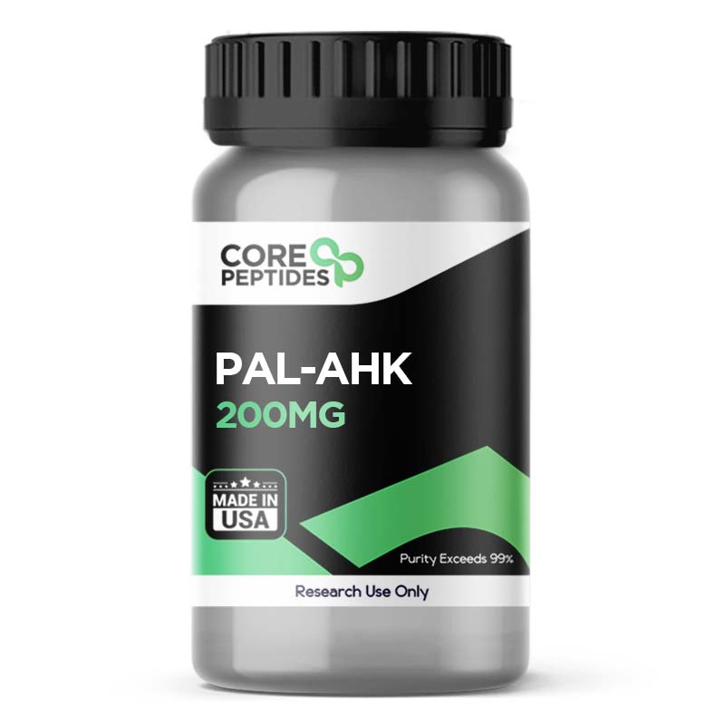
Pal-AHK (200mg)
Pal-AHK is a synthetic peptide composed of amino acids alanine, histidine, and lysine, with the addition of a palmitoyl group. Studies have only investigated the action of non-palmitoylated AHK. Still, Pal-AHK likely exerts similar impacts since adding palmitoyl to the AHK molecule is not likely to significantly alter the compound's mechanism of action. Palmitoylation is typically considered only to enhance a compound’s capacity for cell penetration, which may make it more capable of exerting action. Research suggests one key potential action of AHK and Pal-AHK may be to stimulate the production of collagen and elastin, two proteins considered essential within the extracellular matrix (ECM) of skin cells. Collagen is a major structural protein that lends strength and durability to the skin barrier, while elastin is considered responsible for supporting its elasticity. In addition to its potential for collagen and elastin production, Pal-AHK may also exhibit anti-oxidative action. Oxidative stress is considered to cause quantitative damage to cells and tissues. Antioxidants may help to neutralize free radicals, the main cause of oxidative stress. By scavenging these free radicals, antioxidants may prevent or reduce damage and environmental exposure biomarkers, including limiting wrinkle depth and length along the skin barrier. Studies have suggested that Pal-AHK may also have the potential to induce hair follicle growth due to anti-oxidative action and interaction with various molecules and growth factors. Examples include transforming growth factor-β1 (TGF-β1), Bcl-2/Bax ratio, vascular endothelial growth factor (VEGF), caspase-3, poly-(ADP-ribose) polymerase (PARP), and others. Chemical Makeup Molecular formula: C31H56N6O5 Molecular weight: 592.8 g/mol Other Known Titles: Palmitoyl AHK Research and Clinical Studies Pal-AHK Peptide and Antioxidation Tripeptide AHK was first developed from albumin hydrolysate.(2)(3) The addition of palmitoyl may improve the skin cell permeability of the peptide. Scientists report that the tripeptide may potentially reduce the expression of TGF-β1.(3) TGF-β1 is a molecule that supports the formation of new blood vessels and is considered to stimulate the production of extracellular matrix components, regulating the inflammatory response. Excessive activity may lead to chronic inflammation and tissue damage in certain contexts. Thus, reducing TGF-β1 levels may help to mitigate this action. The potential antioxidant action of Pal-AHK may be attributable to its unique amino acid composition.(4) Researchers suggest that the peptide “provides an increased dermal cell multiplication and viability to help to the production of collagen.” Researchers posit that this supposed action of AHK may directly impact the ECM through cell and protein production. Pal-AHK Peptide and Hair No research exists on Pal-AHK's impact on hair/fur growth. However, derivatives of AHK suggest that similarly structured compounds may promote the growth of hair follicles via the proliferation of dermal papilla cells (DPCs) and blocking their apoptosis.(5) The researchers suggested that AHK-analogs “stimulated the elongation of [...] hair follicles [...] and the proliferation of DPCs.” Pal-AHK may achieve similar potential by elevating the Bcl-2/Bax ratio and reducing negative growth factors such as cleaved caspase-3 and PARP. The Bcl-2/Bax ratio is considered important in regulating apoptosis (programmed cell death). Bcl-2 is classified an anti-apoptotic protein that may inhibit cell death, while Bax is a pro-apoptotic protein that may promote cell death. Thus, a higher Bcl-2/Bax ratio would indicate a predominance of Bcl-2, which may inhibit apoptosis and promote cell survival. The theoretical potential of Pal-AHK to promote hair growth by preventing dermal papilla cell apoptosis is a hypothesis founded on research that suggests the peptide’s involvement in reducing the levels of the cleaved forms of caspase-3 and PARP. Caspase-3 is considered to be a primary player in the process of apoptosis and is believed to contribute to the loss of dermal papilla cells when present in its cleaved, active form. When cleaved by caspase-3, PARP may facilitate cellular disassembly, a hallmark of apoptosis. Thus, inhibiting caspase-3 activation and reducing the cleaved forms of PARP as part of the same apoptotic pathway may plausibly contribute to decreased cell death in the hair follicles, potentially fostering an environment more conducive to hair retention and growth. Research also suggests this action possibly lowered apoptosis levels in these cell types under experimental observation.(6) Furthermore, studies report that Pal-AHK may increase the expression of vascular endothelial growth factor (VEGF), which, combined with reducing TGF-β1, may also help reduce shedding and promote hair growth.(7) VEGF is a protein that is considered to play an important role in angiogenesis. It is also thought to promote the growth of blood vessels around hair follicles, which may help supply nutrients and oxygen to the hair follicle and support growth. Pal-AHK Peptide and Skin Cells Although blocking TGF-β1 tends to have antiproliferative action, researchers suggest that Pal-AHK may stimulate the proliferation of skin cells and collagen synthesis via other pathways. The specific potential of Pal-AHK on the skin is yet to be investigated. Still, researchers have suggested that the main molecule, AHK, may yield positive action in preliminary laboratory research. One such potential is the viability and proliferation of dermal fibroblasts, key cells responsible for producing the essential proteins of the ECM, including collagen. Cell culture studies using normal dermal fibroblasts found that AHK appeared to increase cell growth and viability while potentially promoting the production of collagen type I.(8) This was determined by isolating the amount of collagen type I produced by fibroblasts in cell culture after incubation with various amounts of AHK. Results suggested that AHK may increase collagen type I production, with studies reporting a 300% increase compared to a control group. The findings suggest that AHK may potentially renew the extracellular matrix. Pal-AHK peptide is available for research and laboratory purposes only. Please review and adhere to our Terms and Conditions before ordering. References: Tsuge, N., Eikawa, Y., Nomura, Y., Yamamoto, M., & Sugisawa, K. (1991). The anti-oxidative activity of peptides prepared by enzymatic hydrolysis of egg-white albumin. Journal of the Agricultural Chemical Society of Japan. Shimura, H., Tanaka, R., Shimada, Y., Yamashiro, K., Hattori, N., & Urabe, T. (2017). Glycyl-alanyl-histidine protects PC12 cells against hydrogen peroxide toxicity. BMC biochemistry, 18(1), 14. https://doi.org/10.1186/s12858-017-0089-x Nirmal, B. (2018). Key MessAges. IADVL Textbook of Trichology, 163. Kecel-Gunduza, S., Kocb, E., Bicaka, B., Kokcub, Y., Ozela, A. E., & Akyuzc, S. (2020). IN SILICO ANALYSIS FOR CHARACTERIZING THE STRUCTURE AND BINDING PROPERTIES OF ALA-HIS-LYS (AHK) TRIPEPTIDE. The Online Journal of Science and Technology-July, 10(3). Pyo, H. K., Yoo, H. G., Won, C. H., Lee, S. H., Kang, Y. J., Eun, H. C., Cho, K. H., & Kim, K. H. (2007). The effect of tripeptide-copper complex on human hair growth in vitro. Archives of pharmacal research, 30(7), 834–839. https://doi.org/10.1007/BF02978833 Anastassakis, K. (2022). Copper Tripeptides Copper tripeptides. In Androgenetic Alopecia From A to Z: Vol. 2 Drugs, Herbs, Nutrition and Supplements (pp. 225-230). Cham: Springer International Publishing. Sadgrove, N. J., & Simmonds, M. S. J. (2021). Nutricosmetic products for healthy hair and dermal antiaging using “dual-acting” (2 for 1) plant-based peptides, hormones, and cannabinoids. FASEB bioAdvances, 3(8), 601–610. https://doi.org/10.1096/fba.2021-00022 Patt, L. M., & Procyte, A. (2009). Neova® DNA Repair Factor Nourishing Lotion Stimulates Collagen and Speeds Natural Repair Process. skin, 1, 2. Dr. MarinovDr. Marinov (MD, Ph.D.) is a researcher and chief assistant professor in Preventative Medicine & Public Health. Prior to his professorship, Dr. Marinov practiced preventative, evidence-based medicine with an emphasis on Nutrition and Dietetics. He is widely published in international peer-reviewed scientific journals and specializes in peptide therapy research.

Oxytocin (10mg)
Oxytocin is a naturally occurring cyclic peptide hormone composed of nine amino acids. Scientists consider the peptide hormone to be secreted by the pituitary gland and to act as a neurotransmitter within the brain.(1) The synthetic form of the peptide, Recombinant Oxytocin, is a cyclic nonapeptide developed to be analogous to the naturally occurring peptide.(2) The first isolation of the peptide was carried out in 1920, followed by its structure discovery in the 1950s. Several research studies were conducted to fully explore the peptide's action and characteristics. Scientists consider it to be created by the hypothalamus, and Oxytocin is then secreted and stored in the posterior pituitary gland, potentially released in the event of a specific stimulation.(3) Unlike other hormones, naturally occurring Oxytocin has been posited to work via a positive feedback mechanism. This means that the initial secretion of the hormone peptide may lead to further release of the peptide in higher concentrations and with higher intensity.(3) Both synthetic and natural forms appear to work through the same potential mechanisms. Considered to be especially pivotal in the process of gestation and labor, Oxytocin, once secreted into the system, may bind with the G-protein coupled receptors to potentially increase intracellular calcium levels. This calcium secretion may have various downstream impacts, such as inducing uterine contractions. Once the uterine contractions begin, they may stimulate further release of Oxytocin, leading to higher frequency and intensity of the contractions via a positive feedback mechanism.(3) The peptide appears to contribute to the contractions of myoepithelial cells found in the alveolar ducts of mammary glands. These contractions may stimulate milk ejection from alveolar ducts into the larger sinuses, thereby expelling milk. The positive feedback mechanism may also work here, with initial milk expulsion potentially stimulating increased Oxytocin circulation and continuous milk release.(3) Chemical Makeup Molecular Formula: C43H66N12O12S2 Molecular Weight: 1007.193 g/mol Other Known Titles: endopituitrina Research and Clinical Studies Oxytocin Peptide and Dopamine Signaling Oxytocin might play a role in shaping sexual behavior by possibly influencing the activity of dopamine within central nervous system regions associated with the rewards system, notably the ventral tegmental area (VTA) and the nucleus accumbens.(4) This influence may stem from projections of Oxytocin to the VTA, where the peptide may either augment dopamine release, or enhance the sensitivity of dopaminergic neurons. Such an increase in dopaminergic activity may, in turn, potentially boost sexual motivation and the perception of reward, possibly aiding the anticipatory aspects of mating and copulatory behavior. The hypothesis suggests that Oxytocin may directly interact with dopaminergic neurons within the VTA by activating specific oxytocin receptors, initiating a sequence of events that culminates in heightened dopamine release within the nucleus accumbens. This sequence may include the stimulation of nitric oxide synthesis within the VTA, hinting at an intricate interplay involving Oxytocin, dopamine, and nitric oxide. Additionally, there is the potential for Oxytocin to exert an indirect action on dopaminergic activity across other cerebral locales, including the hippocampus and amygdala, adding further depth to its complex role. Such indirect influences might modulate the activity of either glutamatergic or GABAergic neurons, which may, in turn, influence dopaminergic neurons located within the VTA and nucleus accumbens.(4) Oxytocin Peptide and Neuroplasticity Researchers are actively exploring Oxytocin's impact on developing or maintaining neuroplasticity. The peptide is posited to engage with G-protein coupled receptors, which may increase intracellular calcium levels.(5) This elevation in calcium is thought to play a role in neuronal excitability and synaptic modulation, affecting the transmission of signals across neurons. Oxytocin's potential influence in the brain may extend to neurogenesis and synaptic plasticity, which may impact the intricate dance of neural circuit formation and function. The presence of Oxytocin receptors across various neural cell types hints at the peptide's broad potential impact, possibly modulating the behavior of neural progenitor cells and influencing the fate of these cells. This action might stretch into early brain development, where Oxytocin's presence might subtly shape the emerging neural circuits by modulating neurotransmitter dynamics, including those of glutamate and GABA, which serve as the brain's primary excitatory and inhibitory signals, respectively. At the synaptic level, Oxytocin's actions appear to diverge, potentially enhancing neurotransmitter release in some contexts, while diminishing it in others. This dual action may reflect Oxytocin's potential to modulate the balance between excitation and inhibition within the neural circuits, a balance considered crucial for maintaining the functional integrity of the brain. Such modulation may manifest through alterations in the release of neurotransmitters or changes in membranes, indirectly influencing neuronal excitability and the flow of neural information. The interactions of Oxytocin with glial cells, such as astrocytes, further complicate its role in neurotransmitter dynamics, suggesting a broader regulatory influence that extends beyond the neurons to the supportive environment that nurtures and maintains synaptic connections. This interaction may influence the synaptic plasticity and overall function of neural circuits, offering a glimpse into the complex regulatory roles Oxytocin might play in the neural ecosystem.(5) Oxytocin Peptide and Labor Induction The main aim of this clinical study(6) was to determine the rate of cesarean delivery in pregnant females after removing Oxytocin presence once active labor starts (5 cm cervical dilation) compared to pregnant females where Oxytocin was given until the maintenance level as determined in clinical trial study protocol. A randomized study was conducted on 252 female subjects between 18 to 50 years. The subjects were divided into two groups, with 127 subjects (Group A) continuously given the peptide per study protocol even after the subjects reached active labor, and 125 subjects (Group B) given Oxytocin, discontinued after active labor induction. After a 24-72 hour study period, there were 32 reported cesarean deliveries in Group A, with 10 infants exhibiting one or more abnormalities that warranted further observation. In contrast, there were 24 reported cesarean deliveries in Group B, with 9 infants exhibiting abnormalities. Oxytocin Peptide and Lactation The main goal of this clinical study(7) was to evaluate the potential action of peptide supplementation during the course of breastfeeding. A retrospective study(7) was conducted where 100 pregnant females given Oxytocin during labor were compared with 100 pregnant females not exposed to Oxytocin. Following the study result analysis, the researchers reported that the new mothers exposed to Oxytocin exhibited apparently impaired breastfeeding for the first hour. During the first three months after delivery, subjects from both groups (27% exposed to Oxytocin and 14% without) were not apparently able to breastfeed. Following three months, breastfeeding was reported to significantly increase in the Oxytocin-exposed subjects. The reason suggested by researchers for impaired breastfeeding during the first three months was the high pre-gestational body mass index in the test subjects. Oxytocin Peptide and Vasodilation Vasodilation is the dilation of blood vessels, which is considered to be a mechanism to increase blood flow. The main purpose of this study(8) was to evaluate the potential of Oxytocin on cardiac function and tone in the heart muscles. The study observed a set of pregnant females in the first trimester, divided into two groups, one given Oxytocin and the other a placebo. Heart rate and blood pressure were recorded. The results suggested that the peptide may have exhibited vasodilatory properties on the small and outlying arteries and elevated left ventricular ejection time. Oxytocin Peptide and Autism The main aim of this clinical study(9) was to evaluate what action, if any, the peptide may exert on research models of autism. Autism is considered to be a psychiatric disorder related to poor brain development impacting how the subject socializes and communicates. A total of 59 autistic males, aged 6 to 11 years, were examined in this study. Out of the 59 test subjects, 29 were autistic, and the rest were considered control subjects by the researchers. The plasma levels of Oxytocin were measured in this study, and the researchers reported that autistic subjects exhibited lower levels of peptide hormone than the control subjects. Once the levels of Oxytocin were elevated as part of the study, the VABS score (analytic behavior score) of autistic subjects appeared to improve. Oxytocin peptide is available for research and laboratory purposes only. Please review and adhere to our Terms and Conditions before ordering. References: National Center for Biotechnology Information. "PubChem Compound Summary for CID 439302, Oxytocin" PubChem Recombinant Oxytocin (Code C724). https://ncit.nci.nih.gov/ncitbrowser/ConceptReport.jsp?dictionary=NCI_Thesaurus&ns=NCI_Thesaurus&code=C724 Osilla EV, Sharma S. Oxytocin. [Updated 2021 Jul 27]. In: StatPearls [Internet]. Treasure Island (FL): StatPearls Publishing; 2021 Jan. https://www.ncbi.nlm.nih.gov/books/NBK507848/ Melis, Maria Rosaria, and Antonio Argiolas. “Oxytocin, Erectile Function and Sexual Behavior: Last Discoveries and Possible Advances.” International journal of molecular sciences vol. 22,19 10376. 26 Sep. 2021, doi:10.3390/ijms221910376 Bakos, Jan et al. “Molecular Mechanisms of Oxytocin Signaling at the Synaptic Connection.” Neural plasticity vol. 2018 4864107. 2 Jul. 2018, doi:10.1155/2018/4864107 Diven, Liany C et al. “Oxytocin discontinuation during active labor in women who undergo labor induction.” American journal of obstetrics and gynecology vol. 207,6 (2012): 471.e1-8. doi:10.1016/j.ajog.2012.08.035 Gomes M, Trocado V, Carlos-Alves M, Arteiro D, Pinheiro P. Intrapartum synthetic oxytocin and breastfeeding: a retrospective cohort study. J Obstet Gynaecol. 2018 Aug;38(6):745-749. Epub 2018 Mar 9. https://pubmed.ncbi.nlm.nih.gov/29523035/ Rabow, S., Hjorth, U., Schönbeck, S. et al. Effects of oxytocin and anaesthesia on vascular tone in pregnant women: a randomized, double-blind placebo-controlled study using non-invasive pulse wave analysis. BMC Pregnancy Childbirth 18, 453 (2018). Cochran, D. M., Fallon, D., Hill, M., & Frazier, J. A. (2013). The role of oxytocin in psychiatric disorders: a review of biological and therapeutic research findings. Harvard review of psychiatry, 21(5), 219–247. https://doi.org/10.1097/HRP.0b013e3182a75b7d Dr. MarinovDr. Marinov (MD, Ph.D.) is a researcher and chief assistant professor in Preventative Medicine & Public Health. Prior to his professorship, Dr. Marinov practiced preventative, evidence-based medicine with an emphasis on Nutrition and Dietetics. He is widely published in international peer-reviewed scientific journals and specializes in peptide therapy research.