Medicines & Treatments

GHRP-2 (5mg / 10mg)
GHRP-2 is a synthetic peptide made of five amino acids (pentapeptide) analogous to the endogenous neurotransmitter met-enkephalin.(1) Yet, the peptide appears to lack any neurotransmitter-like properties and instead appears to activate the receptors for the hormone ghrelin. Ghrelin is a naturally occurring hormone that is posited to regulate food intake.(11) Growth hormone-releasing peptide GHRP-2 may induce the secretion of growth hormone (GH) by apparently activating the ghrelin receptors on the pituitary gland, also termed as growth hormone secretagogue receptors (GHS-Rs). Overview of GHRP-2 Mechanisms Bovine studies have suggested that GHRP-2 peptide exhibits multifaceted impacts, but its main mechanism of action appears to involve the GHS-Rs. These receptors are naturally triggered by ghrelin and may be found in various parts of the nervous system and other tissues. In the nervous system, they are located in the hypothalamus and the pituitary, among other structures. When GHRP-2 appears to engage with GHS-Rs, it's believed that a structural transformation might be triggered upon attachment, possibly initiating intracellular signaling networks primarily mediated through G-proteins. The release of Gαq/11, a component of the G-proteins, might commence, potentially setting off subsequent signaling sequences. Phospholipase C (PLC) may split phosphatidylinositol 4,5-bisphosphate (PIP2) into secondary signaling molecules, IP3 and DAG (diacylglycerol). IP3 is thought to prompt the release of calcium ions. In contrast, DAG may activate protein kinase C (PKC), potentially heightening the signaling pathway and possibly facilitating the secretion of growth hormone from pituitary cells. Moreover, this process may also involve the activation of cyclic AMP (cAMP), which plays a crucial role in cellular signaling. Increasing cAMP levels may further enhance the signaling cascade, potentially boosting GH synthesis.(13)(14) Nevertheless, GHRP-2 appears to induce desensitization at these receptors immediately after exposure, potentially reducing sensitivity lasting a complete four hours before reversal.(5) By apparently activating the GHS-Rs on other parts of the nervous system, GHRP-2 might trigger a series of cellular events leading to the enhanced synthesis of hunger-promoting neuropeptides, namely Neuropeptide Y (NPY) and Agouti-related peptide (AgRP). These peptides are thought to be integral to the regulation of energy balance and the control of appetite. At the same time, GHRP-2 is believed to possibly inhibit the secretion of the appetite-reducing hormone, melanocyte-stimulating hormone (α-MSH), thus shifting the equilibrium towards increased hunger and encouraging dietary intake. Moreover, GHRP-2 might affect the mesolimbic reward system, a neural pathway associated with regulating the craving for food, via the activation of GHSR-1a receptors. This action might theoretically heighten the drive for food consumption, potentially through the activation of cyclic adenosine monophosphate (cAMP) pathways, further implicating GHRP-2 in the modulation of feeding behavior and reward-based eating. Chemical Makeup Molecular Formula: C45H55N9O6 Molecular Weight: 817.97 g/mol Other Known Titles: pralmorelin Research and Clinical Studies GHRP-2 Peptide and Growth Hormone Synthesis This study(7) was conducted to understand the action of two synthetic GHRPs, GHRP-2 and Hexarelin (HEX), on growth hormone, prolactin, adrenocorticotropic hormone (ACTH), and cortisol concentrations. Two groups of various stages of mature development were evaluated, and both groups experienced a higher GH increase compared to the physiological increase induced by native growth hormone-releasing hormone (GHRH). Unfortunately, GHRP-2 also may induce an apparent increase in the levels of ACTH and cortisol. Further studies have reported that exposure to GHRP-2 in laboratory settings may induce a significant increase in peak GH levels, mean pulsatile GH secretion by the anterior pituitary gland cells, and may potentially upregulate mediators of GH’s anabolic actions such as insulin-like growth factor-1 (IGF-1): In one trial, GHRP-2 was suggested to induce up to 181-fold higher spike in GH production by the anterior pituitary gland cells.(17) Another experiment posited that the peptide may induce a 47-fold increase in mean 2.5-hour pulsatile GH secretion by the anterior pituitary gland cells compared to placebo.(18) A third trial on prolonged GHRP-2 exposure reported that the peptide may have induced an apparent increase in GH levels between 3-fold and 5-fold. Moreover, IGF-1 levels apparently increased from an average of 100mcg/l at baseline to a plateau value of around 180mcg/l.(19) GHRP-2 Peptide and Appetite In one study,(2) two groups were evaluated, one following GHRP-2 exposure and the other with saline. They were then taken to a buffet meal to measure their food intake. The researchers reported that the GHRP-2 models ate an average of 36% more than the saline group, with each model reportedly exhibiting increased food intake when measured against their respective body weight. The researchers reported that the energy intake per kilogram of body weight increased up to 136.0±13.0 kJ/kg vs 101.3±10.5 kJ/kg for the placebo group. Furthermore, the GH levels also presented significant incremental increases in GHRP-2 models compared to saline. The levels of the hormone measured as “area under the curve” (AUC) increased up to 5550±1090 μg/L/240 min vs. 412±161 μg/L/240 min. The researchers concluded that “GHRP-2, like ghrelin, increases food intake, suggesting that GHRP-2 [may be] a valuable tool for investigating ghrelin effects on eating behavior.” GHRP-2 Peptide and Muscle Tissues Murine models of thermal injury have suggested that GHRP-2 may significantly decrease in proinflammatory markers such as IL-6 and E3 ubiquitin ligases (MuRF-1 and MAFbx), which are associated with muscle wasting in critical conditions. The authors also posited that the peptide may directly reduce total muscle protein breakdown in the experimental models, therefore suggesting a muscle-sparing action GHRP-2.(20) Case studies have also suggested that the peptide may induce muscle and weight gain.(21) GHRP-2 Peptide and Antioxidative Actions Studies indicate that GHRP-2 may display antioxidative potential, with researchers noting its possible affinity for CD36, a receptor thought to play a key role in capturing oxidized low-density lipoprotein (OxLDL). This interaction could potentially limit the cellular absorption of OxLDL, which is reputed to contribute to atherogenic processes. In experiments conducted on mouse models lacking the ApoE gene (ApoE(-/-)), GHRP-2 exposure over 12 weeks seemed to elevate circulating levels of IGF-I, with increases observed to be between 1.2 to 1.6 times the initial measurements. Additionally, a reduction of approximately 66% in circulating interferon-gamma levels was reported. Although GHRP-2's introduction did not appear to significantly alter the extent of atherosclerotic plaque coverage, it is suggested to have reduced superoxide production in the aorta, as indicated by dihydroethidium staining. Furthermore, GHRP-2 is reported to have notably decreased, by about 92%, the aortic gene expression of 12/15-lipoxygenase, and also diminished the aortic expressions of interferon-gamma and macrophage migration inhibitory factors. Observations in cultured aortic smooth muscle cells suggested that GHRP-2 may counter the production of peroxides triggered by OxLDL, prevent the suppression of the IGF-I receptor, and potentially block apoptosis. In macrophages exposed to OxLDL, GHRP-2 is hypothesized to reduce lipid accumulation, further illustrating its potential antioxidative and protective roles against proatherogenic agents.(15) GHRP-2 Peptide and Inflammation To further elucidate the potential action of GHRP-2 on oxidative stress and inflammation, a study on murine models with artificially induced acute lung injury suggested that exposure to GHRP-2 may reduce lung edema, neutrophil infiltration, and levels of pro-inflammatory cytokines. The peptide was also posited to have suppressed nuclear factor-kappaB activation, a major mediator of inflammation. The cascades that follow its activation often lead to tissue damage.(16) GHRP-2 Peptide and GH Deficiency The most common diagnostic tool for GH deficiency is the insulin tolerance test (ITT), which may cause contraindications. A study(8) aimed to explore the potential of GHRP-2 as a diagnostic resource for GH deficiency. The study evaluated research models first tested via ITT. Of these, the study indicated 77 models exhibited normal insulin tolerance and 58 exhibited GH peak levels of less than three. Post overnight fasting, all research models were presented GHRP-2. After 2 hours, their blood samples were collected and tested. Upon analysis, the researchers reported that the GH levels peaked after one hour of GHRP-2 in all cases. These results were reportedly reproducible upon repetition of the tests. An additional study(9) was conducted to research the diagnostic properties of GHRP-2 on still-developing research models of GH deficiency (GHD) in comparison to a conventionally-used compound. Research models of GHD were enrolled in this study, presented with at least one conventional compound, and later presented with GHRH and GHRP-2. All models were first given GHRP-2. When measured, the serum GH levels appeared to have significantly increased. Models who demonstrated a robust response to the peptide were then presented with GHRP-2 and GHRH in combination. All models exhibited an apparent positive response in their GH levels, according to the researchers. Combination Studies with TRH and GnRH This study(10) was conducted on research models of prolonged hypo-somatotropism, hypogonadism, or hypothyroid complications to evaluate the action of compounds of GHRP 2, Thyrotropin-releasing hormone (TRH), and Gonadotropin-releasing hormone (GnRH) with GHRP-2 alone and with GHRP-2 and TRH in combination. Over 5 days, one group was presented with a placebo, one group with GHRP-2 every hour, another group with GHRP-2 + TRH every hour, and the remaining group with GHRP-2 + TRH + GnRH every 90 minutes. Serum samples were collected on the first and last night of the study. After the results were analyzed, researchers suggested that the combination of GHRP-2 + GnRH + TRH induced the greatest apparent activation of growth hormones along thyroid stimulating hormone and luteinizing hormone axes, along with other possible metabolic effects. These actions were reported to be absent with GHRP-2 solo presentation and only partially seen with GHRP-2 and TRH combination. GHRP 2 peptide is available for research and laboratory purposes only. Please review and adhere to our Terms and Conditions before ordering. References: Garcia JM, Merriam GR, Kargi AY. Growth Hormone in Aging. In: Feingold KR, Anawalt B, Boyce A, et al., editors. Endotext. South Dartmouth (MA): MDText.com https://www.ncbi.nlm.nih.gov/books/NBK279163/ Laferrère, Blandine et al. “Growth hormone releasing peptide-2 (GHRP-2), like ghrelin, increases food intake in healthy men.” The Journal of clinical endocrinology and metabolism vol. 90,2 (2005): 611-4. https://www.ncbi.nlm.nih.gov/pmc/articles/PMC2824650/ Bowers CY. History to the discovery of ghrelin. Methods Enzymol. 2012;514:3-32. doi: 10.1016/B978-0-12-381272-8.00001-5. PMID: 22975043. https://pubmed.ncbi.nlm.nih.gov/22975043/ Roh SG, He ML, Matsunaga N, Hidaka S, Hidari H. Mechanisms of action of growth hormone-releasing peptide-2 in bovine pituitary cells. J Anim Sci. 1997 Oct;75(10):2744-8. doi: 10.2527/1997.75102744x. PMID: 9331879. https://pubmed.ncbi.nlm.nih.gov/9331879/ Asad Rahim, Stephen M. Shalet, in Growth Hormone Secretagogues, 1999. Does desensitization to growth hormone secretagogues occur? https://www.sciencedirect.com/topics/medicine-and-dentistry/pralmorelin Furuta S, Shimada O, Doi N, Ukai K, Nakagawa T, Watanabe J, Imaizumi M. General pharmacology of KP-102 (GHRP-2), a potent growth hormone-releasing peptide. Arzneimittelforschung. 2004;54(12):868-80. doi: 10.1055/s-0031-1297042. PMID: 15646371. https://pubmed.ncbi.nlm.nih.gov/15646371/ Emanuela Arvat, Lidia Di Vito, Barbara Maccagno, Fabio Broglio, Muni F Boghen, Romano Deghenghi, Franco Camanni, Ezio Ghigo, Effects of GHRP-2 and Hexarelin, Two Synthetic GH-Releasing Peptides, on GH, Prolactin, ACTH and Cortisol Levels in Man. Comparison with the Effects of GHRH, TRH and hCRH, Peptides, Volume 18, Issue 6, 1997, Pages 885-891, ISSN 0196-9781, https://doi.org/10.1016/S0196-9781(97)00016-8 Chihara K, Shimatsu A, Hizuka N, Tanaka T, Seino Y, Katofor Y; KP-102 Study Group. A simple diagnostic test using GH-releasing peptide-2 in adult GH deficiency. Eur J Endocrinol. 2007 Jul;157(1):19-27. doi: 10.1530/EJE-07-0066. https://pubmed.ncbi.nlm.nih.gov/17609397/ Pihoker C, Middleton R, Reynolds GA, Bowers CY, Badger TM. Diagnostic studies with intravenous and intranasal growth hormone-releasing peptide-2 in children of short stature. J Clin Endocrinol Metab. 1995 Oct;80(10):2987-92. https://pubmed.ncbi.nlm.nih.gov/7559885/ Van den Berghe G, Baxter RC, Weekers F, Wouters P, Bowers CY, Iranmanesh A, Veldhuis JD, Bouillon R. The combined administration of GH-releasing peptide-2 (GHRP-2), TRH and GnRH to men with prolonged critical illness evokes superior endocrine and metabolic effects compared to treatment with GHRP-2 alone. Clin Endocrinol (Oxf). 2002 May;56(5):655-69. https://pubmed.ncbi.nlm.nih.gov/12030918/ GHRP 2, GPA 748, Growth Hormone-Releasing Peptide 2, KP-102 D, KP-102 LN, KP-102D, KP-102 LN. https://link.springer.com/article/10.2165/00126839-200405040-00011# Phung LT, Sasaki A, Lee HG, Vega RA, Matsunaga N, Hidaka S, Kuwayama H, Hidari H. Effects of the administration of growth hormone-releasing peptide-2 (GHRP-2) orally by gavage and in feed on growth hormone release in swine. Domest Anim Endocrinol. 2001 Jan;20(1):9-19. https://pubmed.ncbi.nlm.nih.gov/11164330/ Yin, Y., Li, Y., & Zhang, W. (2014). The growth hormone secretagogue receptor: its intracellular signaling and regulation. International journal of molecular sciences, 15(3), 4837–4855. https://doi.org/10.3390/ijms15034837 Sinha, D. K., Balasubramanian, A., Tatem, A. J., Rivera-Mirabal, J., Yu, J., Kovac, J., Pastuszak, A. W., & Lipshultz, L. I. (2020). Beyond the androgen receptor: the role of growth hormone secretagogues in the modern management of body composition in hypogonadal males. Translational andrology and urology, 9(Suppl 2), S149–S159. https://doi.org/10.21037/tau.2019.11.30 Titterington JS, Sukhanov S, Higashi Y, Vaughn C, Bowers C, Delafontaine P. Growth hormone-releasing peptide-2 suppresses vascular oxidative stress in ApoE-/- mice but does not reduce atherosclerosis. Endocrinology. 2009 Dec;150(12):5478-87. doi: 10.1210/en.2009-0283. Epub 2009 Oct 9. PMID: 19819949; PMCID: PMC2795722. Li, G., Li, J., Zhou, Q., Song, X., Liang, H., & Huang, L. (2010). Growth hormone releasing peptide-2, a ghrelin agonist, attenuates lipopolysaccharide-induced acute lung injury in rats. The Tohoku journal of experimental medicine, 222(1), 7–13. https://doi.org/10.1620/tjem.222.7 Veldhuis, J. D., Keenan, D. M., Bailey, J. N., Adeniji, A. M., Miles, J. M., & Bowers, C. Y. (2009). Novel relationships of age, visceral adiposity, insulin-like growth factor (IGF)-I and IGF binding protein concentrations to growth hormone (GH) releasing-hormone and GH releasing-peptide efficacies in men during experimental hypogonadal clamp. The Journal of clinical endocrinology and metabolism, 94(6), 2137–2143. https://doi.org/10.1210/jc.2009-0136 Veldhuis, J. D., & Keenan, D. M. (2008). Secretagogues govern GH secretory-burst waveform and mass in healthy eugonadal and short-term hypogonadal men. European journal of endocrinology, 159(5), 547–554. https://doi.org/10.1530/EJE-08-0414 Bowers, C. Y., Granda, R., Mohan, S., Kuipers, J., Baylink, D., & Veldhuis, J. D. (2004). Sustained elevation of pulsatile growth hormone (GH) secretion and insulin-like growth factor I (IGF-I), IGF-binding protein-3 (IGFBP-3), and IGFBP-5 concentrations during 30-day continuous subcutaneous infusion of GH-releasing peptide-2 in older men and women. The Journal of clinical endocrinology and metabolism, 89(5), 2290–2300. https://doi.org/10.1210/jc.2003-031799 Sheriff, S., Joshi, R., Friend, L. A., James, J. H., & Balasubramaniam, A. (2009). Ghrelin receptor agonist, GHRP-2, attenuates burn injury-induced MuRF-1 and MAFbx expression and muscle proteolysis in rats. Peptides, 30(10), 1909–1913. https://doi.org/10.1016/j.peptides.2009.06.029 Sigalos, J. T., & Pastuszak, A. W. (2018). The Safety and Efficacy of Growth Hormone Secretagogues. Sexual medicine reviews, 6(1), 45–53. https://doi.org/10.1016/j.sxmr.2017.02.004 Dr. MarinovDr. Marinov (MD, Ph.D.) is a researcher and chief assistant professor in Preventative Medicine & Public Health. Prior to his professorship, Dr. Marinov practiced preventative, evidence-based medicine with an emphasis on Nutrition and Dietetics. He is widely published in international peer-reviewed scientific journals and specializes in peptide therapy research.

LL-37 (5mg)
LL-37, also known as Cathelicidin, is a cationic peptide composed of 37 amino acids and is primarily found in neutrophils.(1) The peptide appears to be produced by the extracellular breakdown of the hCAP18 proteins caused by protease enzymes. Researched for its potential antimicrobial characteristics, the peptide appears to form agglomerates and lipid bilayers, which may prevent it from easily degrading and protect it from enzymatic action.(1) Overview Antimicrobial peptides are structured with the potential to fight against bacteria, fungi, and some virus strains. These peptides may interact with targets in a non-specific fashion, which supports researchers' belief that once the peptide is introduced, pathogens are unable to develop resistance against these peptides.(2) LL-37 is a α-helical peptide that scientists believe is required to maintain immunity against all microbes.(3) To understand the functioning of the peptide, a peptide model was created as part of a study(4) based on the assumption that the peptide might interact directly with the bacterial membrane. This study suggested that the peptide first interacts with the lipids on the bacterial membrane via electrostatic characteristics, followed by lateral diffusion and consequent assembly of the peptide on the membrane. This potential interaction may lead to membrane interference and degradation of the bacterial cell. Several other studies hypothesize how the peptide interacts with microbial membranes, including pore formation on the membrane(3)(5) and extreme membrane disruption caused by the peptide and lipid complexes.(6) These studies universally suggest that peptides have the potential to interact with the microbial membrane, leading to membrane breakdown. Chemical Makeup Molecular Formula: C205H340N50O53 Molecular Weight: 4493.34 g/mol Other Known Titles: CAP-18 Research and Clinical Studies LL-37 Peptide and Inflammatory Response The main aim of this study(7) was to determine the inflammatory potential of this peptide. Tissue culture was used, half without alteration and the other half with added U1 RNA. U1 RNA is a non-coding RNA released upon tissue injury. LL-37 peptide was then added to both cultures. Upon genetic analysis, it was suggested by the researchers that the culture that was given both U1 RNA and LL-37 peptide stimulated a reportedly significant response towards epidermal inflammation and defense response. The study proposes that the peptide might potentially enhance the immune system's response to damaged cells by influencing how self-nucleic acids (DNA and RNA) are recognized. This recognition is apparently facilitated when the peptide interacts with specific cellular receptors, including scavenger receptors (SRs), which may lead to clathrin-dependent endocytosis. This process appears critical for the subsequent activation of inflammatory pathways within the cells. Moreover, the study indicates that LL-37 might enable the binding of dsRNA (double-stranded RNA) to these scavenger receptors, which in turn might lead to a series of signaling events culminating in cytokine expression. Notably, the interaction between LL37 and scavenger receptors such as SR-A6 and SR-B1 may be essential for this process, as blocking these receptors with a competitive inhibitor like fucoidan or silencing their expression significantly reduced cytokine production. Another interesting aspect of the study is the hypothesis that LL-37 may modulate the immune system by potentially altering how intracellular signaling pathways, such as those involving Toll-like receptors (TLR) and the interferon regulatory factors, and may be activated in response to foreign nucleic acids. As detailed in the study, the involvement of clathrin-mediated endocytosis suggests that LL-37 may help orchestrate the entry of these immune-modulating molecules into cells, which is a vital step for triggering an immune response. LL-37 Peptide and Autoimmunity Models The main aim of this study(1) was to understand the role of LL-37 in models of autoimmunity such as psoriasis. This disease pathogenesis study suggested that endogenous peptide may form complex DNA, increasing interferon mechanisms and more inflammatory responses. This study theorized that LL-37 may be favorable for tissue and wound injury; however, in some cases, LL-37 levels appeared to indicate psoriasis presence. In fact, it might potentially exert anti-apoptotic actions on keratinocytes, which might be linked to the observed cellular proliferation in psoriatic lesions. LL-37 is a naturally occurring antimicrobial peptide that forms part of the immune system, playing various roles in immune responses. This action may potentially contribute to developing the thick, scaly skin that is a hallmark of psoriasis. While LL-37 has been implicated in promoting inflammation via type I interferon (IFN) pathways, it appears to simultaneously offer a protective action against the activation of the AIM2 inflammasome by cytosolic double-stranded DNA (dsDNA). Cytosolic dsDNA often triggers immune responses that may lead to inflammation. In cases where LL-37 forms complexes with DNA, these complexes seemingly do not promote the production of interleukin-1β (IL-1β), a pro-inflammatory cytokine, nor activate the inflammasome. This implies that LL37 might protect keratinocytes from the inflammatory responses typically triggered by the AIM2 inflammasome in the presence of dsDNA. This dual potential of LL-37 underscores its complex involvement in immune regulation and inflammatory processes. LL-37 Peptide and Arthritis The main objective of this study(1)(8) was to evaluate the potential of LL-37 in arthritic joints. A group of rats were used in this study,(8) with one control group and one group experimentally induced with rheumatoid arthritis. Upon inducing the condition, researchers reported an apparent increased regulation of rCRAMP, the rat analog of LL-37 peptide, in inflammatory cells. Researchers suggested that LL-37 peptide might further induce apoptosis of osteoblasts, thereby leading to decreased bone formation in the joints. This study suggested that increased LL-37 levels are characteristic of joint aches and arthritis and may potentially be used for diagnostic purposes. Besides these, study(1) has suggested that LL-37 elevation is seen in other inflammatory circumstances, such as arteriosclerosis. Scientists report that LL-37 activation and the consequent upregulation of interferons are characteristic of arteriosclerosis-induced cells. The researchers of this study suggested that the peptide may have potential as an immunomodulatory agent. LL-37 Peptide and Tissue Repair In this study,(9) mice presented with an anti-inflammatory compound were then presented with LL-37 to study the potential of this peptide on angiogenesis and wound healing. The researchers suggested that the peptide mice exhibited an apparent increase in vascularization and skin cell formation. This study suggested that LL-37 has the potential to induce endothelial skin cell proliferation and formation of tubule-like structures, which are both required in angiogenesis mechanisms. Further, the study indicates that LL37 possibly counteracts the activation of macrophages triggered by lipopolysaccharide (LPS), a component known to provoke immune responses. Furthermore, it seems to foster endothelial cell behaviors essential for wound healing, such as proliferation, migration, and the formation of tubule-like structures, all indicative of angiogenesis. Experiments on catabolism-induced murine models, utilizing both synthetic and recombinant forms of LL-37, suggest that exposure to the peptide may enhance vascularization and re-epithelialization. These observations collectively lead to the hypothesis that the peptide might be crucial in promoting wound regeneration, potentially through its actions on vascularization. LL-37 Peptide and Cancer Cells Studies(10) are ongoing to explore the potential of the peptide in cancer cell development. These studies have suggested that the peptide may inhibit gastric cancer cell proliferation by activating the bone morphogenetic protein signaling system. The main aim of this research was to consider the potential of LL-37 as an immunotherapeutic agent or consider the potential of LL-37 peptide as an adjuvant in eliminating cancer cells from the host system. CpG oligodeoxynucleotides are widely considered to be immunotherapeutic compounds as they appear to promote the tumor-suppressing activity. When presented with LL-37, researchers reported that the peptide LL-37 appeared to increase the CpG oligodeoxynucleotides sensitivity in lymphocytes. LL-37 Peptide and GI Tract LL-37 may potentially impact ailments associated with the gastrointestinal (GI) tract. There appears to be an increased expression of LL-37 in research models of gastrointestinal ulcers. This upregulation might be mediated through the activation of Toll-like receptor 3 (TLR-3) by its ligand, polyinosinic-polycytidylic acid (poly(I)). Poly(I) stimulation possibly enhances LL-37 expression by triggering intracellular signaling cascades involving proteins such as Toll/IL-1R domain-containing adaptor-inducing interferon (TRIF), tumor necrosis factor receptor-associated factor 6 (TRAF6), and transforming growth factor β-activated kinase 1 (TAK1). Owing to its antimicrobial potential, LL-37 may be capable of protecting GI mucosa from microbial damage.(12) The protective potential of this peptide in the gastrointestinal tract may involve its interaction with lipopolysaccharide (LPS), a component of bacterial cell walls. LL-37 may be able to suppress LPS-induced secretion of pro-inflammatory cytokines such as interleukin-6 (IL-6) and IL-8 in colonic subepithelial myofibroblasts (SEMFs), which possibly contributes to a protective action by moderating local inflammation. LL-37 peptide is available for research and laboratory purposes only. Please review and adhere to our Terms and Conditions before ordering. References: Kahlenberg, J Michelle, and Mariana J Kaplan. “Little peptide, big effects: the role of LL-37 in inflammation and autoimmune disease.” Journal of immunology (Baltimore, Md. : 1950) vol. 191,10 (2013): 4895-901. https://www.ncbi.nlm.nih.gov/pmc/articles/PMC3836506/ Seil, M., Nagant, C., Dehaye, J. P., Vandenbranden, M., & Lensink, M. F. (2010). Spotlight on Human LL-37, an Immunomodulatory Peptide with Promising Cell-Penetrating Properties. Pharmaceuticals, 3(11), 3435–3460. h https://www.ncbi.nlm.nih.gov/pmc/articles/PMC4034075/ Zeth, Kornelius, and Enea Sancho-Vaello. “The Human Antimicrobial Peptides Dermcidin and LL-37 Show Novel Distinct Pathways in Membrane Interactions.” Frontiers in chemistry vol. 5 86. 7 Nov. 2017. https://www.ncbi.nlm.nih.gov/pmc/articles/PMC5681987/ Brogden KA. Antimicrobial peptides: pore formers or metabolic inhibitors in bacteria? Nat Rev Microbiol. 2005 Mar;3(3):238-50. https://pubmed.ncbi.nlm.nih.gov/15703760/ Ludtke SJ, He K, Heller WT, Harroun TA, Yang L, Huang HW. Membrane pores induced by magainin. Biochemistry. 1996 Oct 29;35(43):13723-8. https://pubmed.ncbi.nlm.nih.gov/8901513/ Bechinger B, Lohner K. Detergent-like actions of linear amphipathic cationic antimicrobial peptides. Biochim Biophys Acta. 2006 Sep;1758(9):1529-39. https://pubmed.ncbi.nlm.nih.gov/16928357/ Takahashi, T., Kulkarni, N.N., Lee, E.Y. et al. Cathelicidin promotes inflammation by enabling binding of self-RNA to cell surface scavenger receptors. Sci Rep 8, 4032 (2018). https://doi.org/10.1038/s41598-018-22409-3 Hoffmann MH, Bruns H, Bäckdahl L, Neregård P, Niederreiter B, Herrmann M, Catrina AI, Agerberth B, Holmdahl R. The cathelicidins LL-37 and rCRAMP are associated with pathogenic events of arthritis in humans and rats. Ann Rheum Dis. 2013 Jul;72(7): https://pubmed.ncbi.nlm.nih.gov/23172753/ Ramos R, Silva JP, Rodrigues AC, Costa R, Guardão L, Schmitt F, Soares R, Vilanova M, Domingues L, Gama M. Wound healing activity of the human antimicrobial peptide LL37. Peptides. 2011 Jul;32(7):1469-76. doi: 10.1016/j.peptides.2011.06.005. Epub 2011 Jun 13. https://pubmed.ncbi.nlm.nih.gov/21693141/ Wu, W. K., Wang, G., Coffelt, S. B., Betancourt, A. M., Lee, C. W., Fan, D., Wu, K., Yu, J., Sung, J. J., & Cho, C. H. (2010). Emerging roles of the host defense peptide LL-37 in human cancer and its potential therapeutic applications. International journal of cancer, 127(8), 1741–1747. https://www.ncbi.nlm.nih.gov/pmc/articles/PMC2930073/ Wang, C., Wang, S., Li, D., Chen, P., Han, S., Zhao, G., Chen, Y., Zhao, J., Xiong, J., Qiu, J., Wei, D. Q., Zhao, J., & Wang, J. (2021). Human Cathelicidin Inhibits SARS-CoV-2 Infection: Killing Two Birds with One Stone. ACS infectious diseases, 7(6), 1545–1554. Kusaka; et al. Expression of human cathelicidin peptide LL-37 in inflammatory bowel disease. Clin Exp Immunol. 2018 Jan;19(11). Epub 2017 Sep 28. https://pubmed.ncbi.nlm.nih.gov/28872665/ Dr. MarinovDr. Marinov (MD, Ph.D.) is a researcher and chief assistant professor in Preventative Medicine & Public Health. Prior to his professorship, Dr. Marinov practiced preventative, evidence-based medicine with an emphasis on Nutrition and Dietetics. He is widely published in international peer-reviewed scientific journals and specializes in peptide therapy research.

Hexarelin (5mg)
Hexarelin is a growth hormone-releasing peptide (GHRP) that researchers consider may host a potential action parallel tothat of peptide GHRP-6.(1) Hexarelin, or Examorelin, is a synthetic peptide composed of six amino acids.(2) Regarding its mechanism of action, Hexarelin is believed to work by mimicking the naturally occurring peptide ghrelin, which contains 28 amino acids and is considered by scientists to stimulate the release of growth hormone (GH) and induce hunger. Several synthetic compounds, including Hexarelin, which appears to exhibit actions similar to those of ghrelin, were developed more than 25 years ago.(3) Since their development, continuous studies and research have been conducted to fully examine the actions and potential of these peptides. Overview Hexarelin is hypothesized to operate by imitating ghrelin's role in activating ghrelin receptors throughout the organism, notably those within the pituitary gland and hypothalamus. Known as growth hormone secretagogue receptors (GHS-Rs), more specifically the GHSR-1a, their stimulation may lead to the secretion of GH, positioning Hexarelin as a potential growth hormone secretagogue (GHS). This appears to be an alternative mechanism regulating the synthesis of GH by the anterior pituitary cells, compared to the direct stimulation of the hypothalamus via the native growth hormone-releasing hormone (GHRH). The GHSR-1a is found on the hypothalamus and pituitary gland and across various parts of the nervous system and other tissues. Thus, Hexarelin's action is posited to encompass both direct and indirect triggers of GH release, potentially impacting GHS-Rs in the pituitary and indirectly affecting the hypothalamus.(4) When Hexarelin engages GHS-Rs, it is hypothesized that it might cause a structural change, possibly activating intracellular signaling pathways that largely depend on G-proteins, such as the potential activation of protein kinase C (PKC), possibly amplifying the signaling pathway and facilitating GH release from pituitary cells. However, exposure to Hexarelin has also been posited to lead to transient receptor desensitization that may last for days or weeks.(5) Furthermore, Hexarelin has been posited to be unselective towards the apparent synthesis of GH, and researchers suggest the peptide may also induce the production of other pituitary hormones. Namely, these hormones may include the adrenocorticotropic hormone (ACTH) and prolactin.(6) In general, the upregulation of these hormones in experimental settings is undesired by researchers. Researchers note that Hexarelin's potential to activate GHS-Rs also in other nervous system areas might initiate cellular processes that increase the production of hunger-related neuropeptides, such as Neuropeptide Y (NPY) and Agouti-related peptide (AgRP). These are considered critical in managing energy balance and appetite control. Concurrently, Hexarelin might also reduce the secretion of the appetite-suppressing hormone, melanocyte-stimulating hormone (α-MSH), thus tipping the balance towards increased hunger and encouraging food intake. Hexarelin may also influence the mesolimbic reward system, associated with craving regulation for flavorful food, through potential GHSR-1a activation. This could theoretically amplify the motivation for eating, possibly by activating cyclic adenosine monophosphate (cAMP) pathways, thereby suggesting Hexarelin's potential role in altering feeding behavior and reward-driven eating practices.(7) Chemical Makeup Molecular Formula: C47H58N12O6 Molecular Weight: 887.05 g/mol Other Known Titles: examorelin Research and Clinical Studies Hexarelin Peptide and Growth Hormone Release A study(8) was conducted on three groups of research models at the adolescent, maturation, and elderly stages of development to evaluate the GH-releasing potential of the peptide. All models were presented with Hexarelin, GHRH alone, or GHRH combined with arginine. In adolescent models, the GH levels were reportedly elevated by GHRH + arginine combination, while GH levels did not appear to be raised by Hexarelin alone. On the other hand, Hexarelin appeared to induce higher GH levels than the increased GH levels induced by GHRH alone and GHRH + arginine combination in adolescent and mature models. In elderly models, Hexarelin reportedly induced higher GH levels in comparison to GHRH. However, the GH levels were reportedly lower than GHRH + arginine combination. These results suggest that Hexarelin may potentially elevate GH levels in adolescent and mature models. While it might increase GH levels in the elderly and adolescent groups, no significant effects were reported. Hexarelin Peptide and Potential GHRH Synergism The main aim of this study(9) was to determine the potential of Hexarelin on the GH1 murine tumor cell line, which may be insensitive towards GHRH. Furthermore, this study monitored the potential involvement of GHRH in the possible action of Hexarelin on the GH1 murine tumor cells. Hexarelin was presented in normal control murine pituitary cells and GH1 murine cells. Researchers reported that GHRH appeared to increase GH levels in the control rat cells without presenting any apparent effects on GH1 cells. Moreover, when presented with Hexarelin, GHRH appeared to cause no impact on GH release. These results suggest that GHRPs and GHRHs may act on two distinct sites, and GHRPs, such as Hexarelin, may have the potential to act on cells that are not sensitive to GHRH effects. Most importantly, the researchers commented, "In this latter cell model, GHRH and Hexarelin were [posited] to have additive stimulatory effects on GH secretion.” Thus, it may be suggested that due to the distinct pathways of GH-stimulation by Hexarelin and GHRH, they may have complementary action on the GH synthesis and potentially result in greater stimulation than either agent alone. Hexarelin Peptide and Cardiovascular Activity Studies(10) have suggested that the acute presentation of Hexarelin may induce positive inotropic activity in the cardiovascular system. When presented to research models, the peptide reportedly increased the left ventricular ejection fraction (LVEF), possibly without affecting blood pressure. When presented to research models of disrupted coronary artery flow, the Hexarelin reportedly exhibited the potential to increase cardiac output and arterial pressure without changing heart rate. Furthermore, when presented in ischemic rat hearts, Hexarelin appeared to have restored the electrophysiological properties of the heart cells, and inhibited cell apoptosis, thereby producing positive inotropic activity and potentially promoting heart cell survival.(11) Another study(12) was conducted in which Hexarelin was presented daily to murine heart cells that were experimentally induced to undergo myocardial infarction. The results suggested that the peptide had the potential to increase stroke volume and cardiac output while decreasing peripheral resistance. Hexarelin Peptide and Composition One study(13) aimed to determine the possible fluctuation of sex-based weight composition with the growth hormone-releasing potential of Hexarelin. This study evaluated the impact of Hexarelin exposure, and upon analyzing the test samples, researchers suggested that the stimulus of Hexarelin and the consequent peak growth hormone release appeared to be negatively correlated to fat mass. Increased fat mass may lead to a reduction in GH release following Hexarelin exposure. Gender reportedly exhibited no significant impact on GH release. Hexarelin Peptide and Muscle Tissue Preliminary experiments suggest that Hexarelin may have sparing effects on muscle tissue in research models exposed to catabolic conditions. Several such experiments have reported an apparent reduction in muscle mass loss and muscle strength loss, potentially due to the action of Hexarelin. For example, one of the studies suggested that a group of research models exposed to catabolic agents lost 12% muscle mass. In comparison, the addition of Hexarelin may have lowered that loss to 7%.(14) A similar experiment also commented that the peptide may have attenuated the reduction in strength levels associated with exposure to catabolic agents.(15) Hexarelin peptide is available for research and laboratory purposes only. Please review and adhere to our Terms and Conditions before ordering. References: Giustina A, Bonfanti C, Licini M, Ragni G, Stefana B. Hexarelin, a novel GHRP-6 analog, stimulates growth hormone (GH) release in a GH-secreting rat cell line (GH1) insensitive to GH-releasing hormone. Regul Pept. 1997 May 14;70(1):49-54. https://pubmed.ncbi.nlm.nih.gov/9250581/ National Center for Biotechnology Information (2021). PubChem Compound Summary for CID 6918297, Examorelin. Retrieved August 19, 2021 from https://pubchem.ncbi.nlm.nih.gov/compound/Examorelin Fabio Broglio et al, Ghrelin: Much more than a natural growth hormone secretagogue. Division of Endocrinology and Metabolism, Department of Internal Medicine; Division of Pathological Anatomy, Department of Biomedical Sciences and Oncology https://www.ima.org.il/FilesUploadPublic/IMAJ/0/56/28152.pdf Torsello A, Grilli R, Luoni M, Guidi M, Ghigo MC, Wehrenberg WB, Deghenghi R, Müller EE, Locatelli V. Mechanism of action of Hexarelin. I. Growth hormone-releasing activity in the rat. Eur J Endocrinol. 1996 Oct;135(4):481-8. https://pubmed.ncbi.nlm.nih.gov/8921832/ Rahim, A., O'Neill, P. A., & Shalet, S. M. (1998). Growth hormone status during long-term hexarelin therapy. The Journal of clinical endocrinology and metabolism, 83(5), 1644–1649. https://doi.org/10.1210/jcem.83.5.4812 Massoud, A. F., Hindmarsh, P. C., & Brook, C. G. (1996). Hexarelin-induced growth hormone, cortisol, and prolactin release: a dose-response study. The Journal of clinical endocrinology and metabolism, 81(12), 4338–4341. https://doi.org/10.1210/jcem.81.12.8954038 Bresciani, E., Pitsikas, N., Tamiazzo, L., Luoni, M., Bulgarelli, I., Cocchi, D., Locatelli, V., & Torsello, A. (2008). Feeding behavior during long-term hexarelin administration in young and old rats. Journal of endocrinological investigation, 31(7), 647–652. https://doi.org/10.1007/BF03345618 Bellone J, Bartolotta E, Sgattoni C, Aimaretti G, Arvat E, Bellone S, Deghenghi R, Ghigo E. Hexarelin, a synthetic GH-releasing peptide, is a powerful stimulus of GH secretion in pubertal children and in adults but not in prepubertal children and in elderly subjects. J Endocrinol Invest. 1998 Sep;21(8):494-500. https://pubmed.ncbi.nlm.nih.gov/9801989/ Giustina A, Bonfanti C, Licini M, Ragni G, Stefana B. Hexarelin, a novel GHRP-6 analog, stimulates growth hormone (GH) release in a GH-secreting rat cell line (GH1) insensitive to GH-releasing hormone. Regul Pept. 1997 May 14;70(1):49-54. https://pubmed.ncbi.nlm.nih.gov/9250581/ Mao, Yuanjie et al. “The cardiovascular action of hexarelin.” Journal of geriatric cardiology : JGC vol. 11,3 (2014): 253-8. https://www.ncbi.nlm.nih.gov/pmc/articles/PMC4178518/ Ma Y, Zhang L, Edwards JN, Launikonis BS, Chen C. Growth hormone secretagogues protect mouse cardiomyocytes from in vitro ischemia/reperfusion injury through regulation of intracellular calcium. PLoS One. 2012;7(4):e35265. https://pubmed.ncbi.nlm.nih.gov/22493744/ Tivesten A, Bollano E, Caidahl K, Kujacic V, Sun XY, Hedner T, Hjalmarson A, Bengtsson BA, Isgaard J. The growth hormone secretagogue hexarelin improves cardiac function in rats after experimental myocardial infarction. Endocrinology. 2000 Jan;141(1):60-6. https://pubmed.ncbi.nlm.nih.gov/10614623/ Rahim A, O'Neill P, Shalet SM. The effect of body composition on hexarelin-induced growth hormone release in normal elderly subjects. Clin Endocrinol (Oxf). 1998 Nov;49(5):659-64. https://pubmed.ncbi.nlm.nih.gov/10197083/ Bresciani, E., Rizzi, L., Molteni, L., Ravelli, M., Liantonio, A., Ben Haj Salah, K., Fehrentz, J. A., Martinez, J., Omeljaniuk, R. J., Biagini, G., Locatelli, V., & Torsello, A. (2017). JMV2894, a novel growth hormone secretagogue, accelerates body mass recovery in an experimental model of cachexia. Endocrine, 58(1), 106–114. https://doi.org/10.1007/s12020-016-1184-2 Conte, E., Camerino, G. M., Mele, A., De Bellis, M., Pierno, S., Rana, F., Fonzino, A., Caloiero, R., Rizzi, L., Bresciani, E., Ben Haj Salah, K., Fehrentz, J. A., Martinez, J., Giustino, A., Mariggiò, M. A., Coluccia, M., Tricarico, D., Lograno, M. D., De Luca, A., Torsello, A., … Liantonio, A. (2017). Growth hormone secretagogues prevent dysregulation of skeletal muscle calcium homeostasis in a rat model of cisplatin-induced cachexia. Journal of cachexia, sarcopenia and muscle, 8(3), 386–404. https://doi.org/10.1002/jcsm.12185 Dr. MarinovDr. Marinov (MD, Ph.D.) is a researcher and chief assistant professor in Preventative Medicine & Public Health. Prior to his professorship, Dr. Marinov practiced preventative, evidence-based medicine with an emphasis on Nutrition and Dietetics. He is widely published in international peer-reviewed scientific journals and specializes in peptide therapy research.
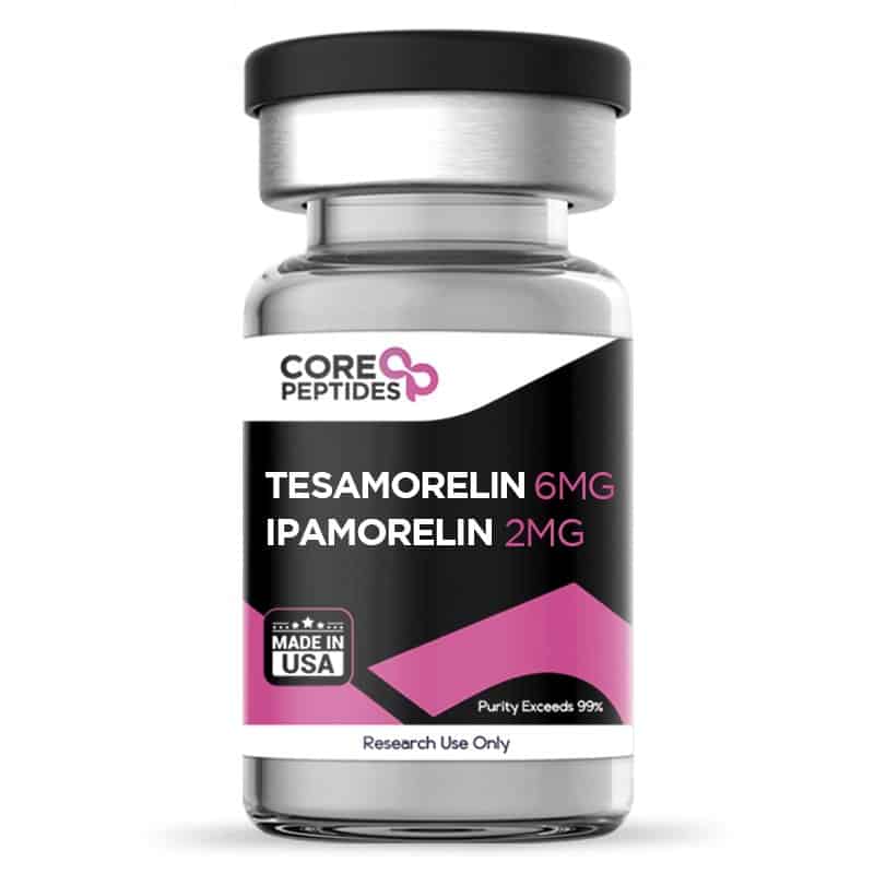
Tesamorelin & Ipamorelin Blend (8mg)
The Tesamorelin and Ipamorelin blend consists of two peptides that appear to share the potential to stimulate the growth hormone axis, albeit through differing mechanisms. This combination may synergistically activate the pituitary gland, which research suggests results in the release of endogenous growth hormone. The blend seems to offer a potential means to optimize growth hormone levels and may elicit effects on sleep, metabolic function, cognition, muscle tissue, lean mass, and lipid profiles.(1)(2) It is possible that the combination of Tesamorelin and Ipamorelin may yield a range of impacts, which may include improved deep sleep, reduced levels of triglycerides, visceral adipose tissue (VAT), and carotid intima-media thickness (cIMT), enhanced cognition, and possible overall optimization of metabolic function. This blend appears to present an opportunity to harness the synergistic actions of both to potentially augment natural growth hormone levels. Chemical Makeup(3)(4) Molecular Formula Tesamorelin: C221H366N72O67S Ipamorelin: C38H49N9O5 Molecular Weight Tesamorelin: 5136 g/mol Ipamorelin: 711.8 g/mol Sequence Tesamorelin: Unk-Tyr-Ala-Asp-Ala-Ile-Phe-Thr-Asn-Ser-Tyr-Arg-Lys-Val-Leu-Gly-Gln-Leu-Ser-Ala-Arg-Lys-Leu-Leu-Gln-Asp-Ile-Met-Ser-Arg-Gln-Gln-Gly-Glu-Ser-Asn-Gln-Glu-Arg-Gly-Ala-Arg-Ala-Arg-Leu-NH2 Ipamorelin: H-Aib-His-D-2Nal-D-Phe-Lys-NH2 Other Known Titles Tesamorelin: (3E)-hex-3-enoylsomatoliberin Ipamorelin: Ipamorelin Acetate, Aib-His-D-2-Nal-D-Phe-Lys-NH2 Research and Clinical Studies Tesamorelin & Ipamorelin Blend and Growth Hormone Deficiency The Tesamorelin and Ipamorelin blend appears to exert its potential through distinct yet complementary proposed mechanisms of action. Tesamorelin, a growth hormone-releasing hormone (GHRH) analog, appears to act by binding to and activating the GHRH receptor on somatotrophs in the pituitary gland. This stimulation potentially triggers the synthesis and secretion of endogenous growth hormone (GH) in a pulsatile manner. By promoting GH release, Tesamorelin may possibly enhance lipolysis, reduce visceral adipose tissue, and also potentially improve glucose metabolism. Ipamorelin, a growth hormone secretagogue receptor (GHSR) agonist, by contrast, appears to activate GHSR in the hypothalamus and peripheral tissues, potentially leading to the release of GH. This peptide appears to exhibit a high selectivity for GHSR, potentially without interfering with other hormones. This GHSR activation by Ipamorelin may result in increased GH secretion, which, in turn, might promote protein synthesis, lipolysis, and insulin-like growth factor-1 (IGF-1) production, as suggested by researchers. When combined, the Tesamorelin and Ipamorelin blend may provide a synergistic impact by targeting different components of the growth hormone axis. Tesamorelin has been suggested to enhance GHRH-mediated GH release, while Ipamorelin has been suggested to directly stimulate GHSR to increase GH secretion. This dual action appears to amplify the overall GH, such as improved body composition, lipid profile, insulin sensitivity, and overall metabolic function in laboratory test models. By seemingly targeting the growth hormone axis through their supposed distinct mechanisms of action, Tesamorelin and Ipamorelin may collectively stimulate the pituitary gland, thereby potentially enhancing the release of endogenous growth hormone.(5) Tesamorelin & Ipamorelin Blend and the Pituitary Gland The Tesamorelin and Ipamorelin blend appears to exhibit some impact on the pituitary gland, a critical endocrine organ considered responsible for the regulation of growth hormone secretion. Studies suggest that Tesamorelin may specifically target the growth hormone-releasing hormone receptor (GHRHR), potentially activating the signaling cascade that leads to growth hormone synthesis and subsequent release. As per the researchers, “Tesamorelin is a synthetic growth hormone-releasing hormone that acts on the anterior pituitary gland to stimulate the endogenous growth hormone secretion.”(6) Studies suggest that the peptide may potentially cause a 69% increase in total growth hormone levels (apparently assessed via area under the curve - AUC) and a purported 55% increase in the mean pulse area of the growth hormone. The peptide was not reported to affect growth hormone pulse frequency or peak growth hormone levels.(7) Ipamorelin, on the other hand, studies suggest may act as a potent agonist for the growth hormone secretagogue receptor (GHSR), seemingly promoting growth hormone secretion.(8) As stated in the studies, “Ipamorelin is the first GHRP-receptor agonist with a selectivity for GH release similar to that displayed by GHRH. The specificity of ipamorelin makes this compound a very interesting candidate for future clinical development.”(8) Ipamorelin may have exhibited a propensity to elevate growth hormone levels, potentially reaching heights of up to 80mIU/l (equivalent to an approximate concentration of 26.6ng/ml). When expressed as a percentage increase in comparison to a placebo (1.31mIU/l or 0.4ng/ml), this augmentation appears to surpass 60-fold.(9) When combined, these peptides appear to exert a synergistic action on the pituitary gland, which might result in enhanced growth hormone production. This proposed synergistic interaction between Tesamorelin and Ipamorelin may offer a promising avenue for research in optimizing growth hormone levels. Tesamorelin & Ipamorelin Blend and Lipodystrophy Research studies frequently indicate that lipodystrophy is often accompanied by insulin resistance, dyslipidemia, and an increased risk of cardiovascular complications. Clinical studies have suggested that Tesamorelin presentation in lipodystrophic test subjects might lead to a reduction in visceral adipose tissue (VAT) and improvements in insulin sensitivity and lipid profiles. Tesamorelin appears to act through the activation of the growth hormone-releasing hormone receptor (GHRHR), potentially stimulating endogenous growth hormone secretion and promoting lipolysis; and potentially preserving “abdominal subcutaneous adipose tissue, improving body image and lipids”.(10) Similarly, Ipamorelin has been reported to exhibit some promise in influencing adipose tissue metabolism. By combining Tesamorelin and Ipamorelin, it is hypothesized by researchers that the synergistic potential of these peptides may enhance the reduction of VAT and improve metabolic parameters in lipodystrophy test models.(10) Tesamorelin & Ipamorelin Blend and Type 2 Diabetes Preclinical and clinical studies have suggested that both Tesamorelin and Ipamorelin hold promise in improving glycemic control and mitigating the metabolic abnormalities associated with T2DM. Tesamorelin appears to stimulate endogenous growth hormone secretion, which has been long considered by scientists to enhance insulin sensitivity and glucose utilization. Ipamorelin studies also indicate that the peptide may influence glucose metabolism and insulin sensitivity. Combining Tesamorelin and Ipamorelin may provide complementary effects, which could also include reductions in hemoglobin A1c (HbA1c) levels, possible improvements in insulin sensitivity, and possible reductions in visceral adiposity in individuals with Type II Diabetes Mellitus.(11) Tesamorelin & Ipamorelin and Cognitive Improvement Studies have indicated that growth hormone and its secretagogues, such as Tesamorelin and Ipamorelin, might play a role in neuroplasticity, neuronal survival, and synaptic plasticity, all of which are considered critical for optimal cognitive performance. Preclinical research has further suggested that Tesamorelin introduction may improve memory and learning abilities, possibly through its proposed impact on neurogenesis and synaptic plasticity. Furthermore, Ipamorelin has been suggested to enhance spatial memory and cognitive function in animal models. The combined influence of Tesamorelin and Ipamorelin may potentially amplify these cognitive benefits through their complementary mechanisms of action. By stimulating the growth hormone axis and modulating neurotrophic factors, this blend holds promise in cognitive improvement research. Tesamorelin & Ipamorelin Blend and Muscle Density In one of the aforementioned scientific examinations, the potential effects of Tesamorelin on muscle tissue integrity were assessed utilizing computed tomography (CT) scans. The results posited that there might be a relationship between Tesamorelin and enhancements in muscle density and size. Interestingly, certain muscle groups, notably the rectus abdominis, psoas major, and paraspinal muscles, appeared to display more pronounced changes, which were typified by either an augmentation in muscle density and size or a diminution in fat content. Statistically, these changes were distinct when compared with placebo. Yet, while it is believed that Tesamorelin's mechanism might be interconnected with molecules such as IGF-1, the study found no significant linkage between IGF-1 level shifts and modifications in muscle dimensions or density.(6) In parallel, early explorations employing experimental frameworks have posited that Ipamorelin may potentially manifest actions akin to those observed with Tesamorelin in relation to skeletal muscle and bone structures. Yet, these observations are yet to be definitively corroborated. Delving deeper, it was suggested that Ipamorelin might interface with, and perhaps elevate, IGF-I concentrations. Such interactions seemed to correlate with a surge in muscle fiber dimensions, overall muscle volume, and consequently, a potential amplification in skeletal muscle robustness during this murine investigation.(12) Tesamorelin & Ipamorelin Blend and Bone Density Ipamorelin seems to have a positive impact on bone health, potentially stimulating bone formation and fostering an enhancement in bone mass. The research posited a possible augmentation in bone mineral content that could be related to Ipamorelin. Various murine trials have alluded to the favorable effects of Ipamorelin on bone tissue.(13) (14) One specific murine study delved into the effects of Ipamorelin on bone mineral content (BMC). The findings proposed that there might be a surge in the test animals’ body weight and BMC, possibly discerned through dual X-ray absorptiometry. Yet, when standardized for body weight variations, the BMC to body weight ratio appeared consistent. An in vitro examination also indicated that the rise in cortical BMC could be attributed to an enlarged bone area, while the volumetric BMD seems to be stable. Tesamorelin & Ipamorelin Blend on Appetite and Digestion The interaction of Ipamorelin with ghrelin receptors appears to have implications for appetite modulation, potentially leading to augmented weight gain. One investigation posited that subjects exposed to Ipamorelin observed a roughly 15% ascent in body weight. It is suggested that this compound might have selectively amplified fat pad weights in relation to total body mass. Consequently, dual-energy X-ray absorptiometry assessments could reveal a nuanced increase in body fat composition. Emerging data also hints that Ipamorelin might elevate serum leptin levels, a hormone integral to energy and appetite regulation. This prompts the scientific community to consider the possibility that heightened food consumption may play a role in the documented weight augmentation within the Ipamorelin cohorts.(15) In addition, activating the ghrelin receptor also appears to affect digestion. Thus, researchers delved into the possible effects of Ipamorelin on gastric functions compared to placebo, with a spotlight on its purported ability to expedite gastric emptying. To gauge this, they employed an intricate technique that tracked the residual radioactivity in the stomach 15 minutes after the introduction of a specific substance through intragastric gavage. It was posited that abdominal surgical interventions might have played a role in decelerating gastric emptying, a phenomenon seemingly prominent in the placebo control cohort. In contrast, Ipamorelin appeared to expedite this emptying process relative to the control group. These observations suggested the potential of Ipamorelin in augmenting the rate of gastric emptying.(16) Diving deeper, the team sought to unravel the effects of this compound on the contractile dynamics of gastric smooth muscles when exposed to acetylcholine and electrical field stimulation. Data hinted that surgical manipulations of the intestines might markedly dampen the contractile responses to both inducements. Yet, intriguingly, this inhibitory effect seemed to be mitigated when Ipamorelin was co-presented with ghrelin. This introduces the tentative proposition that Ipamorelin might not just enhance gastric muscle contractility, but also possibly counterbalance the repressive outcomes instigated by specific surgical measures. (16) Tesamorelin & Ipamorelin blend is available for research and laboratory purposes only. Please review and adhere to our Terms and Conditions before ordering. References: Adrian S, Scherzinger A, Sanyal A, Lake JE, Falutz J, Dubé MP, Stanley T, Grinspoon S, Mamputu JC, Marsolais C, Brown TT, Erlandson KM. The Growth Hormone Releasing Hormone Analogue, Tesamorelin, Decreases Muscle Fat and Increases Muscle Area in Adults with HIV. J Frailty Aging. 2019;8(3):154-159. doi: 10.14283/jfa.2018.45. PMID: 31237318; PMCID: PMC6766405. https://pubmed.ncbi.nlm.nih.gov/31237318/ Clemmons DR, Miller S, Mamputu JC. Safety and metabolic effects of tesamorelin, a growth hormone-releasing factor analogue, in patients with type 2 diabetes: A randomized, placebo-controlled trial. PLoS One. 2017 Jun 15;12(6):e0179538. doi: 10.1371/journal.pone.0179538. PMID: 28617838; PMCID: PMC5472315. https://pubmed.ncbi.nlm.nih.gov/28617838/ National Center for Biotechnology Information (2023). PubChem Compound Summary for CID 16137828, Tesamorelin. https://pubchem.ncbi.nlm.nih.gov/compound/Tesamorelin National Center for Biotechnology Information (2023). PubChem Compound Summary for CID 9831659, Ipamorelin. https://pubchem.ncbi.nlm.nih.gov/compound/Ipamorelin Rogério G. Gondo et al, Growth Hormone-Releasing Peptide-2 Stimulates GH Secretion in GH-Deficient Patients with Mutated GH-Releasing Hormone Receptor, The Journal of Clinical Endocrinology & Metabolism, Volume 86, Issue 7, 1 July 2001, Pages 3279–3283, https://doi.org/10.1210/jcem.86.7.7694 Adrian S, Scherzinger A, Sanyal A, Lake JE, Falutz J, Dubé MP, Stanley T, Grinspoon S, Mamputu JC, Marsolais C, Brown TT, Erlandson KM. The Growth Hormone Releasing Hormone Analogue, Tesamorelin, Decreases Muscle Fat and Increases Muscle Area in Adults with HIV. J Frailty Aging. 2019;8(3):154-159. doi: 10.14283/jfa.2018.45. PMID: 31237318; PMCID: PMC6766405. https://www.ncbi.nlm.nih.gov/pmc/articles/PMC6766405/ Stanley TL, Chen CY, Branch KL, Makimura H, Grinspoon SK. Effects of a growth hormone-releasing hormone analog on endogenous GH pulsatility and insulin sensitivity in healthy men. J Clin Endocrinol Metab. 2011 Jan;96(1):150-8. doi: 10.1210/jc.2010-1587. Epub 2010 Oct 13. PMID: 20943777; PMCID: PMC3038486. https://www.ncbi.nlm.nih.gov/pmc/articles/PMC3038486/ Raun K, Hansen BS, Johansen NL, Thøgersen H, Madsen K, Ankersen M, Andersen PH. Ipamorelin, the first selective growth hormone secretagogue. Eur J Endocrinol. 1998 Nov;139(5):552-61. doi: 10.1530/eje.0.1390552. PMID: 9849822. https://pubmed.ncbi.nlm.nih.gov/9849822/ Gobburu, J. V., Agersø, H., Jusko, W. J., & Ynddal, L. (1999). Pharmacokinetic-pharmacodynamic modeling of ipamorelin, a growth hormone releasing peptide, in human volunteers. Pharmaceutical research, 16(9), 1412–1416. https://doi.org/10.1023/a:1018955126402 Falutz J, Mamputu JC, Potvin D, Moyle G, Soulban G, Loughrey H, Marsolais C, Turner R, Grinspoon S. Effects of tesamorelin (TH9507), a growth hormone-releasing factor analog, in human immunodeficiency virus-infected patients with excess abdominal fat: a pooled analysis of two multicenter, double-blind placebo-controlled phase 3 trials with safety extension data. J Clin Endocrinol Metab. 2010 Sep;95(9):4291-304. doi: 10.1210/jc.2010-0490. Epub 2010 Jun 16. PMID: 20554713. https://pubmed.ncbi.nlm.nih.gov/20554713 Clemmons DR, Miller S, Mamputu JC. Safety and metabolic effects of tesamorelin, a growth hormone-releasing factor analogue, in patients with type 2 diabetes: A randomized, placebo-controlled trial. PLoS One. 2017 Jun 15;12(6):e0179538. doi: 10.1371/journal.pone.0179538. PMID: 28617838; PMCID: PMC5472315. https://www.ncbi.nlm.nih.gov/pmc/articles/PMC5472315/ Andersen, N. B., Malmlöf, K., Johansen, P. B., Andreassen, T. T., Ørtoft, G., & Oxlund, H. (2001). The growth hormone secretagogue ipamorelin counteracts glucocorticoid-induced decrease in bone formation of adult rats. Growth hormone & IGF research : official journal of the Growth Hormone Research Society and the International IGF Research Society, 11(5), 266–272. https://doi.org/10.1054/ghir.2001.0239 Johansen, P. B., Nowak, J., Skjaerbaek, C., Flyvbjerg, A., Andreassen, T. T., Wilken, M., & Orskov, H. (1999). Ipamorelin, a new growth-hormone-releasing peptide, induces longitudinal bone growth in rats. Growth hormone & IGF research : official journal of the Growth Hormone Research Society and the International IGF Research Society, 9(2), 106–113. https://doi.org/10.1054/ghir.1999.9998 Svensson, J., Lall, S., Dickson, S. L., Bengtsson, B. A., Rømer, J., Ahnfelt-Rønne, I., Ohlsson, C., & Jansson, J. O. (2000). The GH secretagogues ipamorelin and GH-releasing peptide-6 increase bone mineral content in adult female rats. The Journal of endocrinology, 165(3), 569–577. https://doi.org/10.1677/joe.0.1650569 Lall, S., Tung, L. Y., Ohlsson, C., Jansson, J. O., & Dickson, S. L. (2001). Growth hormone (GH)-independent stimulation of adiposity by GH secretagogues. Biochemical and biophysical research communications, 280(1), 132–138. https://doi.org/10.1006/bbrc.2000.4065 Greenwood-Van Meerveld, B., Tyler, K., Mohammadi, E., & Pietra, C. (2012). Efficacy of ipamorelin, a ghrelin mimetic, on gastric dysmotility in a rodent model of postoperative ileus. Journal of experimental pharmacology, 4, 149–155. https://doi.org/10.2147/JEP.S35396 { "@context": "https:\/\/schema.org", "@type": "Product", "name": "Tesamorelin & Ipamorelin Blend (8mg)", "description": "Tesamorelin & Ipamorelin blend for sale (8mg). Purchase peptide blends at 99+% purity with unmatched customer service and free shipping.", "image": "https://www.painandanxietymeds.shop/wp-content/uploads/2023/06/Tesamorelin-Ipamorelin-6-2-MG-300x300.jpg", "offers": [ { "@type": "Offer", "priceCurrency": "USD", "price": "90", "availability": "https:\/\/schema.org\/InStock", "itemCondition": "https:\/\/schema.org\/NewCondition", "seller": { "@type": "Organization", "name": "painandanxietymeds.shop" }, "url": "https:\/\/www.painandanxietymeds.shop\/tesamorelin-ipamorelin-blend-8mg/", "hasMerchantReturnPolicy": { "@type": "MerchantReturnPolicy", "applicableCountry": "US", "returnPolicyCategory": "https:\/\/schema.org\/MerchantReturnNotPermitted" }, "shippingDetails": { "@type": "OfferShippingDetails", "shippingRate": { "@type": "MonetaryAmount", "minValue": 0, "maxValue": 9.25, "currency": "USD" }, "shippingDestination": { "@type": "DefinedRegion", "addressCountry": "US" }, "deliveryTime": { "@type": "ShippingDeliveryTime", "handlingTime": { "@type": "QuantitativeValue", "minValue": 1, "maxValue": 2, "unitCode": "d" }, "transitTime": { "@type": "QuantitativeValue", "minValue": 1, "maxValue": 5, "unitCode": "d" } } }, "priceValidUntil": "2027-12-20T15:11:59+00:00" } ], "url": "https:\/\/www.painandanxietymeds.shop\/tesamorelin-ipamorelin-blend-8mg/", "aggregateRating": { "@type": "AggregateRating", "ratingValue": 98, "bestRating": 100, "reviewCount": 524 }, "review": [] } Dr. MarinovDr. Marinov (MD, Ph.D.) is a researcher and chief assistant professor in Preventative Medicine & Public Health. Prior to his professorship, Dr. Marinov practiced preventative, evidence-based medicine with an emphasis on Nutrition and Dietetics. He is widely published in international peer-reviewed scientific journals and specializes in peptide therapy research.

PTD-DBM (5mg)
PTD-DBM peptide, short for Protein Transduction Domain-fused Dishevelled Binding Motif peptide, has been studied for its potential to stimulate follicle growth. PTD is a short sequence of amino acids that may allow the peptide to enter cells, while DBM is a sequence considered to act as a binding agent with Dishevelled (Dvl) proteins, considered highly involved in the Wnt/β-catenin signaling pathway. More specifically, PTD-DBM has been hypothesized by researchers to disrupt the interaction between CXXC5 and Dishevelled (Dvl) proteins, which appears to potentially enhance the activity of the Wnt/β-catenin signaling pathway. Studies hypothesize that this disruption might facilitate an increase in β-catenin activity. This augmented activity was observed to potentially promote hair follicle growth and induce the anagen phase, which is the growth phase of the follicle, in murine models. More precisely, the exposure of PTD-DBM in conjunction with either Wnt3a or valproic acid (VPA)—both agents believed to activate the Wnt/β-catenin pathway—exhibited an apparent synergistic action. This action was speculated by the increased expression levels of β-catenin, alkaline phosphatase (ALP), and proliferating cell nuclear antigen (PCNA). These markers are intrinsically associated with cell proliferation and are indicators of follicle function.(1) PTD-DBM peptide appears to activate the Wnt and β-catenin signaling pathway, a complex network of proteins that may be critical in regulating cell growth, migration, and differentiation during embryonic development and the maintenance of tissues. It is also deemed crucial in processes such as cell fate determination, and appears to play a role in follicle development and cell regeneration. It may stimulate the stem cells in hair follicles, leading to the regeneration of hair and the growth of new follicles. The peptide may also increase blood microcirculation, which is believed to enhance the delivery of nutrients and oxygen to follicles to promote increased rates of growth.(1) Chemical Makeup(2) Molecular Formula: C124H223N61O28S2 Molecular Weight: 3080.7 g/mol Other known titles: Protein Transduction Domain-fused Dishevelled Binding Motif peptide, Hair growth peptide Research and Clinical Studies PTD-DBM and Hair Follicles In an investigation into the biological mechanisms underlying hair loss, coupled with an assessment of potential exposure to compounds structurally similar to PTD-DBM, the study proposed that Prostaglandin D2 (PGD2) may act as a catalyst for hair loss.(3) It purportedly induces this by enhancing the expression of CXXC5, a negative regulator of the Wnt/β-catenin signaling pathway, which plays a pivotal role in developing and regenerating hair follicles. The interaction between CXXC5 and the protein Dishevelled (Dvl) appears to impede this pathway, potentially leading to alopecia. The hypothesis suggests that PGD2 may increase CXXC5 levels through the Bone Morphogenetic Protein (BMP) signaling pathway, which is implicated in the regression of hair follicles during the catagen phase—the transitional stage in hair cycling. This upregulation of CXXC5 by PGD2 seems to inhibit Wnt/β-catenin signaling, which, in turn, may suppress follicle growth. In contrast, PTD-DBM, believed to disrupt the binding between CXXC5 and Dvl, appears to restore the function of the Wnt/β-catenin pathway and thereby may inadvertently promote hair growth. Data supporting this restoration has been published in murine model experiments where the gene for CXXC5 was disabled (knocked out) or the animals were exposed to PTD-DBM, indicating that inhibiting CXXC5 may have counterbalanced the hair loss induced by PGD2. Further, the research points to dihydrotestosterone (DHT), another significant contributor to androgenetic alopecia, intensifying hair loss by stimulating CXXC5 expression through the PGD2-mediated pathways. This suggests a complex interaction where DHT and PGD2 may collaboratively impede hair growth by a concomitant modulation of signaling pathways that involve CXXC5. Intriguingly, the study notes that concurrently inhibiting both CXXC5 and a key enzyme in the β-catenin destruction complex, Glycogen Synthase Kinase-3 beta (GSK-3β), may offer potential in promoting hair growth rather than targeting either component alone. Further studies have investigated the mechanisms behind the peptide’s potential to suppress the interaction between CXXC5 and Dvl proteins.(4) This is presumably achieved by PTD-DBM binding to the PDZ domain of Dvl, thereby possibly preventing the suppressive action of CXXC5 on Wnt/β‐catenin signaling. Furthermore, experimental observations from the study indicated that exposure to PTD-DBM in murine models appeared to have led to hair regrowth and possibly the initiation of wound-induced hair follicle neogenesis (WIHN). In this phenomenon, new hair follicles form as part of the wound-healing process. These outcomes suggest that PTD-DBM might not only promote the anagen phase of the hair cycle but also contribute to the creation of new hair follicles in response to skin injury. PTD-DBM and Bone Regeneration A recent study was conducted on a murine model exposed to PTD-DBM routinely over the course of four weeks.(5) After the completion of the study, it was suggested by the researchers that the peptide may have induced the formation of new bones without histological changes. Hyun-Yi Kim et al. stated, “The inhibitors of Dvl–CXXC5 interaction showed bone-forming effects in ex vivo and in vivo calvaria growth, and […] inhibitor recovered bone loss in postmenopausal model mice. Antibody-based drugs such as anti-Dkk-1 and anti-sclerostin antibodies [...] and PTH-based drugs are expensive [...] Therefore, the development of small-molecule compounds […] would be a valuable addition to osteoporosis [research].” PTD-DBM and Tissue Damage A study presented a detailed examination of the potential mechanisms by which PTD-DBM might facilitate regenerative wound healing, focusing primarily on the modulation of the Wnt/β-catenin signaling pathway.(6) The introduction of PTD-DBM was suggested by researchers to potentially enhance the regenerative healing process. This peptide has been speculated to function by preventing CXXC5 from binding to Dvl proteins, thereby facilitating the activation of the Wnt/β-catenin pathway. Apart from apparent hair growth, such activation is associated with numerous cellular processes deemed crucial for wound healing, including cell proliferation, differentiation, migration, and stem cell activation in tissue regeneration. Moreover, the study notes that the apparent activation of Wnt/β-catenin signaling by PTD-DBM may have led to an increase in markers associated with wound healing and tissue regeneration. This includes the enhancement of fibroblast activity and collagen deposition, which are believed to be essential for the structural rebuilding of damaged tissue. In another study,(7) experimental C3H mice with cutaneous wounds were divided into three groups randomly exposed to the peptide PTD-DBM, valproic acid, or a combination of both. As a control, a separate group of experimental mice was given epidermal growth factor (EGF). After completing the study, it was suggested by the researchers that all three groups appeared to exhibit a gradual improvement in the wound-healing process. However, even more than the EGF alone, the most notable impact was reported in the group presented with the peptide and valproic acid. The research team of Soung-Hoon Lee et al. stated, “Combination [...] with PTD-DBM and VPA significantly induced reepithelialization and enhanced collagen deposition in the large wounds.” PTD-DBM peptide is available for research and laboratory purposes only. Please review and adhere to our Terms and Conditions before ordering. References Lee SH, Seo SH, Lee DH, Pi LQ, Lee WS, Choi KY. Targeting of CXXC5 by a Competing Peptide Stimulates Hair Regrowth and Wound-Induced Hair Neogenesis. J Invest Dermatol. 2017 Nov;137(11):2260-2269. doi: 10.1016/j.jid.2017.04.038. Epub 2017 Jun 6. PMID: 28595998. https://pubmed.ncbi.nlm.nih.gov/28595998/ National Center for Biotechnology Information. "PubChem Substance Record for SID 476298902, PTD-DBM Acetate, Source: CreativePeptides" PubChem, https://pubchem.ncbi.nlm.nih.gov/substance/476298902. Accessed 3 May, 2024. Ryu YC, Park J, Kim YR, Choi S, Kim GU, Kim E, Hwang Y, Kim H, Han G, Lee SH, Choi KY. CXXC5 Mediates DHT-Induced Androgenetic Alopecia via PGD2. Cells. 2023 Feb 9;12(4):555. doi: 10.3390/cells12040555. PMID: 36831222; PMCID: PMC9954685. Ryu YC, Lee DH, Shim J, Park J, Kim YR, Choi S, Bak SS, Sung YK, Lee SH, Choi KY. KY19382, a novel activator of Wnt/β-catenin signalling, promotes hair regrowth and hair follicle neogenesis. Br J Pharmacol. 2021 Jun;178(12):2533-2546. doi: 10.1111/bph.15438. Epub 2021 May 5. PMID: 33751552; PMCID: PMC8251890. Hyun-Yi Kim, Sehee Choi, Ji-Hye Yoon, Hwan Jung Lim, Hyuk Lee, Jiwon Choi, Eun Ji Ro, Jung-Nyoung Heo, Weontae Lee, Kyoung Tai No, Kang-Yell Choi, Small molecule inhibitors of the Dishevelled-CXXC5 interaction are new drug candidates for bone anabolic osteoporosis therapy, EMBO Mol Med (2016)8:375-387. https://doi.org/10.15252/emmm.201505714 Choi S, Yoon M, Choi KY. Approaches for Regenerative Healing of Cutaneous Wound with an Emphasis on Strategies Activating the Wnt/β-Catenin Pathway. Adv Wound Care (New Rochelle). 2022 Feb;11(2):70-86. doi: 10.1089/wound.2020.1284. Epub 2021 Apr 20. PMID: 33573472; PMCID: PMC9831250. Lee SH, Kim MY, Kim HY, Lee YM, Kim H, Nam KA, Roh MR, Min do S, Chung KY, Choi KY. The Dishevelled-binding protein CXXC5 negatively regulates cutaneous wound healing. J Exp Med. 2015 Jun 29;212(7):1061-80. doi: 10.1084/jem.20141601. Epub 2015 Jun 8. PMID: 26056233; PMCID: PMC4493411. https://www.ncbi.nlm.nih.gov/pmc/articles/PMC4493411/ Dr. MarinovDr. Marinov (MD, Ph.D.) is a researcher and chief assistant professor in Preventative Medicine & Public Health. Prior to his professorship, Dr. Marinov practiced preventative, evidence-based medicine with an emphasis on Nutrition and Dietetics. He is widely published in international peer-reviewed scientific journals and specializes in peptide therapy research.

Cardiogen (20mg)
Cardiogen is considered a peptide bioregulator that possibly regulates fibroblasts, which are cells that play a role in scar formation and tissue repair. The peptide has been widely researched for its potential to interact with the tissues in the cardiovascular system. Still, recent research has suggested that it may also be functional in other tissues via influencing fibroblast activities. Furthermore, scientists believe Cardiogen may increase tumor cell apoptosis (programmed cell death). Chemical Makeup Molecular Formula: C18H31N7O9 Molecular Weight: 489.5 g/mol Structure: H-Ala-Glu-Asp-Arg-OH Other Known Titles: SCHEMBL3194515 Research and Clinical Studies Cardiogen Peptide and Cancer Research Researchers consider the peptide to be an apoptotic reductant in cardiac cells via possibly decreasing p53 expression and may exhibit opposite impacts on tumor cells. The p53 gene produces a protein found inside the nucleus of cells and is considered essential in controlling cell division and cell death. An experiment was conducted on murine models with M-1 sarcoma (indicating it has metastasized to other distant tissues and organs) to determine the tumor-modulating potential of Cardiogen, with apoptosis on tumor cells exceeding normal levels and considered uncontrollable.(1) The results indicated a consequence of necrotic and hemorrhagic development and the improvement of tumor cell apoptosis. The results of the experiment suggested that: “The [concentration-dependent] inhibition of M-1 sarcoma growth after [...] cardiogen was caused by the development of hemorrhagic necrosis and stimulation of tumor cell apoptosis. The parameters of proliferative activity indicate that inhibition of tumor growth was not caused by the direct cytostatic effect of the [peptide] on the tumor. Morphological signs indicate a specific mechanism of cardiogen action, realized through the vascular network of the tumor.” (1) The supposed anti-proliferative action of the peptide suggests that tumor growth inhibition may not result from a cytostatic compound on the tumor. According to morphology, tumor growth inhibition may be mediated by a specific mechanism of action related to blood cell supply. This event may be particular to some tumor cells due to their atypical and increased vascular supply. Cardiogen Peptide and Prostate Cell Aging According to researchers, Cardiogen may significantly increase the expression of signaling factors involved in the differentiation of prostate fibroblasts that typically decrease in senescent cultures.(2) Because these signaling factors are considered to decline in aging and senescent fibroblasts, and Cardiogen appears to restore the levels of these factors, especially in senescent cell cultures, it is posited that the peptide may be a relevant candidate for further research in developing methods to address age-related dysfunctions of prostate cells. Cardiogen Peptide and Cardiomyocytes Proliferation Cardiogen may potentially be able to enter different parts of cells, specifically the cytoplasm, nucleus, and nucleolus. Additionally, researchers have hypothesized that Cardiogen might be able to hinder the breakdown of DNA fragments by endonucleases, which are enzymes involved in DNA processing. To explore this, a study was performed using murine embryonic fibroblast cells.(3) These cells were grown in a laboratory setting, in a culture medium known as DMEM, enriched with approximately 10% embryonic calf serum, and kept in a humid environment. After about five days of growth, the cells were separated into two groups. The first group was left as is, serving as a control, while the second group was exposed to Cardiogen for approximately 30 minutes. It appears that in the cells cultivated with Cardiogen, there was a notable increase in the levels of certain proteins within the cytoplasm (such as actin, vimentin, and tubulin) and in the nucleus (specifically nuclear matrix proteins lamin A and C). Actin, vimentin, and tubulin are integral components of the cytoskeleton, the complex network of protein fibers that provide structure and shape to the cells. Actin forms microfilaments, which are thin, flexible fibers crucial for cellular movement and shape. Vimentin, a type of intermediate filament, offers mechanical support and resilience, particularly vital in cells like fibroblasts that endure stress. Tubulin, on the other hand, is the building block of microtubules, which are thicker, hollow tubes essential for maintaining cell shape, enabling intracellular transport, and facilitating chromosome segregation during cell division. Further, lamin A and C are types of nuclear lamins, which are fibrous proteins providing structural support to the nucleus. They form a mesh-like layer called the nuclear lamina on the inner surface of the nuclear envelope. Lamins are involved in organizing the chromatin (DNA and proteins) within the nucleus, and they also play roles in DNA replication, cell division, and regulating gene expression. The protein increases were roughly 2x - 5x higher with Cardiogen than in the control group. These findings suggest that Cardiogen might be activating the expression of these cytoskeletal and nuclear matrix proteins. A possible explanation might be that Cardiogen may potentially influence the proteins associated with DNA, such as enzymes and transcription factors. This influence may improve the way genes related to these cytoskeletal proteins are accessed for transcription, potentially leading to increased cellular metabolism and the stimulation of cell growth and differentiation.(3) Cardiogen Peptide and Cardiomyocytes Apoptosis Researchers have suggested Cardiogen to increase cardiomyocyte proliferation while possibly decreasing fibroblast growth and development and scar formation, resulting in potential long-term and improved cardiac remodeling. Studies suggest that Cardiogen may reduce the expression of the p53 gene, resulting in a lower apoptosis rate of cardiomyocytes after injury.(4,5) Researchers propose Cardiogen's potential in cardiac tissue: “The tetrapeptide cardiogen demonstrated the great stimulating effect on the proliferation both in tissues from young and old rats. The immunohistochemical study demonstrated a decrease of the p53 protein expression by cardiogen action. This fact can testify that cardiogen inhibits the apoptosis process in the myocardial tissue.”(5) More specifically, the synthetic tetrapeptide Cardiogen appears, based on the study's observations, to potentially play a role in enhancing cell proliferation in murine models of both young and aged specimens. This intriguing finding leads to the tentative hypothesis that Cardiogen might have the capacity to stimulate cell growth in heart muscle tissue. As mentioned, Cardiogen might possibly lead to a reduction in the expression of the p53 protein. This protein, often referred to as the "guardian of the genome," is believed to have crucial roles in controlling the cell cycle and may act as a tumor suppressor, thereby making this a significant point of interest for future cancer research. When active, p53 is considered to have the ability to initiate apoptosis, or programmed cell death. Therefore, a potential decrease in p53 expression as a result of Cardiogen might imply that this compound could inhibit apoptosis in the heart's myocardial tissue. This aspect of the study's findings seems to add an extra layer of complexity and potential significance, warranting further investigation.(5) Another experiment investigating the potential of Cardiogen on cardiomyocyte cell apoptosis was conducted in an experimental murine model of myocardial damage, achieved through the deliberate ligation of the coronary artery.(6) There appears to be research data suggesting that the Cardiogen peptide might significantly reduce mortality following this experimentally induced heart damage. Observations point to a threefold decrease in mortality compared to the control group. Additionally, this peptide may play a role in diminishing necrotic zones within the myocardial tissue, which are considered to be areas of cell death due to a suspected lack of blood flow (ischaemia). It is also proposed that the Cardiogen peptide might assist in preserving the glycogen content in the myocardial tissue. Glycogen, considered to be a vital form of energy storage in cells, when preserved, indicates that the cells might have been better able to maintain their energy reserves in the presence of the Cardiogen peptide. This preservation may potentially enhance the survival and function of cells post-ischaemia. The study further suggests that the Cardiogen peptide may exert a protective effect on mitochondria, which are the structures within cells responsible for energy production. Finally, there is a hypothesis that the Cardiogen peptide might stimulate reparative processes, which may theoretically contribute to repairing the damage caused by ischaemia. Such processes might also potentially improve the metabolism of cardiomyocytes, and reduce cell apoptosis.(6) Cardiogen peptide is available for research and laboratory purposes only. Please review and adhere to our Terms and Conditions before ordering. References Levdik NV, Knyazkin IV. Tumor-modifying effect of cardiogen peptide on M-1 sarcoma in senescent rats. Bull Exp Biol Med. 2009 Sep;148(3):433-6. English, Russian Kheĭfets OV, Poliakova VO, Kvetnoĭ IM. [Peptidergic regulation of the expression of signal factors of fibroblast differentiation in the human prostate gland in cell aging]. Adv Gerontol. 2010;23(1):68-70 Khavinson, V. K.h, Lin'kova, N. S., Polyakova, V. O., Kvetnoy, I. M., Benberin, V. V., D'yakonov, M. M., & Titkov, Y. S. (2012). Tetrapeptide H-Ala-Glu-Asp-Arg-OH stimulates expression of cytoskeletal and nuclear matrix proteins. Bulletin of experimental biology and medicine, 153(4), 559–562. https://doi.org/10.1007/s10517-012-1766-9 Grieco P, Gomez-Monterrey I. Natural and synthetic peptides in the cardiovascular diseases: An update on diagnostic and therapeutic potentials. Arch Biochem Biophys. 2019 Feb 15;662:15-32. doi: 10.1016/j.abb.2018.11.021 N. I. Chalisova et al., “[The effect of the amino acids and cardiogen on the development of myocard tissue culture from young and old rats],” Adv. Gerontol. Uspekhi Gerontol., vol. 22, no. 3, pp. 409–413, 2009 Khavinson, V., Linkova, N., Dyatlova, A., Kantemirova, R., & Kozlov, K. (2022). Senescence-Associated Secretory Phenotype of Cardiovascular System Cells and Inflammaging: Perspectives of Peptide Regulation. Cells, 12(1), 106. Dr. MarinovDr. Marinov (MD, Ph.D.) is a researcher and chief assistant professor in Preventative Medicine & Public Health. Prior to his professorship, Dr. Marinov practiced preventative, evidence-based medicine with an emphasis on Nutrition and Dietetics. He is widely published in international peer-reviewed scientific journals and specializes in peptide therapy research.

P21 (5mg)
The P21 (also identified as P021) is a synthetic peptide designed to mimic the activity of ciliary neurotrophic factor (CNTF) on the brain without triggering an allergic response. CNTF is a protein that promotes the survival and differentiation of various neural cells, including neurons and oligodendrocytes. CNTF has been suggested to exhibit possible neuroprotective and neurodegenerative activity in animal models of neurodegenerative diseases and injuries. Scientists identified CNTF's most active regions based on epitope mapping of neutralizing antibodies to CNTF.(1) This led to the development of Peptide 6, made of 11 amino acids (Ac-VGDGGLFEKKL-NH(2)) and a subsequence of just 4 amino acids called Peptide 6c (Ac-DGGL-NH(2)). These amino acids have been theorized to support hippocampus-dependent learning and memory, increase neurogenesis, and potentially boost neuronal plasticity, as posited in murine models. To improve the stability of the peptide and boost its potential to pass through the blood-brain barrier, the scientists report that they "added adamantane building blocks to the C-terminus or both C- and N-termini of Peptide 6c.” The result is P21, which appears to have the potential to mimic the action of CNTF by activating the CNTF receptor complex and downstream signaling pathways, possibly leading to enhanced cognition, increased proliferation, and neuronal differentiation of adult hippocampal progenitors in murine models. Chemical Makeup Molecular Formula:C30H54N6O5 Molecular Weight: 578.3 g/mol Sequence: Ac-DGGL-adamatanylglycine-NH2 Other Known Titles: P021 Research and Clinical Studies P21 Peptide and Cognitive impairment P21 has been extensively researched in experimental models of cognitive impairment. One notable study examined the impact of P21 peptide exposure in 3xTg-AD murine models between 6-9 months prior to the onset of amyloid beta (Aβ) or tau pathology and during the period of synaptic compensation.(2) The study suggested that P21 appeared to rescue dendritic and synaptic deficits, boost neurogenesis, and reverse cognitive impairment in the 3xTg-AD murine models. The study suggests that P21 may contribute to the mitigation of synaptic deficits and cognitive impairment by potentially providing neurotrophic support during synaptic compensation. According to the researchers, this potential may be achieved via the proposed influence of the peptide on synaptic plasticity, as the peptide appears to increase the levels of several markers essential for synaptic function, such as synapsin, PSD-95, and MAP2. Synapsin may regulate neurotransmitter release, which is considered crucial for communication between neurons. PSD-95 is posited to organize components within postsynaptic densities, ensuring signals are received and processed at synaptic junctions. MAP2 is thought to stabilize microtubules in dendrites, supporting their structure and possibly influencing the formation and maintenance of synaptic connections. Overall, these proteins may support the regulation of neurotransmitter release, potentially stabilizing synaptic changes and maintaining the structural integrity of dendrites. These are essentially nerve cell projections that may receive information from other neurons, called presynaptic neurons, or from the environment. By boosting these markers, P21 is posited to help preserve and enhance synaptic functionality. Another study suggested that the P21 may reduce the natural decline in learning and memory in aged Fisher murine models by inhibiting neurogenesis deficit and possibly increasing the expression of brain-derived neurotrophic factor (BDNF) and restoring synaptic deficits in the cortex and hippocampus.(3) BDNF is posited to be a key protein in the survival and differentiation of neurons and the growth of dendrites. BDNF may work by activating pathways that may prevent cell death and promote the differentiation of progenitor cells into mature neurons, especially in the hippocampus, which may aid in ongoing neurogenesis. BDNF is also thought to be instrumental in enhancing synaptic plasticity. It may help strengthen synaptic connections and modulate neurotransmitter release, facilitating better cognition in models of cognitive impairment. BDNF may also contribute to the structural complexity of neurons by encouraging the growth and branching of dendrites, thereby increasing neural connectivity. The study also indicated that P21 appeared to reduce the concentration of myoinositol, a metabolite that naturally increases in murine models. The study suggests that "stimulating endogenous neuroprotective mechanisms using P21 may be [an interesting research] approach for cognitive aging, Alzheimer's disease, and associated neurodegenerative disorders." P21 Peptide and Alzheimer's Disease (AD) Models Experiments are actively investigating the potential research outcomes of P21 in Alzheimer's models. One study focusing on the potential of P21 suggested that chronic exposure of P21 appeared to possibly reduce the brain level of total tau in aged Fisher rats and possibly also reduce tau levels in the cerebrospinal fluid (CSF) to that of young adult rats.(4) The study also observed that "P21 [may be] blood-brain-barrier-permeable and [may] not induce any detectable immune reaction." The exact mechanism for the apparent reduction of tau protein levels remains unknown. Yet, the researchers posit that the increased BDNF activity may modulate downstream effectors such as glycogen synthase kinase-3 (GSK-3). GSK-3 is posited to be a kinase involved in the phosphorylation of tau. Phosphorylated tau is prone to aggregation, forming neurofibrillary tangles, a hallmark of Alzheimer’s (AD) models. By modulating BDNF levels, P21 may indirectly inhibit GSK-3 activity, potentially reducing tau phosphorylation. Another study investigated the potential of P21 on neurobehavior and AD-like pathology in a transgenic murine model of AD.(5) The compound was presented during the mice's prenatal to early postnatal development. Results suggested that the peptide appeared to rescue cognitive deficits, reduce abnormal accumulation of tau and Aβ plaque load, ameliorate certain markers of postsynaptic deficits, and decrease neuroinflammation in the brain. The purported mechanism may have involved improving PSD-95 levels and cAMP response element-binding protein (CREB) activity, which are considered markers of synaptic function and memory formation. Additionally, P21 may have reduced glial fibrillary acidic protein (GFAP) levels, indicating a potential decrease in neuroinflammation. In another experiment, the researchers investigated the neurotrophic potential of the P21 compound in preventing neurodegeneration, amyloid-β, and tau pathologies in 3xTg-AD murine models.(6) The researchers started P21 exposure during the period of synaptic compensation several months before the appearance of any overt pathology. They observed that P21 initiated during this period might have prevented neurodegeneration, Aβ, and tau pathologies, rescued episodic memory impairment, and may have potentially reduced the mortality rate. One trial also investigated the potential of P21 on cognitive function and synaptic plasticity in a transgenic murine model of AD.(7) Results indicated that the peptide might mitigate cognitive impairment, increase expressions of pCREB and BDNF, and ameliorate synaptic protein deficit in the murine models. These actions may lead to the activation of three key signaling pathways: PLC/PKC, MEK/ERK, and PI3K/Akt. These pathways are purported to play a potential role in neuronal survival, growth, and synaptic plasticity. By enhancing these signaling cascades, the peptide is posited to improve synaptic protein expression. The study also suggests that P21 might potentially rescue synaptic deficits and cognitive impairment in familial AD and related tauopathies during early development. P21 Peptide and Macular Degeneration Age-related macular degeneration (AMD) affects the macula, the central part of the retina responsible for central vision. It is considered a common neurodegenerative disease that may lead to vision loss at its end stage. Recent research has suggested that consistent exposure to the neurotrophic peptidergic compound may help prevent the occurrence of AMD pathology. A study conducted in aged and 3xTg-AD murine models reported that chronic presentation of P21 appeared to possibly prevent several pathological changes associated with AMD.(8) The study reported identifying photoreceptor degeneration, lipofuscin granules, vacuoles, atrophy in retinal pigment epithelium (RPE), and Bruch's membrane (BM) thickening. The study also reported a rosette-like structure formation in aged murine models, a hallmark of AMD pathology. Microgliosis and astrogliosis, inflammatory responses in the retina, were also observed in different retinal layers. Furthermore, the study indicated that total tau, phosphorylated tau, Aβ/APP, and VEGF appeared widely distributed in the sub-retina of aged and 3xTg murine models. These molecules are associated with Alzheimer's disease pathology, and their presence suggests that retinal changes associated with aging and Alzheimer's disease may share some common features. Importantly, consistent exposure to P21 for three months in rats and 18 months in 3xTg murine models appeared to ameliorate the pathological changes described above. P21 peptide is available for research and laboratory purposes only. Please review and adhere to our Terms and Conditions before ordering. References Li, B., Wanka, L., Blanchard, J., Liu, F., Chohan, M. O., Iqbal, K., & Grundke-Iqbal, I. (2010). Neurotrophic peptides incorporating adamantane improve learning and memory, promote neurogenesis and synaptic plasticity in mice. FEBS letters, 584(15), 3359–3365. https://doi.org/10.1016/j.febslet.2010.06.025 Baazaoui, N., & Iqbal, K. (2017). Prevention of dendritic and synaptic deficits and cognitive impairment with a neurotrophic compound. Alzheimer's research & therapy, 9(1), 45. https://doi.org/10.1186/s13195-017-0273-7 Bolognin, S., Buffelli, M., Puoliväli, J., & Iqbal, K. (2014). Rescue of cognitive-aging by administration of a neurogenic and/or neurotrophic compound. Neurobiology of aging, 35(9), 2134–2146. https://doi.org/10.1016/j.neurobiolaging.2014.02.017 Khatoon, S., Chalbot, S., Bolognin, S., Puoliväli, J., & Iqbal, K. (2015). Elevated Tau Level in Aged Rat Cerebrospinal Fluid Reduced by Treatment with a Neurotrophic Compound. Journal of Alzheimer's disease : JAD, 47(3), 557–564. https://doi.org/10.3233/JAD-142799 Wei, W., Wang, Y., Liu, Y., Dai, C. L., Tung, Y. C., Liu, F., & Iqbal, K. (2020). Prenatal to early postnatal neurotrophic treatment prevents Alzheimer-like behavior and pathology in mice. Alzheimer's research & therapy, 12(1), 102. https://doi.org/10.1186/s13195-020-00666-7 Baazaoui, N., & Iqbal, K. (2017). Prevention of Amyloid-β and Tau Pathologies, Associated Neurodegeneration, and Cognitive Deficit by Early Treatment with a Neurotrophic Compound. Journal of Alzheimer's disease : JAD, 58(1), 215–230. https://doi.org/10.3233/JAD-170075 Wei, W., Liu, Y., Dai, C. L., Baazaoui, N., Tung, Y. C., Liu, F., & Iqbal, K. (2021). Neurotrophic Treatment Initiated During Early Postnatal Development Prevents the Alzheimer-Like Behavior and Synaptic Dysfunction. Journal of Alzheimer's disease : JAD, 82(2), 631–646. https://doi.org/10.3233/JAD-201599 Liu, Y., Wei, W., Baazaoui, N., Liu, F., & Iqbal, K. (2019). Inhibition of AMD-Like Pathology With a Neurotrophic Compound in Aged Rats and 3xTg-AD Mice. Frontiers in aging neuroscience, 11, 309. https://doi.org/10.3389/fnagi.2019.00309 Dr. MarinovDr. Marinov (MD, Ph.D.) is a researcher and chief assistant professor in Preventative Medicine & Public Health. Prior to his professorship, Dr. Marinov practiced preventative, evidence-based medicine with an emphasis on Nutrition and Dietetics. He is widely published in international peer-reviewed scientific journals and specializes in peptide therapy research.
Melanotan 2 (10mg)
Melanotan 2 peptide is a synthetic compound, which is an analog of the α-melanocyte-stimulating hormone.(1) The peptide is a cyclic heptapeptide that appears to have a modified affinity towards the receptors that the endogenous hormone may interact with. In addition to possibly stimulating melanogenesis (the production of melanin pigment) in dermal cells, this peptide may have increased affinity towards other receptors that play a role in the regulation of appetite and various types of arousal.(2) Overview Melanotan 2 likely serves as a non-selective agonist with the potential to bind with four out of the five different subtypes of melanocortin receptors (MC-R).(3) Depending on the localization, the receptor-Melanotan 2 bond may induce different actions. Namely, the four receptors that Melanotan 2 may interact with include: The melanocortin 1 receptor (MC1R) may be expressed in melanocytes, which are cells found in tissues such as dermal tissues, hair, and possibly cells and tissues found in the eye. The melanocortin 3 receptor (MC3R) might be found in a range of tissues, potentially including the brain and the placenta. Initial observations suggest that MC3R might be involved in modulating appetite under certain experimental conditions. The melanocortin 4 receptor (MC4R) may be localized within the central nervous system, perhaps in the hypothalamus. Some early indications suggest that this receptor may impact neurons that are believed to have some influence over mating behaviors and general arousal. The melanocortin 5 receptor (MC5R) appears to be distributed across multiple tissues, although what role it might serve remains unclear. For example, the potential interaction between Melanotan 2 and the MC1Rs may lead to increased production of eumelanin, which causes darkening of the epidermal layer’s pigment.(4) On the other hand, when Melanotan 2 binds with the MC4R, it may induce supraspinal centers in the brain, which may lead to increased libido. These signals may then be carried to the sympathetic and parasympathetic centers in the spinal cord and thoracolumbar region.(3) Chemical Makeup Other Known Titles: MT-II Molecular Weight: 1024.19 g/mol Molecular Formula: C50H69N15O9 Research and Clinical Studies Melanotan 2 Peptide and Nerve Cell Regeneration Research(5) in a murine model of an induced peripheral nerve injury has been employed to investigate the neurotrophic potential of Melanotan 2. 48 hours after half of the murine models were presented with the peptide, it was noted that the Melanotan 2 research models appeared to indicate a recovery in their sensory function. Furthermore, when the murine models were presented with a chemotherapeutic compound, Melanotan 2 appeared to exhibit neuroprotective properties, which protected the nerves from the compound's induced neurotoxicity to a certain extent. This potential is posited to be mediated via the MC4 receptors, which might even promote neurite outgrowth and possibly support the intrinsic capacity of neuronal tissue to recover after injury. Although the exact signaling pathways are not fully understood, it is often suggested that the pro-opiomelanocortin (POMC)-derived melanocortin peptides, including compounds analogous to a-melanocyte-stimulating hormone (a-MSH), may influence neuronal structures by increasing the number and length of neurites and potentially promoting nerve sprouting in damaged regions. Since Melanotan 2 is considered a potent melanocortin receptor agonist, it may trigger a cascade of intracellular events that theoretically lead to a better-supported ability of nerve fibers to regenerate after various forms of insult, as well as a partial protective response against toxic neuropathic conditions. Therefore, the researchers concluded that they “observed that Melanotan-II also possesses neuroprotective properties, as it partially protected the nerve from a toxic neuropathy induced by cisplatin.” Melanotan 2 Peptide and Arousal Neurosignaling In a clinical study,(6) Melanotan 2 has been suggested to induce increased arousal neuron signaling in more than 80% of cases, compared to only 20% success with a placebo. This research peptide may act via the MC4 receptors and downstream of established neuromodulators, including dopaminergic and oxytocinergic signals, possibly integrating their actions within discrete hypothalamic centers. These regions are thought to coordinate various homeostatic and motivational behaviors, and the introduction of an agonist like Melanotan 2 may potentially reframe the balance of neuronal activity. These researchers also posit that the involvement of MC5 receptors in certain peripheral glands may potentially provide a parallel route that links central neuro signaling with peripheral modulatory factors. However, this remains an area where mechanisms are only hypothesized. Melanotan 2 Peptide and Neurodevelopmental Modulation Researchers have said that Melanotan 2 may potentially influence aberrant neural mechanisms by possibly stimulating populations of neurons that may govern social cognition through endogenous oxytocinergic signaling.(7) These MC4R-sensitive circuits, potentially located in regions such as the paraventricular nucleus of the hypothalamus, might release endogenous oxytocin in response to Melanotan 2, possibly recalibrating imbalanced neurochemical activity thought to underlie key aspects of social impairment. This oxytocin release may, in turn, modulate neurotransmission involving serotonin, glutamate, dopamine, and GABA, all of which are implicated in shaping social adaptation. By engaging these systems, Melanotan 2 may alter the functional connectivity of cortical and subcortical networks—regions including, for instance, the anterior cingulate cortex—where oxytocin receptor distribution may differ in atypical neurodevelopmental contexts. In doing so, researchers have been able to hypothesize that Melanotan 2 might restore or modify synaptic communication and synaptic plasticity, which may go so far as to reshape the underlying neuroarchitecture. Melanotan 2 Peptide and Models of Sunless Tanning Melanotan 2 may increase melanin production and thereby induce darker pigmentation without the need for ultraviolet irradiation by engaging the MC1R on melanocytes.(8) Moreover, the peptide's cyclic structure supports a more prolonged metabotropic activity compared to other MSH analogs. Although the precise intracellular signaling cascades remain incompletely understood, current data suggest that receptor interactions might lead to the elevated synthesis of eumelanin. This might offer a potential pathway for the development of sunless tanning models in a controlled research environment. Specifically, researchers have made comments about their observation of outcomes in research models, such as “increased [darkened] pigmentation in the face, upper body, and buttock” in experimental settings. Melanotan 2 peptide is available for research and laboratory purposes only. Please review and adhere to our Terms and Conditions before ordering. References: Ryakhovsky, Vladimir V et al. “The first preparative solution phase synthesis of Melanotan II.” Beilstein Journal of Organic Chemistry vol. 4 (2008): 39. doi:10.3762/bjoc.4.39. https://pubmed.ncbi.nlm.nih.gov/19043625/ Mac E. Hadley, Discovery that a melanocortin regulates sexual functions in male and female humans, Peptides, Volume 26, Issue 10, 2005, Pages 1687-1689, ISSN 0196-9781, https://doi.org/10.1016/j.peptides.2005.01.023 King, Stephen H et al. “Melanocortin receptors, melanotropic peptides and penile erection.” Current topics in medicinal chemistry vol. 7,11 (2007): 1098-1106. https://www.ncbi.nlm.nih.gov/pmc/articles/PMC2694735/ Peters, Björn, et al. “Melanotan II: a possible cause of renal infarction: review of the literature and case report.” CEN case reports vol. 9,2 (2020): 159-161. doi:10.1007/s13730-020-00447-z. https://www.ncbi.nlm.nih.gov/pmc/articles/PMC7148395/ Ter Laak, Mariël P, et al. “The potent melanocortin receptor agonist melanotan-II promotes peripheral nerve regeneration and has neuroprotective properties in the rat.” European Journal of Pharmacology vol. 462,1-3 (2003): 179-83. doi:10.1016/s0014-2999(02)02945-x. https://pubmed.ncbi.nlm.nih.gov/12591111/ Wessells, H et al. “Melanocortin receptor agonists, penile erection, and sexual motivation: human studies with Melanotan II.” International journal of impotence research vol. 12 Suppl 4 (2000): S74-9. doi:10.1038/sj.ijir.3900582. https://pubmed.ncbi.nlm.nih.gov/11035391/ Minakova E, Lang J, Medel-Matus JS, Gould GG, Reynolds A, Shin D, Mazarati A, Sankar R. Melanotan-II reverses autistic features in a maternal immune activation mouse model of autism. PLoS One. 2019 Jan 10;14(1):e0210389. Doi: 10.1371/journal.pone.0210389. PMID: 30629642; PMCID: PMC6328175. Dorr RT, Lines R, Levine N, Brooks C, Xiang L, Hruby VJ, Hadley ME. Evaluation of melanotan-II, a superpotent cyclic melanotropic peptide in a pilot phase-I clinical study. Life Sci. 1996;58(20):1777-84. doi: 10.1016/0024-3205(96)00160-9. PMID: 8637402. Dr. MarinovDr. Marinov (MD, Ph.D.) is a researcher and chief assistant professor in Preventative Medicine & Public Health. Prior to his professorship, Dr. Marinov practiced preventative, evidence-based medicine with an emphasis on Nutrition and Dietetics. He is widely published in international peer-reviewed scientific journals and specializes in peptide therapy research.

GHK Basic (50mg)
GHK Basic is a tripeptide containing the amino acid sequence glycyl-histidyl-lysine. This short-chain peptide is considered to help cell communication for a wide range of actions. Research on the GHK has yielded a variety of potential biological activities, including possible stimulation of blood vessels and nerve outgrowth, increasing elastin, glycosaminoglycan, collagen synthesis, and possibly promoting dermal fibroblast functions. The tissue repair potential of this peptide has been researched in lung connective tissue, bone tissue, liver, stomach lining, and skin.(1) The peptide has also been suggested to play some role in DNA repair, lung protection, fibroblast reactivation, and possibly in suppressing proinflammatory and pro-aging molecules like NFκB. GHK Basic has also been suggested to have the potential to repair protective proteins in the skin barrier. Chemical Makeup(2) Molecular Formula: C14H24N6O4 Molecular Weight: 340.4 g/mol Research and Clinical Studies GHK Basic and Skin Cell Regeneration According to research, adding plasma to aged liver tissue may cause the aged tissues to produce proteins identical to those seen in plasma.(2,3,4) Several studies have suggested that GHK proteins may increase collagen production, glycosaminoglycans, and small proteoglycans such as decorin. Furthermore, research indicates that GHK might also influence the activities of metalloproteinases – enzymes that initiate extracellular matrix protein breakdown – and antiprotease actions. This GHK action may regulate skin protein breakdown, potentially preventing damaged protein accumulation and excessive proteolysis. GHK Basic and Wound Healing Animal model studies suggest the peptide may promote wound healing through two possible mechanisms. First, GHK, when combined with a high-concentration helium-neon laser, was reported to accelerate wound contraction and granular tissue production while hosting antioxidant enzyme activities and increasing blood vessel growth.(5) Second, wound healing in control and diabetic murine models was reported to be improved by collagen dressing loaded with GHK. The researchers commented that “the wounds were almost closed by 99.39% when [exposed to] biotinylated GHK-loaded films on day 21 when compared to 69.49% wound closure for plain films.” Increased glutathione and ascorbic acid levels were found, as well as enhanced epithelialization and increased collagen synthesis, fibroblasts, and mast cell activation in wounds.(6) GHK and its analogs may also have potential in follicular stimulation and growth. Researchers reported the naturally occurring copper complex GHK-Cu to increase vascular endothelial growth factor production, possibly leading to growth and new blood vessel formation. In one murine experiment, the copper-containing version of GHK appeared to mediate a significant decrease in wound area compared to placebo. Wounds exposed to the copper-containing version of GHK were suggested to have lower concentrations of the pro-inflammatory markers MMP-2, MMP-9, and TNF-alpha.(7) Notably, the copper-containing versions of GHK may also help reduce some potential complications that may negatively impact wound healing, such as colonization of the wound with pathogenic microorganisms. A study of GHK on diabetic ulcers stated that the peptide may have sped up wound closure in larger ulcers and reduced the risk of wound infections compared to the study group that received only a vehicle (placebo). More specifically, the researchers commented that “the enhancement of wound closure was more pronounced (median of 89.2% compared with −10.3% for vehicle; p < 0.01) in larger (greater than 100 mm2 initial area at study entry) plantar ulcers.” The incidence of ulcer infections was also significantly lower, equal to about 7% in the GHK group compared to 34% for vehicle.(8) GHK Basic and Anti-inflammatory, Antioxidant Activities GHK has been suggested to exhibit potential antioxidant action, specifically in its posited interaction with lipid peroxidation, a process which produces adverse free radicals capable of DNA, protein, and cell damage. GHK may help minimize this damage by binding the byproducts of this process.(9) The specific radicals suggested by the scientists include 4-hydroxynoneal, acrolein, malondialdehyde, and others. Further, it has also been proposed that GHK might play a role in diminishing the release of iron from ferritin, an agent that catalyzes lipid peroxidation. More specifically, studies have suggested that GHK could potentially limit the creation of iron compounds in injured tissues, thereby mitigating inflammation.(10) It is believed that GHK's mechanism may involve attaching to ferritin's iron release pathways, potentially decreasing -Fe release by an estimated 87%, which might, in theory, curb inflammation and oxidation in affected tissues. The anti-inflammatory potential of GHK might benefit specific tissues like the lungs. An experiment in murine models examined GHK's impact on lipopolysaccharide-induced lung inflammation.(11) The findings suggest that GHK may lower the generation of reactive oxygen species and inflammatory cytokines while boosting the efficacy of antioxidant enzymes. It is thought to hinder the activation of NF-κB and p38 MAPK signaling pathways, reducing TNF-1 and IL-6 levels. Moreover, the study noted that GHK seemed to mitigate lung tissue alterations and lessen inflammation in murine models suffering from lung damage. More specifically, the peptide was reported to have decreased the influx of inflammatory cells into the lung tissue in cases of experimentally induced acute lung injury in these models. GHK might also contribute to alleviating oxidative stress in lung tissues related to smoke exposure. Research indicates that GHK may suppress oxidative stress in alveolar epithelial cells through the upregulation of Nrf2 expression.(12) GHK Basic and Fibrinogen High fibrinogen levels are considered to increase blood viscosity by increasing rouleaux formation, which research suggests are risk factors for cardiovascular disease.(13) Studies on the peptide generally suggest that it has the potential to inhibit fibrinogen synthesis, thereby possibly lowering the risk of coronary occurrences. GHK Basic peptide is available for research and laboratory purposes only. Please review and adhere to our Terms and Conditions before ordering. References Pickart L, Margolina A. Regenerative and Protective Actions of the GHK-Cu Peptide in the Light of the New Gene Data. Int J Mol Sci. 2018 Jul 7;19(7):1987 National Center for Biotechnology Information (2024). PubChem Compound Summary for CID 378611, Cu-GHK. Retrieved March 17, 2024 from https://pubchem.ncbi.nlm.nih.gov/compound/Cu-GHK. Pickart L, Vasquez-Soltero JM, Margolina A. GHK Peptide as a Natural Modulator of Multiple Cellular Pathways in Skin Regeneration. Biomed Res Int. 2015;2015:648108. DOI: 10.1155/2015/648108. Dou Y, Lee A, Zhu L, Morton J, Ladiges W. The potential of GHK as an anti-aging peptide. Aging Pathobiol Ther. 2020 Mar 27;2(1):58-61. DOI: 10.31491/apt.2020.03.014. Gul, N. Y., Topal, A., Cangul, I. T., & Yanik, K. (2008). The effects of tripeptide copper complex and helium-neon laser on wound healing in rabbits. Veterinary dermatology, 19(1), 7–14. https://doi.org/10.1111/j.1365-3164.2007.00647.x Alven, S., Peter, S., Mbese, Z., & Aderibigbe, B. A. (2022). Polymer-Based Wound Dressing Materials Loaded with Bioactive Agents: Potential Materials for the Treatment of Diabetic Wounds. Polymers, 14(4), 724. https://doi.org/10.3390/polym14040724 Canapp, S. O., Jr, Farese, J. P., Schultz, G. S., Gowda, S., Ishak, A. M., Swaim, S. F., Vangilder, J., Lee-Ambrose, L., & Martin, F. G. (2003). The effect of tripeptide-copper complex on healing of ischemic open wounds. Veterinary surgery : VS, 32(6), 515–523. https://doi.org/10.1111/j.1532-950x.2003.00515.x Mulder, G. D., Patt, L. M., Sanders, L., Rosenstock, J., Altman, M. I., Hanley, M. E., & Duncan, G. W. (1994). Enhanced healing of ulcers in patients with diabetes by treatment with glycyl-l-histidyl-l-lysine copper. Wound repair and regeneration : official publication of the Wound Healing Society [and] the European Tissue Repair Society, 2(4), 259–269. https://doi.org/10.1046/j.1524-475X.1994.20406.x Cebrián, J., Messeguer, A., Facino, R. M., & García Antón, J. M. (2005). New anti-RNS and -RCS products for cosmetic treatment. International journal of cosmetic science, 27(5), 271–278. https://doi.org/10.1111/j.1467-2494.2005.00279.x Miller, D. M., DeSilva, D., Pickart, L., & Aust, S. D. (1990). Effects of glycyl-histidyl-lysyl chelated Cu(II) on ferritin dependent lipid peroxidation. Advances in experimental medicine and biology, 264, 79–84. https://doi.org/10.1007/978-1-4684-5730-8_11 Park, J. R., Lee, H., Kim, S. I., & Yang, S. R. (2016). The tri-peptide GHK-Cu complex ameliorates lipopolysaccharide-induced acute lung injury in mice. Oncotarget, 7(36), 58405–58417. https://doi.org/10.18632/oncotarget.11168 Zhang, Q., Yan, L., Lu, J., & Zhou, X. (2022). Glycyl-L-histidyl-L-lysine-Cu2+ attenuates cigarette smoke-induced pulmonary emphysema and inflammation by reducing oxidative stress pathway. Frontiers in molecular biosciences, 9, 925700. https://doi.org/10.3389/fmolb.2022.925700 Pickart L, Vasquez-Soltero JM, Margolina A. GHK and DNA: resetting the human genome to health. Biomed Res Int. 2014;2014:151479. DOI: 10.1155/2014/151479. Dr. MarinovDr. Marinov (MD, Ph.D.) is a researcher and chief assistant professor in Preventative Medicine & Public Health. Prior to his professorship, Dr. Marinov practiced preventative, evidence-based medicine with an emphasis on Nutrition and Dietetics. He is widely published in international peer-reviewed scientific journals and specializes in peptide therapy research.