Medicines & Treatments

Semax (25mg)
Semax peptide is a synthetic polypeptide analog of an adrenocorticotropic hormone fragment 4-7 (ACTH 4-7),(1) a hormone secreted by the anterior pituitary gland. This fragment has been reported by researchers not to exhibit any action characteristic of ACTH itself but may have a potentially specific action within the brain.(2) Researchers find that naturally occurring peptides may occasionally exhibit instability and easy degradation by biological enzymes, such as those found in gastrointestinal and cerebrospinal fluids. Consequently, researchers aim to synthesize analogs of these naturally occurring but potentially unstable peptides to induce similar biological action with the added value of higher stability. Semax is one such synthetically developed derivative of an endogenous adrenocorticotropic hormone fragment. The Met-Glu-His-Phe sequence in Semax is extended by adding a Pro-Gly-Pro (PGP) sequence at its C-terminal end. This modification with PGP may potentially enhance the molecule's ability to cross the blood-brain barrier (BBB) by increasing its lipophilicity. This increase in lipophilicity may facilitate the molecule’s passive diffusion or uptake through mechanisms such as lipid raft-mediated endocytosis, which may circumvent the tight junctions that typically restrict entry into the brain. Furthermore, the presence of PGP at the peptide's C-terminus might also modify its interactions with specific transporters or receptors on the BBB, potentially favoring its transport via receptor-mediated transcytosis. Moreover, the acetylation of Semax may improve its stability by making it more resistant to enzymatic breakdown, thereby extending its half-life in biological settings. Overview Synthetically developed Semax has been widely researched for its potential mechanism of action and lauded for its apparent biological stability. Researchers suggest it may inhibit select enzymes that regulate the degradation of enkephalins, naturally released neurotransmitters secreted in the brain, which scientists consider may regulate several biological functions. Enkephalins are believed to be significantly involved in nociception (the sensory perception of pain) and stress response. Moreover, an elevation in enkephalin concentrations might potentially impact other neurotransmitter systems due to the intricate interconnections between the opioid system and neurotransmitters such as dopamine and serotonin. This interaction may manifest through modulation or alteration of neurotransmitter release, receptor activity, or signal transduction pathways, indicating a sophisticated and multifaceted relationship that remains an area of active research. In addition to enkephalins, Semax has been evaluated for its potential to inhibit other peptide-degrading secreted enzymes. Based on one specific research study,(4) Semax may induce elevated secretion and release of dopamine as well as possibly increasing the levels of brain-derived neurotrophic factor (BDNF). Based on another study,(5) Semax has been speculated to also host the potential to alter gene expressions that modulate the immune system. In altering gene expression, the levels of immune cells and their mobility may be elevated. Semax was reported by researchers to exhibit altering potential in the encoding of chemokines and immunoglobulins, related to the functioning of the vascular system. Chemical Makeup Molecular Formula: C39H54N10O10S Molecular Weight: 854.99 g/mol Other Known Titles: ACTH (4-7)PGP, HY-P1146 Research Studies and Clinical Trials Semax Peptide and Nootropic Action An initial study(6) was conducted on ACTH hormone and its analogs, including Semax, to determine its nootropic potential in murine models. After peptide exposure in the models, 5-hydroxyindoleacetic acid (5-HIAA) levels were monitored. 5-HIAA levels appeared elevated by 25% after 2 hours of Semax presence. The levels appeared to increase gradually up to a maximum of 180% within 4 hours of peptide exposure. It was noted by researchers that the peptide, when introduced 20 minutes before D-amphetamine, appeared to lead to an elevation of 5-HIAA as compared to exposure to Semax alone. 5-HIAA is a primary metabolite of serotonin, which indicates that Semax might enhance serotonergic activity. This hypothetical influence on serotonin metabolism might enhance the functions of serotonin-dependent pathways, which may affect operations within the central nervous system. This speculative action underscores a possible modulation of neurotransmitter systems that play critical roles in mood, cognition, and overall brain function. Semax Peptide and Neonatal Anxiety Models The main aim of this study(7) was to expose neonatal murine models to an SSRI and then Semax, to evaluate the interaction. Murine models aged between 1 and 14 days received an SSRI, followed by Semax on days 15 to 28. After 28 days, it was noted that upon exposure to the SSRI, the rats exhibited anxiety-like behavior, with an apparently impaired response to stressors and new stimuli during the first 14 days. Following Semax exposure, these SSRI-induced actions appeared to be mitigated, with the rats even exhibiting improved learning abilities and an overall reduction in anxiety-driven behaviors. Researchers posited that Semax might have reestablished normal levels of monoamines in the brain, which may have been initially decreased by the SSRI. By affecting these neurotransmitter systems, Semax might restore or stabilize neural pathways that were previously disrupted. Such changes might balance the excitatory and inhibitory signals in the brain, creating a state more conducive to reduced anxiety. For instance, by potentially increasing serotonin levels, Semax may improve mood and decrease anxiety, while optimizing dopamine might enhance motivation and reward processing. Furthermore, norepinephrine adjustments might improve attention and vigilance. Behavioral assessments conducted in the experiment indicated that these potential actions of Semax appeared temporally stable. The reduction in anxiety-related behaviors was sustained from adolescence through young adulthood, suggesting that Semax might have a lasting influence on neural circuits. This enduring stability implies that Semax might support creating a protective or corrective action on these circuits that persists beyond immediate exposure. Semax Peptide and the Vascular System In this study,(8) researchers evaluated the potential of Semax to protect murine heart models from vascular damage after experimental myocardial infarction (MI). The murine models were induced with myocardial infarction, and a cohort of the models were exposed to Semax in an experimental group for the following 6 days. On the 28th day, it was reported by the researchers that the murine models that served as a control group appeared to have developed cardiac hypertrophy along with decreased arterial blood pressure. The Semax-exposed models exhibited signs indicating preventing diastolic pressure growth in the left ventricle, with apparent left ventricle remodeling. Notably, the peptide may have ameliorated both cardiomyocyte hypertrophy and the imbalance between the growth of contractile and mitochondrial apparatus. Semax Peptide and Neonatal Deprivation Adolescent rats(9) were separated from their mothers for approximately 5 hours per day during postnatal days 1 to 14. From days 15 to 28, these adolescent rats were then exposed to the Semax peptide. After 28 days, it was found that during maternal deprivation, when Semax was not present, there was an apparent increase in anxiety and physical and emotional reactivity in the rats. Upon Semax exposure, researchers reported that reactions and anxiety in the rats appeared restored to control levels. Semax Peptide and Neuroprotection This clinical trial(10) was conducted on 100 models of ischemic stroke. A cohort representing 30% of the models was exposed to Semax, whereas the rest were evaluated as a control group. Following the study, researchers reported that following Semax exposure, there appeared to be an improvement in the rate of restoration of damaged neurological functions. All results were analyzed using EEG mapping. Semax Peptide and Nootropic Properties A small-scale clinical trial(11) was conducted in which research models were given Semax under high-stress conditions, and subsequent brain activity was monitored. At the end of the study, after a total of 24 hours, researchers reported that compared to normal pre-trial thresholds, the models appeared to exhibit increased memory recall and increased focus intervals. Semax peptide is available for research and laboratory purposes only. Please review and adhere to our Terms and Conditions before ordering. References: T. Kolomin et al., A New Generation of Drugs: Synthetic Peptides based on Natural Regulatory peptides. Neuroscience & Medicine, 2013, 223-252. Published Online December 2013. http://dx.doi.org/10.4236/nm.2013.44035 Dornbush RL, Nikolovski O. ACTH 4-10 and short-term memory. Pharmacol Biochem Behav. 1976;5(Suppl 1):69-72. doi: 10.1016/0091-3057(76)90331-2. PMID: 189333. https://pubmed.ncbi.nlm.nih.gov/189333/ Kost NV, Sokolov OIu, Gabaeva MV, Grivennikov IA, Andreeva LA, Miasoedov NF, Zozulia AA. Ingibiruiushchee deĭstvie semaksa i selanka na énkefalindegradiruiushchie fermenty syvorotki krovi cheloveka [Semax and selank inhibit the enkephalin-degrading enzymes from human serum]]. Bioorg Khim. 2001 May-Jun;27(3):180-3. Russian. doi: 10.1023/a:1011373002885. PMID: 11443939. https://pubmed.ncbi.nlm.nih.gov/11443939/ Shih-Jen Tsai, Semax, an analogue of adrenocorticotropin (4–10), is a potential agent for the treatment of attention-deficit hyperactivity disorder and Rett syndrome, Medical Hypotheses, Volume 68, Issue 5, 2007, Pages 1144-1146. https://doi.org/10.1016/j.mehy.2006.07.017 Medvedeva, E.V., Dmitrieva, V.G., Povarova, O.V. et al. The peptide semax affects the expression of genes related to the immune and vascular systems in rat brain focal ischemia: genome-wide transcriptional analysis. BMC Genomics 15, 228 (2014). https://doi.org/10.1186/1471-2164-15-228 Eremin KO, Kudrin VS, Saransaari P, Oja SS, Grivennikov IA, Myasoedov NF, Rayevsky KS. Semax, an ACTH(4-10) analogue with nootropic properties, activates dopaminergic and serotoninergic brain systems in rodents. Neurochem Res. 2005 Dec;30(12):1493-500. doi: 10.1007/s11064-005-8826-8. PMID: 16362768. Nataliya Yu. Glazova, Daria M. Manchenko, Maria A. Volodina, Svetlana A. Merchieva, Ludmila A. Andreeva, Vladimir S. Kudrin, Nikolai F. Myasoedov, Natalia G. Levitskaya, Semax, synthetic ACTH(4–10) analogue, attenuates behavioural and neurochemical alterations following early-life fluvoxamine exposure in white rats, Neuropeptides, Volume 86, 2021, 102114, ISSN 0143-4179. https://doi.org/10.1016/j.npep.2020.102114 Gavrilova SA, Golubeva AV, Lipina TV, Fominykh ES, Shornikova MV, Postnikov AB, Andrejeva LA, Chentsov IuS, Koshelev VB. [Protective effect of peptide semax (ACTH(4-7)Pro-Gly-Pro) on the rat heart rate after myocardial infarction]. Ross Fiziol Zh Im I M Sechenova. 2006 Nov;92(11):1305-21. Russian. PMID: 17385423. https://pubmed.ncbi.nlm.nih.gov/17385423/ Volodina MA, Sebentsova EA, Glazova NY, Levitskaya NG, Andreeva LA, Manchenko DM, Kamensky AA, Myasoedov NF. Semax attenuates the influence of neonatal maternal deprivation on the behavior of adolescent white rats. Bull Exp Biol Med. 2012 Mar;152(5):560-3. English, Russian. doi: 10.1007/s10517-012-1574-2. PMID: 22803132. https://pubmed.ncbi.nlm.nih.gov/22803132/ Gusev EI, Skvortsova VI, Miasoedov NF, Nezavibat'ko VN, Zhuravleva EIu, Vanichkin AV. Effektivnost' semaksa v ostrom periode polusharnogo ishemicheskogo insul'ta (klinicheskoe i élektrofiziologicheskoe issledovanie) [Effectiveness of semax in acute period of hemispheric ischemic stroke (a clinical and electrophysiological study)]. Zh Nevrol Psikhiatr Im S S Korsakova. 1997;97(6):26-34. Russian. PMID: 11517472. https://pubmed.ncbi.nlm.nih.gov/11517472/ Asmarin IP, Nezavibat'ko VN, Miasoedov NF, Kamenskiĭ AA, Grivennikov IA, Ponomareva-Stepnaia MA, Andreeva LA, Kaplan AIa, Koshelev VB, Riasina TV. Nootropnyĭ analog adrenokortikotropina 4-10-semaks (15-letniĭ opyt razrabotki i izucheniia) [A nootropic adrenocorticotropin analog 4-10-semax (l5 years experience in its design and study)]. Zh Vyssh Nerv Deiat Im I P Pavlova. 1997 Mar-Apr;47(2):420-30. Russian. PMID: 9173745. https://pubmed.ncbi.nlm.nih.gov/9173745/ Dr. MarinovDr. Marinov (MD, Ph.D.) is a researcher and chief assistant professor in Preventative Medicine & Public Health. Prior to his professorship, Dr. Marinov practiced preventative, evidence-based medicine with an emphasis on Nutrition and Dietetics. He is widely published in international peer-reviewed scientific journals and specializes in peptide therapy research.

DSIP (5mg)
Delta Sleep-Inducing Peptide, or DSIP, is a naturally-occurring peptide made of nine amino acids, isolated from the central nervous system of experimental models of electrically induced sleep. DSIP is posited to regulate the sleep cycle, as suggested by its name, but may also play a role in regulating various physiological processes. Suggested to induce delta sleep in test models, this neuropeptide also appears to impact electrophysiological activity and possibly regulate the neurotransmitter levels in the brain.(1) DSIP peptide was first characterized and examined from 1963 to 1977 and has since been widely studied by scientists.(2) Initially regarded only for its potential as a sleep-including factor, DSIP was soon suggested to induce other potential actions, such as mitigating pain, sleep cycle regulation, and mitigating withdrawal.(3) Mechanisms of Action DSIP is believed to have the potential to modify the structure and quality of sleep by engaging with the central nervous system. It is thought that DSIP may potentially reduce the time it takes to fall asleep and enhance the overall quality of sleep by influencing the activity of various neurotransmitters within the brain. Despite these possibilities, scientific research has yet to definitively identify the primary mechanisms and pathways through which DSIP exerts its actions. However, it is hypothesized that DSIP may target several specific receptors, which are considered crucial in its function. These include: N-methyl-D-aspartate (NMDA) receptors and gamma-aminobutyric acid (GABA) receptors: NMDA receptors are linked to glutamate, a vital neurotransmitter that facilitates brain excitation, while GABA receptors are associated with inhibitory neurotransmission, playing a significant role in calming the brain. Research conducted on murine models has suggested that DSIP might amplify GABA's calming actions, which assists in reducing brain activity and helps individuals fall asleep more easily. Concurrently, murine studies suggest that DSIP may dampen some of the stimulatory impacts of NMDA receptors, thereby decreasing overall brain stimulation and further aiding in sleep promotion.(4)(5) Opioid receptors: Further research indicates that DSIP might indirectly affect opioid receptors in the brain. This interaction is believed to influence the peptide's ability to modulate sleep and alleviate withdrawal symptoms, highlighting its complex involvement in the brain's signaling systems.(6)(7) Alpha 1-adrenergic receptor: This receptor, apparently found in the pineal gland, has been another focus of DSIP research. An experimental study has suggested that DSIP's modulation of the alpha 1-adrenergic receptor could be a mechanism through which it affects sleep patterns. This interaction also hints at DSIP's potential role in managing stress tolerance, given the significant influence of alpha 1-adrenergic signaling in stress-related processes.(8) These findings underline the complex and multifaceted ways in which DSIP might influence sleep and stress management, although more research is needed to understand its mechanisms of action. Chemical Makeup Molecular Formula: C35H48N10O15 Molecular Weight: 848.82 g/mol Other Known Titles: DSIP nonapeptide; emideltide Research and Clinical Studies DSIP and Sleep Cycles A study(9) was conducted on feline models to examine the potential action of DSIP on sleep patterns. The models were divided into two groups; one was a control, and the other was labeled as the DSIP group. The peptide was presented to the cats and was monitored for 8 hours. Results indicated that there appeared to be a significant increase in total sleep and slow wave sleep (SWS) in the DSIP group. The action of DSIP appeared to be immediate as the amount of SWS sleep elevated within the first hour following study initiation. This increase appeared to be maintained for 7 hours and then decreased in the eighth hour. SWS, often called deep sleep, is suggested as one of the core stages within sleep architecture, broadly categorized into non-rapid eye movement (NREM) sleep and rapid eye movement (REM) sleep. The overall structure of sleep is typically cyclic, alternating between NREM and REM stages multiple times. Apparently, SWS falls under the NREM category and scientists detect it by its low-frequency, high-amplitude delta waves evident in electroencephalogram (EEG) recordings. Sleep begins with NREM sleep, subdivided into three stages: N1, N2, and N3. N1 and N2 are lighter stages of sleep, while N3, synonymous with SWS, is the deepest stage of sleep. Following the deep sleep stage, the cycle progresses into REM sleep, where brain activity increases and dreams occur. A clinical study(10) has suggested that the peptide may lead to an immediate increase in sleep pressure and resulted in a 59% increase in sleep within two hours of initiating DSIP experimentation. The scientists also posited that the peptide may have enhanced sleep efficiency, potentially by shortening sleep onset.(10) DSIP and Endocrine Regulation DSIP has been posited to interact with certain hormonal messengers typically released during sleep. Examples may include the luteinizing hormone (LH), which is considered a crucial hormone in the regulation of reproductive hormones such as testosterone. In a study with murine models,(11) DSIP was examined for its potential actions on the endocrine system. Within 30 minutes, it was noted that the levels of LH appeared to be significantly elevated, whereas there was no perceived impact on another regulatory messenger called follicle-stimulating hormone (FSH). Further studies have suggested that DSIP may lead to increased secretion of growth hormone, positing that the peptide may potentially act on the hypothalamus to regulate hormonal secretion. Utilizing ovariectomized murine models to exclude the actions of gonadal steroids, the research observed an apparent elevation in GH levels caused by DSIP. The potential involvement of a dopaminergic mechanism in this process was inferred from the blocking action of pimozide, a dopamine antagonist, on the DSIP-induced GH increase. Additionally, in vitro studies with pituitary cells suggest a similar increase in GH release upon exposure to DSIP, albeit with a notable decline at higher concentrations. This pattern hints at the complex nature of DSIP's role in GH regulation, potentially linking it to sleep-induced GH release, given the peptide's association with slow-wave sleep patterns and the apparent correlation of such sleep phases with GH secretion.(12) DSIP and Stress Response Researchers have investigated the potential action of DSIP on murine models subjected to experimentally induced stress.(13) The murine models were divided into six groups, where the control group was presented with a placebo and the rest with DSIP. The six groups included (i) control group, (ii) stress group, (iii) group with DSIP one hour before stress experiments, (iv) DSIP 24 hours before stress experiments, (v) DSIP one hour before the last stress experiment and (vi) DSIP 24 hours before the last stress experiment. This research primarily focused on assessing changes in substance P, beta-endorphin, and corticosterone levels, which are critical in understanding the stress response and potential modulatory impacts of DSIP. Initial findings highlighted that DSIP exposure might induce noticeable fluctuations in the levels of these markers, suggesting a possible stress-modulatory role. For instance, there was an initial decrease followed by a dramatic increase in beta-endorphin. This pattern suggests DSIP's potential influence on the opioidergic system, possibly contributing to mechanisms of stress mitigation or adaptation. Regarding corticosterone levels, which directly indicate stress in murine models, a decrease was noted shortly after DSIP exposure. The study posits that DSIP's actions on substance P, beta-endorphin, and corticosterone levels are part of a broader spectrum of biochemical changes, hinting that DSIP might initiate a series of molecular reactions contributing to its stress-modulatory actions.(13) DSIP and Longevity A study(14) was conducted on murine models, equally divided into DSIP and control groups. The researchers suggested that DSIP did not appear to influence food intake, however, it did apparently decrease the weight of the murine models. It appeared to decrease the chromosomal aberrations in the bone marrow by 23% and improve the life span by 24% compared to the control group. Moreover, DSIP also appeared to lead to a 2.5-fold decrease in the occurrence of malignancies. Another study suggests that the apparent protective actions of DSIP may be due to the potential antioxidative properties of the peptide. The study(15) was conducted on murine models, and the researchers posited that DSIP may inhibit the levels of malonic dialdehyde in the models. Malonic dialdehyde is a byproduct of lipid peroxidation, and increased levels of malonic dialdehyde typically induce increased oxidative stress. This suggested that DSIP may prevent lipid peroxidation in the murine models, which may execute antioxidant properties. DSIP may also stimulate the endogenous antioxidant system, influencing various enzymatic levels. The researchers commented that “DSIP exerts stimulating influence upon the superoxid-dismutese, catalase, ceruloplasmin activities as well as the level of nonenzymatic antioxidants--urea and uric acids, because during organism aging the antioxidant defense systems are being suppressed. DSIP increases the volume of tissues and blood endogenous antioxidant defense system mainly by means of enzymatic antioxidant system, especially during later ontogenesis.”(15) DSIP is available for research and laboratory purposes only. Please review and adhere to our Terms and Conditions before ordering. References: National Center for Biotechnology Information. "PubChem Compound Summary for CID 3623358, Emideltide;delta Sleep Inducing Peptide" PubChem, https://pubchem.ncbi.nlm.nih.gov/compound/3623358 Graf MV, Kastin AJ. Delta-sleep-inducing peptide (DSIP): an update. Peptides. 1986 Nov-Dec;7(6):1165-87. https://pubmed.ncbi.nlm.nih.gov/3550726/ Kovalzon VM, Strekalova TV. Delta sleep-inducing peptide (DSIP): a still unresolved riddle. J Neurochem. 2006 Apr;97(2):303-9. https://pubmed.ncbi.nlm.nih.gov/16539679/ Grigor'ev VV, Ivanova TA, Kustova EA, Petrova LN, Serkova TP, Bachurin SO. Effects of delta sleep-inducing peptide on pre- and postsynaptic glutamate and postsynaptic GABA receptors in neurons of the cortex, hippocampus, and cerebellum in rats. Bull Exp Biol Med. 2006 Aug;142(2):186-8. English, Russian. doi: 10.1007/s10517-006-0323-9. PMID: 17369935 Sudakov KV, Umriukhin PE, Rayevsky KS. Delta-sleep inducing peptide and neuronal activity after glutamate microiontophoresis: the role of NMDA-receptors. Pathophysiology. 2004 Oct;11(2):81-86. https://pubmed.ncbi.nlm.nih.gov/15364118/ Nakamura A, Nakashima M, Sakai K, Niwa M, Nozaki M, Shiomi H. Delta-sleep-inducing peptide (DSIP) stimulates the release of immunoreactive Met-enkephalin from rat lower brainstem slices in vitro. Brain Res. 1989 Feb 27;481(1):165-8. doi: 10.1016/0006-8993(89)90498-8. PMID: 2706459. Dick P, Grandjean ME, Tissot R. Successful treatment of withdrawal symptoms with delta sleep-inducing peptide, a neuropeptide with potential agonistic activity on opiate receptors. Neuropsychobiology. 1983;10(4):205-8. doi: 10.1159/000118012. PMID: 6328354. Graf MV, Schoenenberger GA. Delta sleep-inducing peptide modulates the stimulation of rat pineal N-acetyltransferase activity by involving the alpha 1-adrenergic receptor. J Neurochem. 1987 Apr;48(4):1252-7. doi: 10.1111/j.1471-4159.1987.tb05654.x. PMID: 3029331. Susić V, Masirević G, Totić S. The effects of delta-sleep-inducing peptide (DSIP) on wakefulness and sleep patterns in the cat. Brain Res. 1987 Jun 30;414(2):262-70. https://pubmed.ncbi.nlm.nih.gov/3620931/ Schneider-Helmert D, Gnirss F, Monnier M, Schenker J, Schoenenberger GA. Acute and delayed effects of DSIP (delta sleep-inducing peptide) on human sleep behavior. Int J Clin Pharmacol Ther Toxicol. 1981 Aug;19(8):341-5. https://pubmed.ncbi.nlm.nih.gov/6895513/ Iyer KS, McCann SM. Delta sleep inducing peptide (DSIP) stimulates the release of LH but not FSH via a hypothalamic site of action in the rat. Brain Res Bull. 1987 Nov;19(5):535-8. doi: 10.1016/0361-9230(87)90069-4. https://pubmed.ncbi.nlm.nih.gov/3121137/ Iyer KS, McCann SM. Delta sleep-inducing peptide (DSIP) stimulates growth hormone (GH) release in the rat by hypothalamic and pituitary actions. Peptides. 1987 Jan-Feb;8(1):45-8. doi: 10.1016/0196-9781(87)90163-x. PMID: 3575154. Sudakov KV, Coghlan JP, Kotov AV, Salieva RM, Polyntsev YuV, Koplik EV. Delta-sleep-inducing peptide sequels in the mechanisms of resistance to emotional stress. Ann N Y Acad Sci. 1995 Dec 29;771:240-51. https://pubmed.ncbi.nlm.nih.gov/8597403/ Popovich IG, Voitenkov BO, Anisimov VN, Ivanov VT, Mikhaleva II, Zabezhinski MA, Alimova IN, Baturin DA, Zavarzina NY, Rosenfeld SV, Semenchenko AV, Yashin AI. Effect of delta-sleep inducing peptide-containing preparation Deltaran on biomarkers of aging, life span and spontaneous tumor incidence in female SHR mice. Mech Ageing Dev. 2003 Jun;124(6). https://pubmed.ncbi.nlm.nih.gov/12782416/ Bondarenko TI, Maĭboroda EA, Mikhaleva II, Prudchenko IA. [Mechanism of delta-sleep inducing peptide geroprotective activity]. Adv Gerontol. 2011;24(1):80-92. Russian. https://pubmed.ncbi.nlm.nih.gov/21809625/ Dr. MarinovDr. Marinov (MD, Ph.D.) is a researcher and chief assistant professor in Preventative Medicine & Public Health. Prior to his professorship, Dr. Marinov practiced preventative, evidence-based medicine with an emphasis on Nutrition and Dietetics. He is widely published in international peer-reviewed scientific journals and specializes in peptide therapy research.

CJC-1295 NO DAC (Mod GRF 1-29) (5mg)
CJC-1295 is a synthetic peptide derivative of the naturally occurring GHRH hormone and is composed of 29 amino acids.(2) This peptide is also referred to as Modified GRF (1-29) or CJC 1295 without DAC. It is a modified version of the peptide containing four substituted amino groups that are considered to help prevent the peptide's degradation. CJC without DAC does not have a Drug Affinity Complex that is sometimes added to further extend the half-life of the peptide. Modified growth hormone fragments GRF (1-29) were first discovered in the early 1980s, when it was suggested that the first 29 amino acids of the natural growth hormone-releasing hormone might retain all the properties of the full 44 amino acid peptide.(3) One of the potential drawbacks of synthetically developed growth hormone-releasing peptides is a short half-life. Rigorous research has resulted in a supposedly stabilized, longer-lasting CJC-1295 (No DAC) peptide.(1) Overview CJC-1295 without DAC, alternatively known as the tetra-substituted GRF (1-29), represents a synthetic variant of the naturally occurring hormone responsible for the release of growth hormone (GHRH). Scholars have posited that it facilitates the secretion of growth hormone (GH). This compound is essentially constructed from the minimal sequence of amino acids that might bind to GHRH receptors, specifically the initial 29 amino acids of GHRH. There are notable structural differences between CJC-1295 without DAC and the unmodified fragment, primarily due to the alteration of four amino acids within the original 29 amino acids of GHRH. These changes encompass the 2nd, 8th, 15th, and 27th amino acids, and such adjustments might arguably extend the peptide's lifespan against enzymatic breakdown, particularly by enzymes like dipeptidyl peptidase-4 (DPP-4).(4) More specifically, these alterations include: The replacement of L-alanine with D-alanine at the 2nd position, which is thought to bolster resistance against molecular degradation. The substitution of asparagine with glutamine at the 8th position, which could conceivably minimize asparagine reconfiguration and amide hydrolysis. The exchange of glycine for alanine at the 15th position, which is speculated to enhance bioactivity. The alteration from methionine to leucine at the 27th position, which is suggested to prevent methionine oxidation. Chemical Makeup Molecular Formula: C152H252N44O42 Molecular Weight: 3367.9 g/mol Other Known Titles: CJC-1295 Without DAC Research and Clinical Studies CJC-1295 (No DAC) Peptide and the Pituitary Gland CJC-1295 without DAC seems to aim at the GHRH receptor on pituitary cells, potentially engaging with certain binding sites on the receptor molecule. This interaction may induce alterations in the receptor's conformation, possibly sparking a chain of molecular events that might activate signaling pathways within its target cells. The induced structural changes are believed to facilitate the activation of G-proteins, signaling proteins theorized to reside on the cell's interior side of the GHRH receptor.(5) Once activated, these G-proteins might trigger the production of secondary messengers like cAMP or IP3, which are thought to act as intermediary signaling molecules, possibly amplifying the signal inside the cell. Particularly, cAMP may activate protein kinases, which are presumed to be pivotal in phosphorylating specific target proteins.(6) Protein kinases are suggested to have a potential role in managing various cell functions. The stimulation of protein kinases could lead to the phosphorylation of transcription factors, proteins that might influence gene expression regulation. These phosphorylated transcription factors may then enter the nucleus and might alter the transcription of genes linked to the synthesis and secretion of growth hormone. Consequently, the molecular activities seemingly initiated by CJC-1295 no DAC's binding may culminate in the merging of secretory vesicles loaded with growth hormone with the cell membrane. This merger may facilitate the external release of growth hormone from the pituitary cells, potentially enabling it to perform its biological functions. CJC-1295 (No DAC) Peptide and Growth Hormone Pulsatility While no studies directly investigate the potential of CJC-1295 no DAC (tetrasubstituted GRF 1-29), some researchers have conducted extensive experimentations with partially modified versions of GRF 1-29. One notable example is research by Khorram et al. This study on CJC-1295 no DAC explored its potential on growth hormone and insulin-like growth factor 1 (IGF-1), skin cell proliferation, muscle tissue hypertrophy, and other potential outcomes.(7) The investigation highlighted the potential of CJC-1295 no DAC to modulate the growth hormone-IGF-1 axis. More specifically, CJC-1295 no DAC appeared to cause a considerable rise, with an approximate increase of 70-107% higher mean 12-hours release of growth hormone by the somatotroph cells in the anterior pituitary. Concurrently, IGF-1 also experienced an increase, approximately 28%, indicating an enhancement in the growth hormone-IGF-1 axis functionality. This increase was also associated with increases in skin tissue thickness, potentially due to the anabolic actions of growth hormone and IGF-1 on collagen-producing skin cells such as fibroblasts. Furthermore, there was a significant increase in muscle tissue hypertrophy, ultimately resulting in a net lean mass gain of 2.77 lbs. These results imply a potential role of CJC-1295 no DAC in promoting skin cell proliferation and muscle tissue hypertrophy that should be investigated in future experiments. However, the specific mechanisms behind these actions remain to be fully elucidated. CJC-1295 (No DAC) Peptide and Intestinal Studies Studies(8) were conducted in experimental models, which have suggested a potential interaction between GHRH analog peptides and VPAC(1)-R, found on the smooth muscles of the gastrointestinal system. This interaction was suggested to induce bowel movement, though the potential connection between specific peptides and bowel release are still under investigation. CJC-1295 (No DAC) Peptide and Heart Rate Preliminary research(9) in murine models suggested that Modified GRF 1-29 peptide (along with other GHRH derivative analogs) may exhibit some potential to improve heart rate and possibly support the heart's ability to pump blood, particularly following a heart attack. The study(9) commented that GHRH agonist peptides appeared to promote cardiac tissue repair and possibly also improve ejection fraction rates. CJC-1295 (No DAC) Peptide and Combination CJC-1295 NO DAC appears to be a relatively short-lasting GHRH analog when not attached to DAC, compared to other synthetic GHRH peptides. Consequently, combined with other short-acting peptides, it may support longer durations for specific properties. Moreover, the peptide may have synergistic actions when combined with growth hormone secretagogues (GHSs). GHSs are considered to be ghrelin mimetics, which interact with the ghrelin receptors within various tissues. These receptors also happen to be found on the somatotroph cells in the anterior pituitary gland - the same cells that also have the GHRH receptors and are associated with growth hormone synthesis. Activating these receptors may lead to an apparent increase in growth hormone synthesis, and some researchers suggest that the simultaneous activation of the GHRH and ghrelin receptors on these pituitary cells may result in synergistically increased growth hormone elevation.(11) One such highly common peptide combination is with Ipamorelin, a synthetic GHRH pentapeptide. Both Ipamorelin and CJC-1295 peptides have been suggested to stimulate the production of growth hormone by the cells of the pituitary gland, albeit via different modes of action, therefore possibly exerting synergism and stimulating even greater growth hormone secretion. CJC-1295 peptide is available for research and laboratory purposes only. Please review and adhere to our Terms and Conditions before ordering. References: National Center for Biotechnology Information. "PubChem Compound Summary for CID 91976842, CJC1295 Without DAC" PubChem, https://pubchem.ncbi.nlm.nih.gov/compound/CJC1295-Without-DAC Clark, R G, and I C Robinson. “Growth induced by pulsatile infusion of an amidated fragment of human growth hormone releasing factor in normal and GHRF-deficient rats.” Nature vol. 314,6008 (1985): 281-3. https://pubmed.ncbi.nlm.nih.gov/2858818/ The Discovery of Growth Hormone-Releasing Hormone: An Update https://onlinelibrary.wiley.com/doi/full/10.1111/j.1365-2826.2008.01740.x Martin, B., Lopez de Maturana, R., Brenneman, R., Walent, T., Mattson, M. P., & Maudsley, S. (2005). Class II G protein-coupled receptors and their ligands in neuronal function and protection. Neuromolecular medicine, 7(1-2), 3–36. https://doi.org/10.1385/nmm:7:1-2:003 Newton, A. C., Bootman, M. D., & Scott, J. D. (2016). Second Messengers. Cold Spring Harbor perspectives in biology, 8(8), a005926. https://doi.org/10.1101/cshperspect.a005926 Khorram, O., Laughlin, G. A., & Yen, S. S. (1997). Endocrine and metabolic effects of long-term administration of [Nle27]growth hormone-releasing hormone-(1-29)-NH2 in age-advanced men and women. The Journal of clinical endocrinology and metabolism, 82(5), 1472–1479. https://doi.org/10.1210/jcem.82.5.3943 Ito T, Igarashi H, Pradhan TK, Hou W, Mantey SA, Taylor JE, Murphy WA, Coy DH, Jensen RT. GI side-effects of a possible therapeutic GRF analogue in monkeys are likely due to VIP receptor agonist activity. Peptides. 2001 Jul;22(7):1139-51. https://pubmed.ncbi.nlm.nih.gov/11445245/ Schally AV, Zhang X, Cai R, Hare JM, Granata R, Bartoli M. Actions and Potential Therapeutic Applications of Growth Hormone-Releasing Hormone Agonists. Endocrinology. 2019 Jul 1;160(7):1600-1612. https://pubmed.ncbi.nlm.nih.gov/31070727/ Sinha, D. K., Balasubramanian, A., Tatem, A. J., Rivera-Mirabal, J., Yu, J., Kovac, J., Pastuszak, A. W., & Lipshultz, L. I. (2020). Beyond the androgen receptor: the role of growth hormone secretagogues in the modern management of body composition in hypogonadal males. Translational andrology and urology, 9(Suppl 2), S149–S159. https://doi.org/10.21037/tau.2019.11.30 Dr. MarinovDr. Marinov (MD, Ph.D.) is a researcher and chief assistant professor in Preventative Medicine & Public Health. Prior to his professorship, Dr. Marinov practiced preventative, evidence-based medicine with an emphasis on Nutrition and Dietetics. He is widely published in international peer-reviewed scientific journals and specializes in peptide therapy research.

ARA-290 (16mg)
ARA-290 peptide, also known as cibinetide and helix B surface peptide (HBSP) in scientific literature, is an 11 amino acid chain derived from the beta domain of the naturally-occurring protein erythropoietin (EPO). This part of the EPO sequence is considered to assist in tissue regeneration and repair without exerting stimulus on the production of red blood cells.(1) Scientists consider the primary function of the EPO is to stimulate tissues to produce red blood cells. Still, the beta domain of the protein, and consequently ARA-290, do not appear to possess this property. Instead, they may possess certain potential regenerative, anti-inflammatory, anti-nociceptive, and other lesser-known properties of the original naturally occurring EPO protein. Overview Scientific findings(2) thus far suggest that once a tissue undergoes an injury, a tissue-protective receptor (TPR) pathway may be activated. This TPR receptor mainly consists of a beta receptor unit (CD131) and a subunit from the EPO receptor, jointly called the innate repair receptor. Researchers have suggested that the ARA-290 peptide molecule may bind to this innate repair receptor and possibly attenuate nerve and allodynia-led pain (nociception).(3) It is also suggested that the ARA-290 peptide may act primarily via this IRR-mediated pathway. Chemical Makeup Molecular Formula: C51H54N16O21 Molecular Weight: 1257.3 g/mol Other Known Titles: PH-BSP Research and Clinical Studies ARA-290 Peptide and Nociception Scientists consider that Transient Receptor Potential (TRP) channels may be the primary nociceptive stimulating channels, including possible thermal, chemical, and mechanical stimuli. In the presence of such a causative agent, one of the TRP channels, namely TRPV1, may be triggered. Upon activation, it might induce the ejection of neuropeptides, which then are considered to generate action potential in the nervous systems. This action potential is referred to as "nociception."(4) A 2016 study(5) suggested that ARA-290 peptide may have the potential to increase the threshold of this TRPV1 channel, with researchers further implying that the peptide might inhibit the TRPV1 actions and thereby no neuropeptides will be released to spark a nociceptive response. The researchers conducted a study with a C57/BL6 strain of murine models to investigate this hypothesis. They took neurons from two areas of the murine nervous system, the dorsal root ganglion, and the trigeminal ganglion, and studied them using calcium imaging. This involved the introduction of a special dye to the cells that lights up when calcium is present and then examining them under a special microscope. The experiment measured how these neurons potentially reacted to three substances: ARA-290, capsaicin (the compound that makes chili peppers hot), and KCl (a common chemical compound that can be used as a control). The researchers were particularly interested in whether ARA-290 could reduce the reaction of the neurons to capsaicin. They also did some direct tests on the murine models. One test measured how potentially sensitive the murine models' appendages were to nociception. They used different concentrations of ARA-290 or a control solution on the murine models' hind appendages. Then, they exposed them to capsaicin to see if it made them potentially more sensitive to nociception. They used a tool to apply pressure to the appendages and measured the point at which the murine models pulled their appendages away. Another test checked how often the murine models withdrew their appendages over 24 hours after exposure to capsaicin. Here, the researchers were evaluating whether giving the murine models ARA-290 after the capsaicin may have affected their sensitivity to nociception. Thus, the findings suggested that ARA-290 might specifically block the reaction of neurons to capsaicin without affecting other types of heat sensors. This may suggest that ARA-290 targets specifically the TRPV1 channels and might potentially increase the amount of capsaicin needed to activate these channels. ARA-290 is still being actively studied for its possible role in nociception "mitigation". However, researchers have also developed hypotheses about its other potential mechanisms of action, such as potential anti-inflammatory and possible immunomodulatory impact. ARA-290 Peptide and Retinal Ischemia A recent study(6) has suggested that ARA-290 may have the potential to protect the endothelial blood vessels and possibly thereby combat retinal ischemia in models of ischemic retinopathies. A possible mitigation method for retinal ischemia is the restoration of endothelial colony-forming cells (ECFC) in the retinal tissue. This study explored the potential of ARA-290 in reducing the current pro-inflammatory conditions in the ischemic retina and its potential effects on vascular regeneration aided by ECFCs. Experimental murine models induced with retinal ischemia underwent ECFC cell transplantation for this study. To determine the potential of the peptide, transplantation in some murine models occurred in the presence of ARA-290 while the rest was in the absence of peptides. Experimental investigations were conducted to assess the impact of ARA-290 on the survival mechanisms and functionality within ECFC (Endothelial Colony-Forming Cell) cultures. The research suggested that ARA-290 might potentially initiate survival pathways and improve cell survival in ECFCs under oxidative stress caused by H2O2. The study aimed to determine the effectiveness of ECFC transplantation in promoting vascular repair in the retinal area of mice, utilizing the oxygen-induced retinopathy (OIR) model for this purpose. The research compared the effects of transplanting ECFCs with and without ARA-290. Additionally, the study examined the inflammatory cytokine profile and microglia activation, which may act as markers of inflammation. Preliminary results indicated that introducing ECFCs with EPO or ARA-290 before transplantation did not appear to significantly enhance their ability to repair blood vessels in the ischemic retina. However, the systemic introduction of ARA-290 in OIR mice seemed to lower the levels of pro-inflammatory cytokines like IL-1β and TNF-α in the retinal tissue, hinting at its possible anti-inflammatory properties. The introduction of ECFCs into the vitreous humor of the eye led to their integration into the impaired retinal blood vessels and seemed to notably reduce the area without blood vessels. Notably, ARA-290 appeared to enhance the vaso-reparative capacity of the ECFCs, a result not observed with EPO. After the study, it was suggested by the researchers that the peptide exhibited a reduction in the inflammatory expression of interleukin cells in the retina. The scientists commented that: “regulation of the pro-inflammatory milieu of the ischemic retina can be enhanced by ARA290 and may be a useful [research agent] to ECFC-based cell [procedures] for ischemic retinopathies.” ARA-290 Peptide and Inflammatory Cytokine Cells One study(7) was conducted to determine whether the erythropoietin analog, ARA-290 peptide, may possess anti-inflammatory and cell protective potential. More specifically, the researchers investigated the potential of the peptide in pancreatic islet transplantation (PITx) models, which typically demonstrates limited success due to islet damage that occurs during isolation and from the severe inflammatory reactions caused by the transplantation procedure. In this study,(7) experimental murine models, which underwent transplantation intervention for 180+ pancreatic islet cells, were used. The murine models were introduced to the ARA-290 peptide right before, at 0, 6, and 24 hours after intervention. After 12 hours of peptide presence, the liver samples from the murine models were collected and analyzed. Peptide cells appeared to exhibit minimal damage to the islets. The researchers indicated that the peptide may have protected the islet cells from cytokines and subsequent apoptosis. More precisely, the scientists commented that “ARA-290 protected islets from cytokine-induced damage and apoptosis. Secretion of pro-inflammatory cytokines (IL-6, IL-12, and TNF-α) from macrophages was significantly inhibited by ARA-290.” The findings indicated that ARA-290 might potentially maintain the functionality of cultured islets under the stress of proinflammatory cytokines. This hypothesis is supported by observed reductions in caspase 3/7 activity, suggesting a protective effect against apoptosis triggered by cytokines. Additionally, ARA-290 appeared to diminish the production of proinflammatory cytokines in the liver following pancreatic islet transplantation, hinting at its possible role in mitigating inflammatory responses within the liver. Further delving into ARA-290's potential mode of action, it is believed to engage the EPOR-βcR complex, apparently initiating a cascade of signaling events. EPOR-βcR is a complex formed by the erythropoietin receptor (EPOR) and the beta-common receptor (βcR). It plays a key role in cellular signaling, particularly in response to certain growth factors and cytokines. These events might inhibit the transcription of proinflammatory genes and promote cellular survival. It is theorized that this process could involve the activation of the PI3K-Akt and JAK2-STAT5 pathways (pathways with essential roles in cell growth, survival, and metabolism), along with a reduction in NF-κB-mediated gene transcription (proinflammatory pathways that can lead to cell damage and death). ARA-290's interaction with the heightened EPOR-βcR complex typically upregulated in response to cell or tissue damage, is also of interest. Ultimately, the scientists commented that “ARA 290 protected pancreatic islets from cytokine-induced damage and apoptosis and ameliorated the inflammatory response” in these experimental models. Thus, the researchers concluded that this interaction may potentially shield islets against the detrimental effects of proinflammatory cytokines, which might be released by activated macrophages or as a result of the transplantation procedure itself. ARA-290 Peptide and Tissue Protection Based on the available research(8), the ARA-290 peptide is considered to have potential binding capacity to TPRs, which may assist in maintaining the function of the tissues protecting against harmful inflammation and subsequent cellular and tissue death. Researchers have posited the main potential of ARA-290 peptide over the endogenous erythropoietin cells to bind TPR receptor cells without further cardiovascular or muscular impact.(8) This potential of the peptide may help improve tissue regeneration and reduce tissue damage, possibly promoting better wound recovery and healing and decreasing the formation of scars in experimental murine models. ARA-290 Peptide and Immunomodulation via TPR Pathway Scientists posit that the TPR pathway, once stimulated, may express on various immunological cells, including macrophages. With the potential of ARA-290 peptide to bind with the TPR receptor cells, it may restrict the release of certain inflammatory molecules such as interleukin-6 (IL-6). Owing to a reduced secretion, it may lead to reduced severity of immuno-compromising conditions. (8) ARA-290 Peptide and Immunomodulation via Adaptive Immunity Research(8) has suggested that ARA-290 peptide may potentially alter the presentation of antigens by the dendritic cells, thereby possibly altering adaptive immunity in test models. Owing to the peptide's potential to "fine tune" the immune cells, they may not reject transplanted bodies, potentially leading to successful transplantation experiments in laboratory models. ARA-290 peptide is available for research and laboratory purposes only. Please review and adhere to our Terms and Conditions before ordering. References: What is Erythropoietin? Home Health Network. https://www.hormone.org/your-health-and-hormones/glands-and-hormones-a-to-z/hormones/erythropoietin Brines M, Cerami A. The receptor that tames the innate immune response. Mol Med. 2012 May 9;18(1):486-96. https://pubmed.ncbi.nlm.nih.gov/22183892/ Dahan, A., Dunne, A., Swartjes, M., Proto, P. L., Heij, L., Vogels, O., van Velzen, M., Sarton, E., Niesters, M., Tannemaat, M. R., Cerami, A., & Brines, M. (2013). ARA 290 improves symptoms in patients with sarcoidosis-associated small nerve fiber loss and increases corneal nerve fiber density. Molecular medicine (Cambridge, Mass.), 19(1), 334–345. https://www.ncbi.nlm.nih.gov/pmc/articles/PMC3883966/ Jara-Oseguera, A., Simon, S. A., & Rosenbaum, T. (2008). TRPV1: on the road to pain relief. Current molecular pharmacology, 1(3), 255–269. https://doi.org/10.2174/1874467210801030255 Zhang W, Yu G, Zhang M. ARA 290 relieves pathophysiological pain by targeting TRPV1 channel: Integration between immune system and nociception. Peptides. 2016 Feb;76:73-9. https://pubmed.ncbi.nlm.nih.gov/26774587/ O'Leary OE, Canning P, Reid E, Bertelli PM, McKeown S, Brines M, Cerami A, Du X, Xu H, Chen M, Dutton L, Brazil DP, Medina RJ, Stitt AW. The vasoreparative potential of endothelial colony-forming cells in the ischemic retina is enhanced by cibinetide, a non-hematopoietic erythropoietin mimetic. Exp Eye Res. 2019 May;182:144-155. https://pubmed.ncbi.nlm.nih.gov/30876881/ Watanabe M, Lundgren T, Saito Y, Cerami A, Brines M, Östenson CG, Kumagai-Braesch M. A Nonhematopoietic Erythropoietin Analogue, ARA 290, Inhibits Macrophage Activation and Prevents Damage to Transplanted Islets. Transplantation. 2016 Mar;100(3):554-62. https://pubmed.ncbi.nlm.nih.gov/26683514/ Peng, B., Kong, G., Yang, C. et al. Erythropoietin and its derivatives: from tissue protection to immune regulation. Cell Death Dis 11, 79 (2020). https://doi.org/10.1038/s41419-020-2276-8 Dr. MarinovDr. Marinov (MD, Ph.D.) is a researcher and chief assistant professor in Preventative Medicine & Public Health. Prior to his professorship, Dr. Marinov practiced preventative, evidence-based medicine with an emphasis on Nutrition and Dietetics. He is widely published in international peer-reviewed scientific journals and specializes in peptide therapy research.
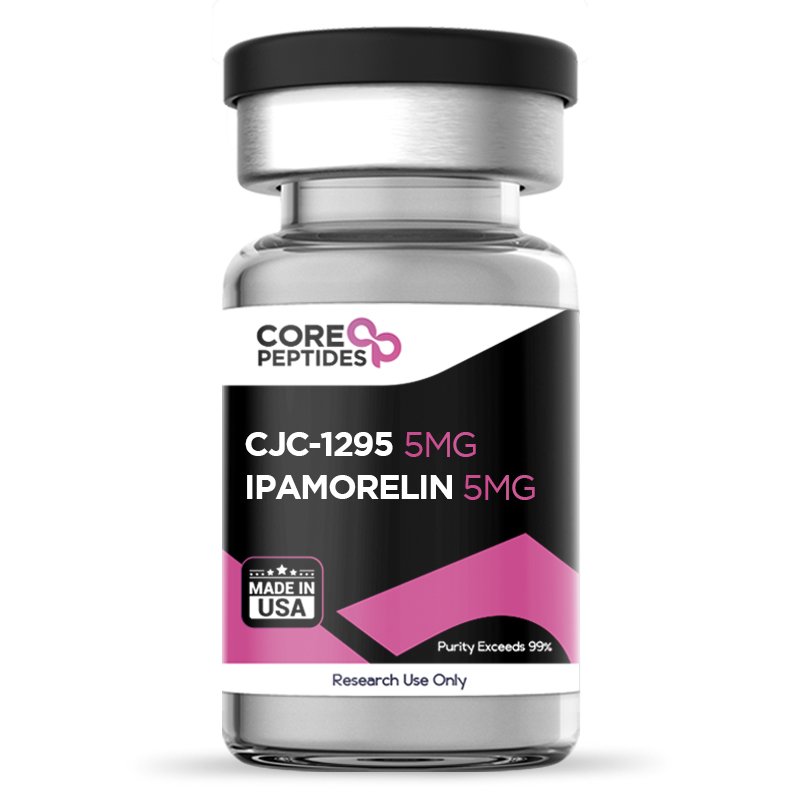
CJC-1295 & Ipamorelin Blend (10mg)
Ipamorelin and CJC-1295 are both considered to be growth hormone secretagogues. Ipamorelin is a synthetic pentapeptide,(1) and CJC-1295 peptide consists of 29 amino acids.(2) Ipamorelin appears to fall into a category of peptides classified as growth hormone secretagogues (GHSs). These are peptides that are assumed to stimulate the release of the growth hormones, however are not considered growth hormone releasing peptides themselves. On the other hand, CJC-1295 has also been suggested by researchers to stimulate the release of growth hormone, primarily by mimicking the actions of the naturally occurring growth hormone-releasing hormone (GHRH). Both Ipamorelin and CJC-1295 peptides have been assigned by researchers to this class, studied for similar potential actions and apparently differing only in terms of their half-life and pharmacokinetic profiles. Overview CJC-1295 & Ipamorelin peptides both are suggested by researchers to augment the levels of the growth hormones through a possible triggering of the anterior pituitary gland. Scientists consider that once triggered, growth hormones may be naturally secreted, maintaining levels of growth hormones in the organism.(3) CJC-1295 peptide is a tetrasubstituted version of GHRH 1-29, developed to represent the shortest functional sequence of GHRH. GHRH 1-29 consists of the first 29 amino acids of the native GHRH peptide, and may potentially stimulate growth hormone production in pituitary gland cells, called somatotrophs. The peptide has four amino acid substitutions in its structure, which scientists suggest may enhance its activity and resistance towards proteolytic enzymes. More specifically, the amino acids which are replaced appear to be the 2nd, 8th, 15th, and 27th amino acids. Owing to these substitutions, the peptide might be able to bind covalently to blood albumin, with trace amounts possibly able to bind to fibrinogen and immunoglobulin G (IgG). As a result, the apparent half-life of the peptide may increase from 10 mins to 30 mins.(4) This may lead to elevated levels of plasma growth hormone and insulin-like growth factor 1 (IGF-1). CJC-1295 might also be linked with the purported drug affinity complex (DAC) element, which may attach to plasma proteins. In particular, the DAC element in CJC-1295 alludes to the connection of N-epsilon-3-maleimidopropionamide derivative of lysine at the C-terminal end. Merging the tetrasubstituted amino acid chain and the DAC element, CJC-1295 appears to display enhanced pharmacokinetics yet retains a comparable attraction to the GHRH receptors in the pituitary gland, similar to natural GHRH.(5) More specifically, researchers comment that when the peptide was “selected for further pharmacokinetic evaluation, where it was found to be present in plasma beyond 72 h.” Ipamorelin is a man-made pentapeptide, also known as NNC 26-0161, that is believed to associate with a specific receptor in the pituitary gland cells, termed the growth hormone secretagogue receptor (GHS-R1a). These receptors are considered to be located in the hypothalamus. Moreover, GHS-R1a is often referred to as the ghrelin receptors because ghrelin seems to be its primary natural ligand. Ipamorelin appears to stand out from other GHSs as a potentially more selective compound, which may possibly stimulate the release of GH levels by somatotroph cells without also increasing other hormones produced by the anterior pituitary gland, such as prolactin. When the peptide blend, sometimes also called the peptide stack, is presented in combination, research studies typically report that the Ipamorelin exerts initial action, exhibiting some sign of impact within the first two hours of presentation, and as it starts to wean off, the CJC-1295 peptide may gradually supplement action.(6) Chemical Makeup Molecular Formula: CJC-1295: C152H252N44O42 Ipamorelin: C38H49N9O5 Molecular Weight: CJC-1295: 3367.9 g/mol Ipamorelin: 711.8 g/mol Other Known Titles CJC-1295: CJC-1295 NO DAC; Mod GRF 1-29 Ipamorelin: NNC 26-0161 Research and Clinical Studies CJC-1295 & Ipamorelin Blend and Half Life Determination Clinical studies have been conducted on test subjects to determine the half life of and individual pharmacokinetic profiles of the two peptides. In one late 1990s study,(6) a clinical trial was conducted on eight male test subjects with a concentration escalation design. The level of growth hormones was monitored after every instance of peptide presentation. At the end of the study, it was suggested by the researchers that there was a single episode of growth hormone release with the highest peak at 0.67 hours, after which there was an exponential decline up to negligible concentrations of the compound. This study concluded that the Ipamorelin peptide appeared to exhibit a short half-life of 2 hours, after which the potential action appears to begin to decline. CJC-1295, by contrast, appears to have a much longer half-life. Researchers comment that a single introduction of the peptide may upregulate growth hormone production by somatotrophs for prolonged periods of time, thus apparently contributing “to an overall increase in [growth hormone] secretion … by 46%” and also potentially upregulating its main anabolic mediator insulin-like growth factor-1 (IGF-1) by 45% on average.(7) Another publication also observes that CJC-1295 may potentially upregulate “[growth hormone] concentrations by 2- to 10-fold,” and estimates that the half-life of the peptide ranges between 5.8 – 8.1 days.(8) CJC-1295 & Ipamorelin Blend General Research In this early 2000s study,(7) a clinical trial was conducted on male test subjects aged between 20 and 40 years old. Test subjects were divided into two groups; one group was presented with the placebo and the other with the peptide. Blood was sampled from the subjects one week before and after the presentation of CJC-1295 peptide (and placebo) to monitor the levels of growth hormone pulsatility. At the end of the study, it was suggested that CJC-1295 contributed to a 7.5-fold increase in the growth hormone pulsatility levels as compared to that of the placebo. Apart from apparently affecting the synthesis of growth hormone, scientists also suggest that CJC-1295 may interact with the survival and proliferation of the cells that synthesize it - the somatotroph cells in the anterior pituitary gland.(9) In one study on murine models, the authors commented that "CJC-1295 caused an increase in total pituitary RNA and GH mRNA, suggesting that proliferation of somatotroph cells had occurred, as confirmed by immunohistochemistry images.” To exert these apparent effects, CJC-1295 appears to interact with specific binding sites on the GHRH receptor protein, leading to conformational changes in the receptor structure and potentially initiating a cascade of molecular events. The binding appears to activate intracellular signaling proteins that potentially act as molecular toggles.(10) These proteins are often referred to as G-proteins, which, upon activation, might drive the generation of secondary messengers like cyclic adenosine monophosphate (cAMP) or inositol trisphosphate (IP3.(11) Secondary messengers such as cAMP may set in motion protein kinases, enzymes believed to alter distinct proteins. These kinases possess a modulatory capacity for cellular activities and might phosphorylate transcription regulators, or proteins overseeing gene modulation. Once phosphorylated, these transcription regulators could migrate into the nucleus of somatotroph cells, possibly impacting genes associated with growth hormone formation.(7) On the other hand, Ipamorelin appears to interact with the anterior pituitary gland cells via the N-terminus of GHS-R1a, which has binding sites that appear to recognize specific sequences in the secretagogue. When Ipamorelin meets this receptor, it may attach in a non-permanent way through forces like hydrogen bonds and forces between molecules called van der Waals forces. This attachment might make the receptor change its shape, which could start cell signals, mainly those involving G-proteins. GHS-R1a might work with a specific part of G-proteins called Gαq/11.(12) A main process started by GHS-R1a involves an enzyme called phospholipase C (PLC). Gαq/11 interacts with PLC, which may split a fat-like molecule, phosphatidylinositol 4,5-bisphosphate (PIP2), into two messaging molecules: IP3 (Inositol trisphosphate) and DAG (Diacylglycerol). IP3 appears to attach to places on a cell part called the endoplasmic reticulum, causing calcium ions (Ca2+) to be released. Also, DAG might turn on an enzyme called protein kinase C (PKC), which may add phosphate groups to other signaling molecules. All these steps might end with the ‘turning on’ of proteins that help release growth hormone from certain cells in the pituitary gland.(13) CJC-1295 & Ipamorelin Blend and Nitrogen Balance The apparent synergistic action of CJC-1295 and Ipamorelin on the production of growth hormone by the somatotroph cells in the anterior pituitary gland appears to result in a positive nitrogen balance and potential increase in lean mass in test models. In a particular study, investigators sought to probe the metabolic capabilities of Ipamorelin within the context of certain hepatic markers related to alpha-amino-nitrogen processing during an artificlaly triggered catabolism. The team evaluated the liver's ability to produce urea-N (CUNS), a potential metric of nitrogen processing within the liver. They examined the observable levels of messenger RNA (mRNA) linked to enzymes of the urea cycle in the liver, gauged the overall nitrogen equilibrium, and postulated the nitrogen quantities in different organs. It was proposed that Ipamorelin might have led to a 20% decline in CUNS, in contrast to the catabolic condition that was artificially prompted by the researchers. Moreover, it could have conceivably decreased the manifestation of urea cycle enzymes, reinstated nitrogen equilibrium, and theoretically adjusted or enhanced the nitrogen values in organs.(14) CJC-1295 & Ipamorelin peptide blend is available for research and laboratory purposes only. Please review and adhere to our Terms and Conditions before ordering. References: Raun K, Hansen BS, Johansen NL, Thøgersen H, Madsen K, Ankersen M, Andersen PH. Ipamorelin, the first selective growth hormone secretagogue. Eur J Endocrinol. 1998 Nov;139(5):552-61. doi: 10.1530/eje.0.1390552. PMID: 9849822. https://pubmed.ncbi.nlm.nih.gov/9849822/ Lucie Jette et al, hGRF1-29-Albumin Bioconjugates Activate the GRF Receptor on the Anterior Pituitary in Rats: Identification of CJC-1295 as a Long Lasting GRF Analog, ResearchGate, January 2005. Raun K, Hansen BS, Johansen NL, Thøgersen H, Madsen K, Ankersen M, Andersen PH. Ipamorelin, the first selective growth hormone secretagogue. Eur J Endocrinol. 1998 Nov;139(5):552-61. doi: 10.1530/eje.0.1390552. PMID: 9849822 https://pubmed.ncbi.nlm.nih.gov/9849822/ The Discovery of Growth Hormone-Releasing Hormone: An Update https://onlinelibrary.wiley.com/doi/full/10.1111/j.1365-2826.2008.01740.x Jetté, L., Léger, R., Thibaudeau, K., Benquet, C., Robitaille, M., Pellerin, I., Paradis, V., van Wyk, P., Pham, K., & Bridon, D. P. (2005). Human growth hormone-releasing factor (hGRF)1-29-albumin bioconjugates activate the GRF receptor on the anterior pituitary in rats: identification of CJC-1295 as a long-lasting GRF analog. Endocrinology, 146(7), 3052–3058. https://doi.org/10.1210/en.2004-1286 Gobburu JV, Agersø H, Jusko WJ, Ynddal L (September 1999). “Pharmacokinetic-pharmacodynamic modeling of ipamorelin, a growth hormone releasing peptide, in human volunteers”. Pharmaceutical Research. 16 (9): 1412–6. doi:10.1023/A:1018955126402 Ionescu M, Frohman LA. Pulsatile secretion of growth hormone (GH) persists during continuous stimulation by CJC-1295, a long-acting GH-releasing hormone analog. J Clin Endocrinol Metab. 2006 Dec;91(12):4792-7. doi: 10.1210/jc.2006-1702. Epub 2006 Oct 3. PMID: 17018654. https://pubmed.ncbi.nlm.nih.gov/17018654/ Teichman SL, Neale A, Lawrence B, Gagnon C, Castaigne JP, Frohman LA. Prolonged stimulation of growth hormone (GH) and insulin-like growth factor I secretion by CJC-1295, a long-acting analog of GH-releasing hormone, in healthy adults. J Clin Endocrinol Metab. 2006 Mar;91(3):799-805. doi: 10.1210/jc.2005-1536. Epub 2005 Dec 13. PMID: 16352683. https://pubmed.ncbi.nlm.nih.gov/16352683/ Alba, M., Fintini, D., Sagazio, A., Lawrence, B., Castaigne, J. P., Frohman, L. A., & Salvatori, R. (2006). Once-daily administration of CJC-1295, a long-acting growth hormone-releasing hormone (GHRH) analog, normalizes growth in the GHRH knockout mouse. American journal of physiology. Endocrinology and metabolism, 291(6), E1290–E1294. https://doi.org/10.1152/ajpendo.00201.2006 Martin, B., Lopez de Maturana, R., Brenneman, R., Walent, T., Mattson, M. P., & Maudsley, S. (2005). Class II G protein-coupled receptors and their ligands in neuronal function and protection. Neuromolecular medicine, 7(1-2), 3–36. https://doi.org/10.1385/nmm:7:1-2:003 Newton, A. C., Bootman, M. D., & Scott, J. D. (2016). Second Messengers. Cold Spring Harbor perspectives in biology, 8(8), a005926. https://doi.org/10.1101/cshperspect.a005926 Yin, Y., Li, Y., & Zhang, W. (2014). The growth hormone secretagogue receptor: its intracellular signaling and regulation. International journal of molecular sciences, 15(3), 4837–4855. https://doi.org/10.3390/ijms15034837 Bill, C. A., & Vines, C. M. (2020). Phospholipase C. Advances in experimental medicine and biology, 1131, 215–242. https://doi.org/10.1007/978-3-030-12457-1_9 Aagaard, N. K., Grøfte, T., Greisen, J., Malmlöf, K., Johansen, P. B., Grønbaek, H., Ørskov, H., Tygstrup, N., & Vilstrup, H. (2009). Growth hormone and growth hormone secretagogue effects on nitrogen balance and urea synthesis in steroid treated rats. Growth hormone & IGF research : official journal of the Growth Hormone Research Society and the International IGF Research Society, 19(5), 426–431. https://doi.org/10.1016/j.ghir.2009.01.001 Dr. MarinovDr. Marinov (MD, Ph.D.) is a researcher and chief assistant professor in Preventative Medicine & Public Health. Prior to his professorship, Dr. Marinov practiced preventative, evidence-based medicine with an emphasis on Nutrition and Dietetics. He is widely published in international peer-reviewed scientific journals and specializes in peptide therapy research.
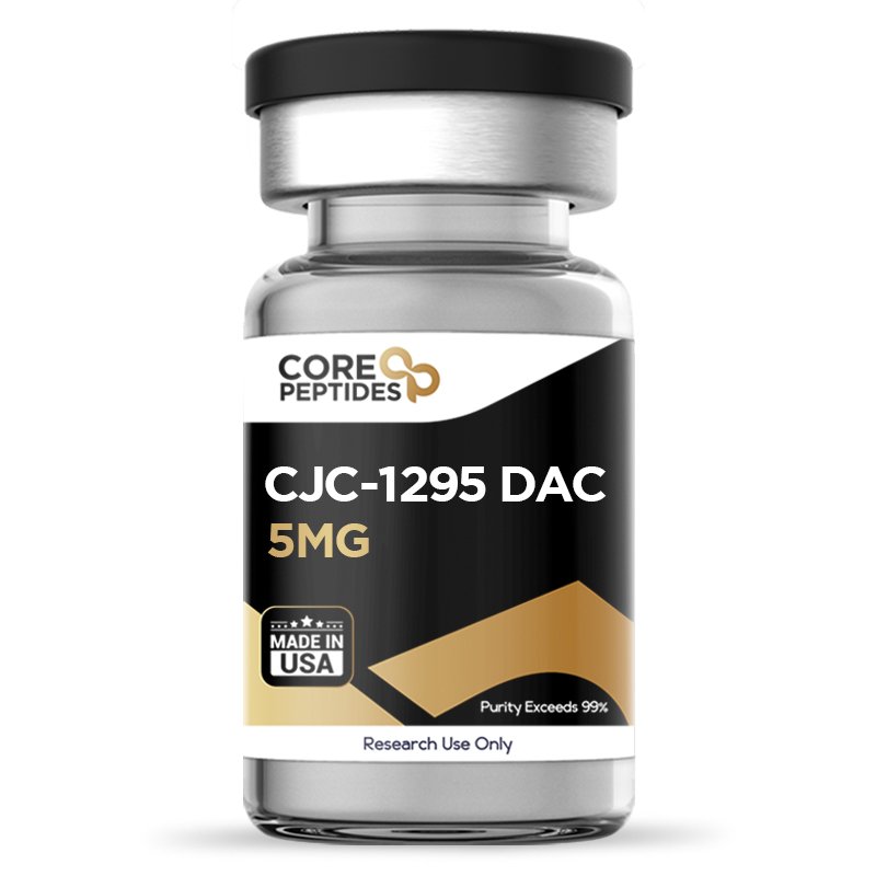
CJC-1295 DAC (5mg)
CJC-1295 DAC is a peptide that researchers posit may function similarly to growth hormone-releasing hormone (GHRH), potentially increasing the endogenous production of growth hormone (GH) within the organism. The CJC-1295 DAC peptide is a synthetic 29 amino acid analog of GHRH.(1) It is the shortest functional analog of GHRH that still has the potential capacity to trigger GH release from the somatotroph cells in the pituitary gland. Moreover, 4 of the original 29 amino acids in this fragment are substituted in CJC-1295 DAC, to possibly improve the pharmacokinetics of the peptide and prolong its half-life. The DAC component is a biochemical complex that may further increase the half-life of the peptide. This phenomenon might be ascribed to the alleged capability of the DAC to bind to proteins present in plasma. More precisely, the DAC component appears to entail the attachment of a lysine derivative, identified as N-epsilon-3-maleimidopropionamide, to the C terminus of CJC-1295 DAC. Integrating this modified amino acid sequence with the DAC component might conceivably enhance the pharmacokinetics of CJC-1295 DAC, extending its half-life to approximately 8 days. Simultaneously, it appears to maintain a significant affinity for GHRH receptors, similar to the affinity exhibited by CJC-1295 without DAC.(2) This does not imply that CJC without DAC has no relevance or use in present day research. For example, CJC-1295 without DAC & Ipamorelin blend has been studied for its suggested synergistic potential, as combining GHRH analogs with peptides that appear to activate the ghrelin receptors in the pituitary gland. It is also posited to result in greater GH-synthesis response from the somatotroph cells.(3) Overview CJC-1295 DAC is recognized by many names: CJC-1295, CJC-1295 with DAC, DAC:GRF, long-acting GHRH analog, and synthetic GHRH analog. Theoretically, as the peptide is considered a releasing factor for growth hormone, CJC-1295 DAC has been studied for its potential role in: The reduction of fat mass by using fat cells as an energy source. It may lead to increased muscle mass via the promotion of protein synthesis. Since growth hormone is considered to promote bone growth and improved joint and connective tissue, CJC-1295 DAC peptide may potentially improve bone mass and thereby reduce the risk of damage. Studies have suggested that GHRH may support the centers in the nervous system for sleep, and potentially this action may be mirrored by analogs like CJC-1295 DAC.(4) Chemical Makeup Molecular Formula: C152H252N44O42 Molecular Weight: 3367.95 g/mol Other Known Titles: Tetrasubsituted GRF 1-29 with DAC Research and Clinical Studies CJC-1295 DAC Peptide Mechanism of Action Researchers conducted two clinical studies in 2006 to examine the action of CJC-1295 DAC. In the first study, CJC-1295 DAC or a placebo was presented in one of four ascending concentrations. In the second study, CJC-1295 DAC was presented repeatedly at a single concentration. According to the results, after the introduction of CJC-1295 DAC, there appeared to be an increase in GH and insulin-like growth factor-1 (IGF-I) levels among the research models.(5)CJC-1295 DAC is thought to elevate levels of IGF-1 by increasing growth hormone production, which in turn may bind to receptors on liver cells, potentially triggering a cascade of intracellular signaling processes. This binding might activate the Janus kinase-signal transducer and activator of the transcription (JAK-STAT) signaling pathway. Subsequently, the activated STAT proteins might migrate to the nucleus, where they may attach to specific DNA sequences considered to be response elements, potentially resulting in the transcription of the IGF-I gene. It is theorized that the IGF-I produced in liver cells may be transported to various target tissues. Additionally, it is believed that many tissues possess GH receptors, which, upon activation, may lead to the production of IGF-I within the tissues themselves. IGF-I is considered a potent hormone that may play a key role in promoting growth, suggesting it mediates many growth and anabolic effects of growth hormone. It is hypothesized to encourage cell growth and proliferation, as well as the enlargement and strengthening of tissues and organs, possibly aiding in protein synthesis and cellular expansion. Preliminary exposure to CJC-1295 DAC in experimental models has been suggested to significantly affect average growth hormone levels, with studies reporting an apparent increase of 2- to 10-fold for 6 days or possibly longer. Moreover, CJC-1295 DAC has been suggested to lead to dependent increases in average IGF-I levels by 1.5- to 3-fold for about 9–11 days, with suggestions that IGF-I levels may remain high for at least two weeks in experimental models. Following repeated exposure to CJC-1295 DAC, average IGF-I levels appear to remain elevated above baseline for up to 28 days. Notably, data suggests a cumulative effect following repeated exposure of the compound.(5) In 2006, another group of scientists assessed the GH pulsatility after a single occurrence of CJC-1295 DAC. They found out that there appeared to be an increase of about 50% in mean GH secretion and IGF-I levels after a single presentation of CJC-1295 DAC.(6) Researchers have suggested that the peptide might contribute to an increase in peak growth hormone levels by as much as 7.5 times in the models studied.(6) It seems that CJC-1295 DAC may interact with certain binding sites on the growth hormone-releasing hormone (GHRH) receptor protein. This interaction may lead to changes in the receptor's structure, potentially triggering a series of molecular processes. This interaction is believed to stimulate specific intracellular signaling proteins, often referred to as G-proteins.(7) Upon activation, these proteins may promote the production of secondary messengers, such as cyclic adenosine monophosphate (cAMP) or inositol trisphosphate (IP3), which are considered to play crucial roles in cellular signaling pathways.(8) Secondary messengers, including cAMP, are thought to activate protein kinases, which are enzymes considered capable of modifying specific proteins. These kinases are considered able to regulate cellular functions by phosphorylating transcription regulators, the proteins responsible for controlling gene expression. Once phosphorylated, these transcription regulators are speculated to move into the nucleus of somatotroph cells, where they might influence the genes involved in the production of growth hormone. This intricate cascade of events highlights the potential of CJC-1295 DAC to modulate growth hormone levels through a complex network of molecular interactions. Additional animal studies were conducted to evaluate the potential of CJC-1295 DAC. One study evaluated murine models presented with either the peptide or a placebo. The researchers concluded that exposing the murine models daily CJC-1295 DAC might completely normalize growth. Another finding was that CJC-1295 DAC presented every 2 or 3 days appeared to produce intermediate results, indicating a probable interval-dependent action.(9) Furthermore, this study suggests that CJC-1295 DAC might potentially impact body composition, seemingly by increasing muscle tissue hypertrophy while not impacting, or even possibly reducing, fat tissue levels. The murine models in this study appeared to have a deletion of the GHRH gene (referred to as GHRHKO); observations suggested that CJC-1295 DAC may boost GH synthesis, leading to a beneficial alteration in body composition. Exposure to CJC-1295 DAC in these GHRHKO murine models appeared to preserve normal levels of lean mass, unlike in models that were not exposed and exhibited suboptimal lean mass levels. Furthermore, the amount of subcutaneous fat mass stayed consistent with control levels in all groups associated with the peptide, whereas GHRHKO murine models without CJC-1295 DAC exposure exhibited signs of increased fat levels. This indicates that CJC-1295 DAC might positively affect muscle and bone structure without promoting an increase in fat accumulation. Additionally, the study noted a possible increase in pituitary RNA and GH mRNA levels following CJC-1295 DAC exposure, suggesting an enhanced presence of somatotroph cells—those believed to produce growth hormone in the pituitary gland. The authors commented that "CJC-1295 caused an increase in total pituitary RNA and GH mRNA, suggesting that proliferation of somatotroph cells had occurred, as confirmed by immunohistochemistry images.” (9) CJC-1295 DAC Peptide Half-life In its original form, CJC-1295 DAC uses a technology referred to as Drug Affinity Complex (DAC).(1) Contrary to GHRH, which is considered to boast a half-life of approximately 7 minutes, researchers report CJC-1295 without DAC to exhibit a longer half-life of 30 minutes due to its truncated 29 amino acid fragment, and 4 of the original amino acids in this fragment are replaced. Alterations to the peptide structure, specifically at the 2nd, 8th, 15th, and 27th amino acid positions, are thought to potentially improve the peptide's stability against breakdown by the enzyme dipeptidyl peptidase-4. These alterations are detailed as follows: At the 2nd position, L-alanine is substituted by D-alanine, a change believed to bolster resistance against enzymatic degradation. At the 8th position, asparagine is replaced with glutamine, a modification that might reduce the risk of asparagine rearrangement and amide hydrolysis. The substitution of glycine with alanine at the 15th position is theorized to enhance the peptide's bioactivity. The alteration from methionine to leucine at the 27th position is considered to potentially prevent methionine oxidation. These modifications aim to enhance the peptide's resilience and functional efficacy by mitigating enzymatic degradation and improving stability under physiological conditions. In addition, the half-life of the peptide appears to be additionally extended to 6-8 days due to the DAC technology.(10) CJC-1295 DAC Peptide Ancillary Studies In 2005, a clinical study aimed to evaluate the mechanism of action of CJC-1295 DAC in models of immunodeficiency virus (HIV) associated with visceral obesity. In this study, the models would be presented with CJC-1295 DAC for 3 months, followed by a 6-week follow-up period. However, this study was terminated during recruitment, and no related results were posted.(11) According to a 2009 Norwegian study, a compound was submitted for analysis to evaluate whether it contained prohibited substances or not. The researchers of the Norwegian Doping Control Laboratory and School of Pharmacy reported that this substance was CJC-1295 DAC. In their published article they concluded that "CJC-1295 DAC is a releasing factor for growth hormone".(1) CJC-1295 DAC peptide is available for research and laboratory purposes only. Please review and adhere to our Terms and Conditions before ordering. References: Henninge J, Pepaj M, Hullstein I, Hemmersbach P. Identification of CJC-1295, a growth-hormone-releasing peptide, in an unknown pharmaceutical preparation. Drug Testing and Analysis. 2010 Nov-Dec;2(11-12):647-650. DOI: 10.1002/dta.233. Jetté L, Léger R, Thibaudeau K, Benquet C, Robitaille M, Pellerin I, Paradis V, van Wyk P, Pham K, Bridon DP. Human growth hormone-releasing factor (hGRF)1-29-albumin bioconjugates activate the GRF receptor on the anterior pituitary in rats: identification of CJC-1295 as a long-lasting GRF analog. Endocrinology. 2005 Jul;146(7):3052-8. doi: 10.1210/en.2004-1286. Epub 2005 Apr 7. PMID: 15817669. Sinha DK, Balasubramanian A, Tatem AJ, Rivera-Mirabal J, Yu J, Kovac J, Pastuszak AW, Lipshultz LI. Beyond the androgen receptor: the role of growth hormone secretagogues in the modern management of body composition in hypogonadal males. Transl Androl Urol. 2020 Mar;9(Suppl 2):S149-S159. doi: 10.21037/tau.2019.11.30. PMID: 32257855; PMCID: PMC7108996. Steiger A, Holsboer F. Neuropeptides and human sleep. Sleep. 1997 Nov;20(11):1038-52. PMID: 9456470. Teichman SL, Neale A, Lawrence B, Gagnon C, Castaigne JP, Frohman LA. Prolonged stimulation of growth hormone (GH) and insulin-like growth factor I secretion by CJC-1295, a long-acting analog of GH-releasing hormone, in healthy adults. J Clin Endocrinol Metab. 2006 Mar;91(3):799-805. doi: 10.1210/jc.2005-1536. Epub 2005 Dec 13. PMID: 16352683. Ionescu M, Frohman LA. Pulsatile secretion of growth hormone (GH) persists during continuous stimulation by CJC-1295, a long-acting GH-releasing hormone analog. J Clin Endocrinol Metab. 2006 Dec;91(12):4792-7. doi: 10.1210/jc.2006-1702. Epub 2006 Oct 3. PMID: 17018654. Martin, B., Lopez de Maturana, R., Brenneman, R., Walent, T., Mattson, M. P., & Maudsley, S. (2005). Class II G protein-coupled receptors and their ligands in neuronal function and protection. Neuromolecular medicine, 7(1-2), 3–36. https://doi.org/10.1385/nmm:7:1-2:003 Newton, A. C., Bootman, M. D., & Scott, J. D. (2016). Second Messengers. Cold Spring Harbor perspectives in biology, 8(8), a005926. https://doi.org/10.1101/cshperspect.a005926 Alba M, Fintini D, Sagazio A, Lawrence B, Castaigne JP, Frohman LA, Salvatori R. Once-daily administration of CJC-1295, a long-acting growth hormone-releasing hormone (GHRH) analog, normalizes growth in the GHRH knockout mouse. Am J Physiol Endocrinol Metab. 2006 Dec;291(6):E1290-4. doi: 10.1152/ajpendo.00201.2006. Epub 2006 Jul 5. PMID: 16822960. Van Hout MC, Hearne E. Netnography of Female Use of the Synthetic Growth Hormone CJC-1295: Pulses and Potions. Subst Use Misuse. 2016 Jan 2;51(1):73-84. doi: 10.3109/10826084.2015.1082595. Epub 2016 Jan 15. PMID: 26771670. ClinicalTrials.gov, A service of the US National Institutes of Health. Available at: http://clinicaltrials.gov/ct2/show/NCT00267527 (27 June 2010). Dr. MarinovDr. Marinov (MD, Ph.D.) is a researcher and chief assistant professor in Preventative Medicine & Public Health. Prior to his professorship, Dr. Marinov practiced preventative, evidence-based medicine with an emphasis on Nutrition and Dietetics. He is widely published in international peer-reviewed scientific journals and specializes in peptide therapy research.

Sermorelin (5mg)
Sermorelin is a 29 amino acid peptide, the shortest synthetically developed peptide that may potentially induce biological activity at the receptors for the growth hormone-releasing hormone (GHRH).(2) Sermorelin polypeptide is an analog of the GHRH factor consisting of GHRH (1-29 acid)-amide. Due to this structural and functional mimicry, Sermorelin has been studied across multiple branches of scientific research involving growth hormone deficiency models.(3) It was in the early 1980s that the action of Sermorelin, classified as a growth hormone-releasing fragment GHRF (1-29) amide, was first explored. Several research studies were conducted on rats where exogenous GHRF (1-29) amide was introduced in conscious and anesthetized rats. It was observed that the presence of GHRF appeared to stimulate the pituitary gland and promote growth. Following this theory, Sermorelin and similar compounds have become the subject of further research in growth hormone deficiency models.(4) Overview Sermorelin is suggested to be a growth hormone analog constituting the first 29 amino acids out of the usual 44 amino acids found in growth hormone-releasing hormone (GHRH). Researchers posit that Sermorelin binds with the GHRH receptors found on the pituitary gland and suggest further that the synthetic peptide may stimulate secretion of growth hormone (hGH). Thus, Sermorelin is believed to maintain the fundamental function of GHRH, possibly stimulating the GHRH receptors in the pituitary gland and leading to sporadic release of growth hormone despite its reduced amino acid sequence. This mechanism is thought to result in increased levels of insulin-like growth factor-1 (IGF-1), primarily recognized for its role in the anabolic actions of growth hormone. The estimated half-life of Sermorelin is around 11 to 12 minutes. A major potential advantage of the peptide is that due to its apparent GHRH receptor specificity, it may not induce any significant change in the levels of other endocrine markers such as prolactin, insulin, cortisol, glucose, or thyroid hormones.(6) Chemical Makeup Molecular Formula: C149H246N44O42S Molecular Weight: 3357.93 g/mol Other Known Titles: GRF 1-29 Research and Clinical Studies Sermorelin and GHRH Receptors Sermorelin is thought to interact with GHRH receptors through complex molecular mechanisms, possibly triggering various cellular signaling pathways. It is hypothesized that upon binding to the GHRH receptor, Sermorelin may alter the receptor's structure, potentially initiating a series of intracellular signaling events.(12) Some researchers propose that Sermorelin might enhance the production of cyclic adenosine monophosphate (cAMP) in specific cells. This enhancement may occur through the activation of adenylate cyclase, which is suggested to convert ATP into cAMP. Higher levels of cAMP might lead to the activation of protein kinase A (PKA), a key enzyme in cellular signaling processes. PKA might phosphorylate various target proteins, thereby initiating further cellular responses. The potential activation of the GHRH receptor by Sermorelin, along with the ensuing cAMP-PKA signaling cascade, is thought to possibly promote the secretion and distribution of growth hormone (hGH) from somatotroph cells in the pituitary gland. The secreted hGH is also believed to contribute to the synthesis of insulin-like growth factor-1 (IGF-1).(12) Sermorelin Peptide and Growth Velocity Researchers reported positive results in the idiopathic GH deficiency when Sermorelin was presented to underdeveloped animal models. Increased growth and height velocity rate was observed within 12 months of consistent, continuous peptide presence. These elevated levels were reported to be sustained for an average of 36 months after continuous presence.(7) Sermorelin Peptide and Anabolic Research Outcomes Preliminary findings from one investigation indicate that Sermorelin may lead to an 82% enhancement in average growth hormone levels, with actions persisting for approximately two hours.(13) A separate study conducted over 16 weeks hypothesizes that Sermorelin might elevate growth hormone levels by as much as 107%, and increase IGF-1 levels by about 28%.(14) The research further suggests a possible increase in lean body mass of approximately 2.78 lbs (1.26 kg), with no significant change in fat mass. These actions are tentatively attributed to the peptide's capacity to boost growth hormone levels, and in turn, IGF-1, which is considered a potential anabolic agent influencing growth hormone activity. The most noteworthy outcomes identified by the researchers include observations that there was “a gain of 1.26 ± 0.52 kg (P < 0.05) in LBM” and that “skin thickness increased significantly.” Sermorelin Peptide and Lipodystrophy Scientists carried out a controlled clinical study involving 31 HIV-positive subjects with lipodystrophy, to investigate the potential impact of Sermorelin.(8) All 31 subjects were divided into two groups, where one was presented with Sermorelin, and the other group with a placebo for 12 weeks. Following the study, it was suggested by the research team that growth hormone levels appeared significantly increased in Sermorelin subjects as compared to the ones given a placebo. Levels of insulin-like growth factor (IGF-1) had apparently increased – resulting in increased lean body mass in the peptide group. Abdominal visceral fat and the ratio of trunk to lower extremity fat were reported by the researchers to be significantly reduced. There was no other reported change in the glucose or insulin levels.(8) Sermorelin Peptide and Cognition In the early 2000s, clinical research studies were conducted with 89 subjects between 68 and 69 years of age to explore the correlation (if any) between tapering growth hormone release and impaired cognition. Scientists consider that with increasing age, levels of growth hormone naturally decline, which may result in reduced physiological functions, including cognition (i.e. ability to collect, process, and recollect information). Following the introduction of Sermorelin, it was observed that there was an apparent improved performance in the Wechsler Adult Intelligence Scale (WAIS) – i.e. improved IQ levels, picture arrangement tests, and verbal tests - amongst the test subjects.(9) Sermorelin Peptide and Tumor Cells A clinical study was designed where 1,018 glioma subjects were presented with over 4,000 compounds each, and following each presentation, a DRS was determined for all compounds, for each subject. Following the results of the study, it appeared that the Sermorelin compound reportedly induced the most sensitivity in the test subjects. Upon analysis, it was suggested by the researchers that this may be due to the potential of Sermorelin to block the tumor cell cycles, thereby possibly preventing tumor cell proliferation.(10) Sermorelin Peptide and Hypogonadism Initial research into the peptide suggested that Sermorelin might be impactful in increasing lean mass. One study sought to explore if Sermorelin had potential in hypogonadism (which is considered to stem from additional fat mass). Test models were divided into two groups where one group was presented with Sermorelin followed by GHRH 1-40, with a one week interval between the two compounds, whereas the other group was given the same combination in reverse order. Following the study, it was reported by the researchers that for both groups, the Sermorelin appeared to stimulate the release of FSH and LH, which might stimulate testosterone production. This initial research spawned additional studies, including one clinical study that included 19 male subjects, 9 of whom were aged between 22 and 33 years of age, and 10 were aged between 60 and 78 years of age. The more elderly subjects were presented with one of two concentrations of Sermorelin for a period of 28 days, with an interval of 14 days in between the two instances. Testosterone levels in the elderly subjects reportedly increased after the presentation of Sermorelin; however, it should be noted that the levels were not statistically significant. Furthermore, researchers suggested that elevated levels of growth hormones, possibly induced by Sermorelin presence, appeared to be at peak during the night time, for all test subjects, as compared during the day.(11) Sermorelin peptide is available for research and laboratory purposes only. Please review and adhere to our Terms and Conditions before ordering. References: Garcia JM, Merriam GR, Kargi AY. Growth Hormone in Aging. [Updated 2019 Oct 7]. In: Feingold KR, Anawalt B, Boyce A, et al., editors. Endotext [Internet]. South Dartmouth (MA): MDText.com, Inc.; 2000. https://www.ncbi.nlm.nih.gov/books/NBK279163/?report=reader Prakash, A, and K L Goa. “Sermorelin: a review of its use in the diagnosis and treatment of children with idiopathic growth hormone deficiency.” BioDrugs : clinical immunotherapeutics, biopharmaceuticals and gene therapy vol. 12,2 (1999): 139-57. https://pubmed.ncbi.nlm.nih.gov/18031173/ National Center for Biotechnology Information. "PubChem Compound Summary for CID 16129620, Sermorelin" PubChem Clark, R G, and I C Robinson. “Growth induced by pulsatile infusion of an amidated fragment of human growth hormone releasing factor in normal and GHRF-deficient rats.” Nature vol. 314,6008 (1985): 281-3. https://pubmed.ncbi.nlm.nih.gov/2858818/ Drugs at FDA: FDA Approved Drugs. https://www.accessdata.fda.gov/scripts/cder/daf/index.cfm?event=overview.process&ApplNo=020443 Junichi I. et al, Growth hormone secretagogues: history, mechanism of action, and clinical development, JSCM Rapid Communications Vol. 3 Issue 1, 09 February 2020. https://onlinelibrary.wiley.com/doi/full/10.1002/rco2.9 Prakash, A, and K L Goa. “Sermorelin: a review of its use in the diagnosis and treatment of children with idiopathic growth hormone deficiency.” BioDrugs : clinical immunotherapeutics, biopharmaceuticals and gene therapy vol. 12,2 (1999): 139-57. https://pubmed.ncbi.nlm.nih.gov/18031173/ Koutkia, Polyxeni et al. “Growth hormone-releasing hormone in HIV-infected men with lipodystrophy: a randomized controlled trial.” JAMA vol. 292,2 (2004): 210-8. https://pubmed.ncbi.nlm.nih.gov/15249570/ Vitiello, Michael V et al. “Growth hormone releasing hormone improves the cognition of healthy older adults.” Neurobiology of aging vol. 27,2 (2006): 318-23. https://pubmed.ncbi.nlm.nih.gov/16399214/ Chang, Yuanhao et al. “A potentially effective drug for patients with recurrent glioma: sermorelin.” Annals of translational medicine vol. 9,5 (2021): 406. doi:10.21037/atm-20-6561. https://www.ncbi.nlm.nih.gov/pmc/articles/PMC8033379/ Sinha, Deepankar K et al. “Beyond the androgen receptor: the role of growth hormone secretagogues in the modern management of body composition in hypogonadal males.” Translational andrology and urology vol. 9,Suppl 2 (2020): S149-S159. doi:10.21037/tau.2019.11.30. https://www.ncbi.nlm.nih.gov/pmc/articles/PMC7108996/ Zhou, F., Zhang, H., Cong, Z., Zhao, L. H., Zhou, Q., Mao, C., Cheng, X., Shen, D. D., Cai, X., Ma, C., Wang, Y., Dai, A., Zhou, Y., Sun, W., Zhao, F., Zhao, S., Jiang, H., Jiang, Y., Yang, D., Eric Xu, H., … Wang, M. W. (2020). Structural basis for activation of the growth hormone-releasing hormone receptor. Nature communications, 11(1), 5205. https://doi.org/10.1038/s41467-020-18945-0 Vittone, J., Blackman, M. R., Busby-Whitehead, J., Tsiao, C., Stewart, K. J., Tobin, J., Stevens, T., Bellantoni, M. F., Rogers, M. A., Baumann, G., Roth, J., Harman, S. M., & Spencer, R. G. (1997). Effects of single nightly injections of growth hormone-releasing hormone (GHRH 1-29) in healthy elderly men. Metabolism: clinical and experimental, 46(1), 89–96. https://doi.org/10.1016/s0026-0495(97)90174-8 Khorram, O., Laughlin, G. A., & Yen, S. S. (1997). Endocrine and metabolic effects of long-term administration of [Nle27]growth hormone-releasing hormone-(1-29)-NH2 in age-advanced men and women. The Journal of clinical endocrinology and metabolism, 82(5), 1472–1479. https://doi.org/10.1210/jcem.82.5.3943 Dr. MarinovDr. Marinov (MD, Ph.D.) is a researcher and chief assistant professor in Preventative Medicine & Public Health. Prior to his professorship, Dr. Marinov practiced preventative, evidence-based medicine with an emphasis on Nutrition and Dietetics. He is widely published in international peer-reviewed scientific journals and specializes in peptide therapy research.
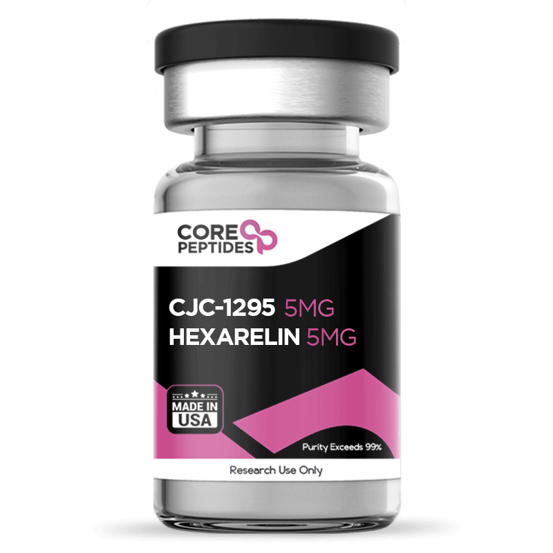
CJC-1295 (Mod GRF 1-29) & Hexarelin Blend (10mg)
CJC-1295 (no DAC) and Hexarelin are research peptides that have been studied by researchers in the interest of learning more about growth hormone synthesis by pituitary cells. Many relevant scientific observations have been drawn from such studies. Specifically, these peptides appear to interact with different receptors on anterior pituitary gland cells and stimulate the synthesis and release of this hormone. Rather than interacting via the same receptors, CJC-1295 (no DAC) and Hexarelin belong to different classes of research peptides that interact with pituitary cells via different receptors. Previous research has suggested that simultaneous activation of both receptor types may initiate synergistic actions; hence, the combination of CJC-1295 (no DAC) and Hexarelin may be of interest to scientists. Chemical Makeup Other Known Titles CJC-1295 (no DAC): Modified GRF 1-29 Hexarelin: Examorelin, P-23905, and MF-6003 Molecular Weight: CJC-1295 (no DAC):9 g/mol Hexarelin:05 g/mol Molecular Formula: CJC-1295 (no DAC): C152H252N44O42 Hexarelin: C47H58N12O6 Research and Clinical Studies CJC-1295 (no DAC) Interactions with GHRH Receptors CJC-1295 (no DAC) and Hexarelin appear to interact with pituitary cells via different receptors. CJC-1295 (no DAC) may interact with the cells via the GHRH receptor, which is posited to be the main type of receptor on these cells regulating growth hormone synthesis. Normally, these receptors respond to the endogenous growth hormone-releasing hormone (GHRH), which has a 44-amino-acid structure. Researchers believe CJC-1295 (no DAC) may mimic that structure. This is suspected in part because the peptide consists of the first 29 amino acids from GHRH with four specific modifications. The first 29 amino acids are posited to retain a GHRH-receptor affinity while being a shorter molecule. Specifically, this is considered by said researchers to be the shortest molecule that may stimulate growth hormone release via the GHRH receptors. To further support the potential of CJC-1295 (no DAC), the peptide bears four modifications, which, according to researchers such as Jette et al., involve the amino acids at positions 2, 8, 15, and 27.(1) Specifically, the modifications involve replacements at: At position 2, alanine is replaced with D-alanine, which may make the peptide less susceptible to common proteolytic enzymes. At position 8, asparagine is replaced with lysine, introducing an extra positive charge that may subtly change the surface charge and potentially support stronger interaction with the GHRH receptor.s At position 15, histidine is replaced with D-phenylalanine, adding a second D-amino acid that may further slow enzymatic degradation. At position 27, cysteine is replaced with N-methylglycine (sarcosine), which may hinder specific peptidases from rapidly dismantling the peptide backbone. This may allow the peptide to remain active for longer and to produce a more sustained receptor signal, possibly. Despite these changes, the downstream signaling is thought to resemble that of endogenous GHRH. The peptide is hypothesized to activate adenylyl cyclase, leading to the conversion of ATP into cyclic AMP (cAMP). Rising cAMP levels may then activate protein kinase A, which is proposed to phosphorylate targets that open voltage-dependent calcium channels on pituitary somatotroph membranes. The resulting calcium influx may trigger vesicle fusion and promote the release of stored growth hormone. Apart from upregulating growth hormone release, the peptide may also upregulate the overall capacity for growth hormone synthesis. An experiment by Alba et al. suggests the peptide "caused an increase in total pituitary RNA and GH mRNA, suggesting that proliferation of somatotroph cells had occurred, as confirmed by immunohistochemistry images.”(2) This pattern suggests that the peptide may also potentially support somatotroph number and GH gene transcription. Hexarelin Interactions with GHS-R1a Rather than a GHRH analog, Hexarelin has been referred to by experts as a growth hormone secretagogue (GHS). This is a class of peptides that react with pituitary cells via a different set of receptors called the growth hormone secretagogue receptors 1a (GHS-R1a). Experimental work undertaken in laboratory settings by Bowers et al. and Yin et al. has suggested that GHS-R1a is also the main receptor for ghrelin, also referred to as the hunger hormone.(3)(4) When Hexarelin binds to these receptors, it may trigger a signaling cascade that looks different from GHRH-type peptides. One potential pathway involves activation of phospholipase C, which may then generate two second messengers: IP₃ and diacylglycerol (DAG). IP₃ is thought to release calcium from intracellular stores. At the same time, diacylglycerol may activate protein kinase C. Higher intracellular calcium, together with PKC activation, is then posited to support growth hormone release from somatotroph cells. Thu, the peptide appears to induce the release of growth hormone by a different mechanism, potentially complementary to those of GHRH-analogs like CJC-1295 (no DAC). CJC-1295 (no DAC) & Hexarelin Potential on Hormone Synthesis Experiments suggest that the growth hormone release induced by CJC-1295 (no DAC) & Hexarelin from pituitary cells may surpass the peaks typically observed in control settings. For example, an experiment with CJC-1295 (no DAC) by Khorram et al. suggests that this kind of peptide may raise growth hormone output from pituitary cells by roughly 70–100% over a 12-hour window in laboratory settings.(5) The authors observed the most pronounced peak in the first two hours after exposure. In their report, 2-hour integrated growth hormone secretion seemed to rise approximately 6-fold from about 200–300 to roughly 1,100–1,600 µg/L·min. Growth hormone released in this way may then bind to GH receptors on peripheral cells and possibly drive IGF-1 production, which is viewed as a key downstream mediator of growth hormone’s anabolic actions. In that experiment, mean IGF-1 levels appeared to increase by around 27% after prolonged CJC-1295 (no DAC) exposure. Similarly, research by Imbimbo et al. has suggested that Hexarelin itself may drive an increase in growth hormone release from pituitary cells. Compared with a baseline near 3.9 ng/mL under placebo conditions, Hexarelin seemed to push peak growth hormone levels up to about 55 ng/mL, where the response plateaued. The maximal stimulus appeared within 30–40 minutes, followed by a gradual fall back toward baseline over about four hours, with an estimated half-life in the range of 50–60 minutes. This pattern suggests a strong but short-lived growth hormone pulse after Hexarelin exposure. The scientists also observed that "plasma glucose, luteinising hormone, follicle-stimulating hormone, thyroid-stimulating hormone” were unaffected. CJC-1295 (no DAC) & Hexarelin Synergistic Potential Currently, there are no experiments conducted in laboratory settings that have evaluated a combination of CJC-1295 (no DAC) & Hexarelin. Yet, the research that is available has investigated blends between Hexarelin and other GHRH-analogs, such as the full-length GHRH, from which CJC-1295 (no DAC) is derived at length. Notably, several studies by Arvat et al. evaluated GHRH and Hexarelin individually or in a blend, and suggest that when pituitary cells are exposed simultaneously to Hexarelin and GHRH, activating both types of receptors may exert synergistic actions.(7) When each compound was exposed to research models alone, Hexarelin appeared to generate a much larger growth hormone synthesis measured as an area under the curve (about 2,200.8 ± 256.9 µg/L·h) than GHRH by itself (around 792.2 ± 117.6 µg/L·h). However, when both were combined, the reported growth hormone area under the curve rose to roughly 4,259.2 ± 308.0 µg/L·h. This is higher than the sum of the two single-agent responses, which the authors interpreted as a true synergistic potential. Taken together, findings like this are often viewed by researchers as potentially suggestive that a GHRH-type peptide and a GHS like Hexarelin may provide a stronger pituitary GH signal when both receptor systems are engaged at the same time. CJC-1295 (no DAC) & Hexarelin blend is available for research and laboratory purposes only. Please review our Terms and Conditions before ordering. References: Jetté L, Léger R, Thibaudeau K, Benquet C, Robitaille M, Pellerin I, Paradis V, van Wyk P, Pham K, Bridon DP. Human growth hormone-releasing factor (hGRF)1-29-albumin bioconjugates activate the GRF receptor on the anterior pituitary in rats: identification of CJC-1295 as a long-lasting GRF analog. Endocrinology. 2005 Jul;146(7):3052-8.DOI: 10.1210/en.2004-1286. Epub 2005 Apr 7. PMID: 15817669. Alba M, Fintini D, Sagazio A, Lawrence B, Castaigne JP, Frohman LA, Salvatori R. Once-daily administration of CJC-1295, a long-acting growth hormone-releasing hormone (GHRH) analog, normalizes growth in the GHRH knockout mouse. Am J Physiol Endocrinol Metab. 2006 Dec;291(6):E1290-4. doi: 10.1152/ajpendo.00201.2006. Epub 2006 Jul 5. PMID: 16822960. Bowers CY. History of the discovery of ghrelin. Methods Enzymol. 2012;514:3-32. doi: 10.1016/B978-0-12-381272-8.00001-5. PMID: 22975043. Yin Y, Li Y, Zhang W. The growth hormone secretagogue receptor: its intracellular signaling and regulation. Int J Mol Sci. 2014 Mar 19;15(3):4837-55. doi: 10.3390/ijms15034837. PMID: 24651458; PMCID: PMC3975427. Khorram O, Laughlin GA, Yen SS. Endocrine and metabolic effects of long-term administration of [Nle27]growth hormone-releasing hormone-(1-29)-NH2 in age-advanced men and women. J Clin Endocrinol Metab. 1997 May;82(5):1472-9. doi: 10.1210/jcem 82.5.3943. PMID: 9141536. Imbimbo BP, Mant T, Edwards M, Amin D, Dalton N, Boutignon F, Lenaerts V, Wüthrich P, Deghenghi R. Growth hormone-releasing activity of hexarelin in humans. A dose-response study. Eur J Clin Pharmacol. 1994;46(5):421-5. doi: 10.1007/BF00191904. PMID: 7957536. Arvat E, Di Vito L, Gianotti L, Ramunni J, Boghen MF, Deghenghi R, Camanni F, Ghigo E. Mechanisms underlying the negative growth hormone (GH) autofeedback on the GH-releasing effect of hexarelin in man. Metabolism. 1997 Jan;46(1):83-8. doi: 10.1016/s0026-0495(97)90173-6. PMID: 9005975. Arvat, E., Gianotti, L., Di Vito, L., Imbimbo, B. P., Lenaerts, V., Deghenghi, R., Camanni, F., & Ghigo, E. (1995). Modulation of growth hormone-releasing activity of hexarelin in man. Neuroendocrinology, 61(1), 51–56. https://doi.org/10.1159/000126827 Dr. MarinovDr. Marinov (MD, Ph.D.) is a researcher and chief assistant professor in Preventative Medicine & Public Health. Prior to his professorship, Dr. Marinov practiced preventative, evidence-based medicine with an emphasis on Nutrition and Dietetics. He is widely published in international peer-reviewed scientific journals and specializes in peptide therapy research.

NAD+ (100mg / 250mg / 750mg)
NAD+ is an acronym for Nicotinamide Adenine Dinucleotide, an endogenous nucleotide that is considered to regulate primary functions such as metabolism, energy production, and DNA repair. It is also considered to act as a secondary messenger via calcium-dependent signaling mechanisms, possibly serving as an immunoregulatory component.(1)(2) NAD+ is considered by researchers to be naturally synthesized via the de novo mechanism of converting the amino acid tryptophan through several enzymatic steps. Researchers posit that there are five components to NAD+ synthesis, including tryptophan, nicotinamide, nicotinic acid, nicotinamide riboside, and nicotinamide mononucleotide.(3) Once synthesized, research suggests it exerts over 500 enzymatic reactions and cellular processes(12) to aid metabolic activities. Essentially, it is suggested to act as a coenzyme in redox functions, converted to NADH (the energy-carrying form of NAD+), which may involve other metabolic pathways. Overview Researchers have suggested Nicotinamide Adenine Dinucleotide (NAD+) to act as a coenzyme, with three major classes of enzymes including: Deacetylase enzymes in the sirtuin class (SIRTs) Poly ADP ribose polymerase (PARPs) enzymes, and Cyclic ADP ribose synthetase (cADPRS) Research suggests that each class of enzymes interacts with NAD+ in the following possible respects: SIRTs may stimulate mitochondrial homeostasis, stem cell regeneration, loss of stem cells, and nerve degeneration. PARPs, composed of 17 different enzymes, may act alongside NAD+ enzymes and synthesize poly ADP ribose polymers, which may lead to genome stability. cADPRS include CD38 and CD157, which are considered to be key immunological cells. cADPRS appears to hydrolyze NAD+ and thereby may stimulate stem cell regeneration and DNA repair, which may be important for maintaining cell cycles. Researchers suggest the above-mentioned enzymes to be NAD+ dependent enzymes, possibly acting based on the presence of Nicotinamide Adenine Dinucleotide (NAD). Researchers suggest that should all three enzymes be dependent on NAD+, they may potentially compete amongst themselves for bioavailability. It has been posited that the potential function of SIRTs, for instance, may lead to reduced PARPs activity and, thereby, potentially lead to weakened systems. Hence, it may be critical to maintain a balance between the availability and consumption of NAD+ to obtain optimal potential impact.(5) Chemical Makeup Molecular Formula: C21H27N7O14P2 Molecular Weight: 663.43 g/mol Other Known Titles: nicotinamide adenine dinucleotide Research and Clinical Studies NAD+ Peptide and Productive Aging Researchers suggest that NAD+ has two key intermediates: nicotinamide riboside (NR) and nicotinamide mononucleotide (NMN). Studies have indicated that these intermediates may be potent agents for promoting 'productive aging.' In a study,(7) normal-aging mice were exposed to the NMN intermediate for 12 months. Following the study, the researchers suggested that NMN may promote NAD+ synthesis in the mice. Peptide exposure may have been the catalyst for the observed reduced weight gain, increased energy metabolism, enhanced physical activity, improved lipid profile, and other physiological impacts in the mice. NAD+ Peptide and Neurodegenerative Activity Scientists consider mitochondrial dysfunction to lead to various functional limitations in the electron transport chain and ATP (energy) synthesis, possibly resulting in various neurodegenerative diseases. A study(8) was conducted where aged mice were exposed to NMN, a NAD+ intermediate, for 3 to 12 months. The study's main aim was to evaluate the potential impact of the peptide on mitochondrial respiratory processes, for which fluorescent NMN protein was presented to the mice models. After peptide introduction, the mitochondrial oxygen consumption rates in the nerve and brain cells of the mice were studied. Upon analysis, it was suggested that mitochondrial functions had been restored in the aged mice, suggesting that NMN may be immediately utilized by the cells to produce NAD+, exerting a possible positive impact. NAD+ Peptide and DNA Repair After Ischemic Stress The main aim of this study(11) was to determine the neuroprotective potential of Nicotinamide Adenine Dinucleotide against ischemic stress induced in mice. For this study, ischemic stress was induced in the neuronal cultures in rats via deprivation of oxygen and glucose for about 2 hours. NAD+ was directly replenished into the culture medium before or after the induced ischemic stress. After 72 hours of introducing NAD+ into the cultures, it was reported by the researchers that the DNA base excision repair activity (DNA BER), cell viability, and oxidative DNA damage repair appeared to be significantly improved, irrespective of whether Nicotinamide Adenine Dinucleotide was added before or after inducing the ischemic stress. Indeed, NAD+ appears crucial for DNA integrity, with studies focusing on the enzyme poly(ADP-ribose) polymerase (PAR polymerase or PARP), which might depend on NAD+ for activating DNA repair. In the event of DNA damage, it is thought that PARP might be triggered, potentially attaching itself to the DNA's damaged parts. Researchers suggest that PARP might utilize NAD+ molecules to add ADP-ribose units to itself and other proteins in a process known as PARylation, potentially aiding in the attraction and activation of other DNA repair proteins and thereby assisting in repairing DNA damage.(15) This PARylation may lead to the formation of PAR chains, which might signal the DNA repair systems to identify and address DNA damage. PARP is also considered to have a role in detecting and mending single-strand DNA breaks. If NAD+ is confirmed as a necessary cofactor, PARP may be important in preserving genomic stability by initiating DNA repair mechanisms. However, this activity could also reduce NAD+ levels within cells, potentially affecting other NAD+-dependent processes like energy production and cellular signaling. It has been noted that DNA damage may cause a rapid increase in PAR synthesis, possibly using up significant amounts of NAD+. Consequently, researchers are exploring the idea that NAD+ depletion, triggered by PAR polymerase activation, might influence the NAD+/SIRT1 pathway, potentially affecting mitochondrial function, ROS production, DNA repair, and cell survival.(16) Consequently, the reintroduction of NAD+ in such settings may compensate for this and may be posited to support the process of DNA repair and cell survival. NAD+ Peptide and the Liver, Kidney Upon introducing experimental mice with the NAD+ peptide and stimulating an increase in Nicotinamide Adenine Dinucleotide levels up to normal concentrations, researchers suggested the peptide exhibited positive potential in preventing obesity and alcoholic hepatitis while possibly improving glucose homeostasis and overall liver function. When aged mice kidney cells were supplemented with NAD+, the results indicated that adding the peptide possibly promoted SIRTs activity, which exhibited neuroprotective potential against glucose-induced kidney cell hypertrophy. Furthermore, when presented with NMN, NAD+ intermediate, it appeared to promote neuroprotective impact against cisplatin-induced kidney injury.(12) NAD+ Peptide and Skeletal Function Upon presenting aged mice with NMN daily for 7 days, researchers suggested that the peptide possibly increased ATP production, reduced inflammation, and elevated mitochondrial functions.(13) The researchers considered this may have been due to the role that NAD+ appears to play in cellular respiration and energy production, specifically acting as a helper molecule in redox reactions, which may be vital to converting nutrients into energy. This process, known as cellular respiration, is thought to allow cells to produce usable energy through a series of steps, and NAD+ is believed to play a key role in two specific phases: glycolysis and the citric acid cycle (or Krebs cycle). In glycolysis, the initial breakdown of glucose into pyruvate is suggested to be produced by a small amount of ATP and NADH. Here, NAD+ is thought to accept electrons and a hydrogen ion from glucose, turning into NADH. This transformation might allow NADH to transport these high-energy electrons to a later stage of energy production. Following glycolysis, pyruvate is further broken down in the citric acid cycle, potentially releasing more energy. NAD+ is implicated in various reactions during this cycle, possibly accepting electrons and hydrogen ions to form NADH. These crucial steps are considered to happen directly within the mitochondria. The NADH formed during both glycolysis and the citric acid cycle is presumed to carry high-energy electrons to the electron transport chain, the last step of cellular respiration. At this stage, NADH may give up its electrons, creating an electrochemical gradient and a chain reaction that drives protons across the mitochondrial membrane. This action appears to lead to the combination of electrons and protons with oxygen to produce water, and the energy released during this process may be used to generate ATP through oxidative phosphorylation. As NADH relinquishes its electrons, it may be transformed back into NAD+, ready to assist in another glycolysis and citric acid cycle. This regeneration of NAD+ is considered to be crucial for the ongoing production of ATP, thus maintaining the cell's energy supply. NAD+ Peptide and Cardiac Functions Researchers have suggested Nicotinamide Adenine Dinucleotide deficiency may lead to reduced SIRT activity, which may in turn cause reduced energy production and aortic constriction. When mice were exposed to NMN 30 minutes prior to induced-ischemia, the peptide reportedly produced a cardioprotective function against ischemic injury.(14) NAD+ peptide is available for research and laboratory purposes only. Please review and adhere to our Terms and Conditions before ordering. References: Schultz, Michael B, and David A Sinclair. "Why NAD(+) Declines during Aging: It's Destroyed." Cell metabolism vol. 23,6 (2016): 965-966. https://www.ncbi.nlm.nih.gov/pmc/articles/PMC5088772/ Braidy N, Liu Y. NAD+ therapy in age-related degenerative disorders: A benefit/risk analysis. Exp Gerontol. 2020 Apr;132:110831. doi: 10.1016/j.exger.2020.110831. https://pubmed.ncbi.nlm.nih.gov/31917996/ Johnson, Sean, and Shin-Ichiro Imai. "NAD + biosynthesis, aging, and disease." F1000Research vol. 7 132. 1 Feb 2018. https://www.ncbi.nlm.nih.gov/pmc/articles/PMC5795269/ Bieganowski P, Brenner C. Discoveries of nicotinamide riboside as a nutrient and conserved NRK genes establish a Preiss-Handler independent route to NAD+ in fungi and humans. Cell. 2004 May 14;117(4):495-502. https://pubmed.ncbi.nlm.nih.gov/15137942/ Fang, E. F., Lautrup, S., Hou, Y., Demarest, T. G., Croteau, D. L., Mattson, M. P., & Bohr, V. A. (2017). NAD+ in Aging: Molecular Mechanisms and Translational Implications. Trends in molecular medicine, 23(10), 899–916. https://www.ncbi.nlm.nih.gov/pmc/articles/PMC7494058/ Harden, A; Young, WJ (24 October 1906). "The alcoholic ferment of yeast-juice Part II.--The coferment of yeast-juice". Proceedings of the Royal Society of London. Series B, Containing Papers of a Biological Character. 78 (526): 369–375. https://royalsocietypublishing.org/doi/10.1098/rspb.1906.0070 Mills KF, Yoshida S, Stein LR, Grozio A, Kubota S, Sasaki Y, Redpath P, Migaud ME, Apte RS, Uchida K, Yoshino J, Imai SI. Long-Term Administration of Nicotinamide Mononucleotide Mitigates Age-Associated Physiological Decline in Mice. Cell Metab. 2016 Dec 13;24(6):795-806. https://pubmed.ncbi.nlm.nih.gov/28068222/ Long AN, Owens K, Schlappal AE, Kristian T, Fishman PS, Schuh RA. Effect of nicotinamide mononucleotide on brain mitochondrial respiratory deficits in an Alzheimer's disease-relevant murine model. BMC Neurol. 2015 Mar 1;15:19. https://pubmed.ncbi.nlm.nih.gov/25884176/ Safety & Efficacy of Nicotinamide Riboside Supplementation for Improving Physiological Function in Middle-Aged and Older Adults. https://clinicaltrials.gov/ct2/show/NCT02921659 Braidy N, Liu Y. NAD+ therapy in age-related degenerative disorders: A benefit/risk analysis. Exp Gerontol. 2020 Apr;132:110831. https://pubmed.ncbi.nlm.nih.gov/31917996/ Wang S, Xing Z, Vosler PS, Yin H, Li W, Zhang F, Signore AP, Stetler RA, Gao Y, Chen J. Cellular NAD replenishment confers marked neuroprotection against ischemic cell death: role of enhanced DNA repair. Stroke. 2008 Sep;39(9):2587-95. https://pubmed.ncbi.nlm.nih.gov/18617666/ Rajman, Luis et al. "Therapeutic Potential of NAD-Boosting Molecules: The In Vivo Evidence." Cell metabolism vol. 27,3 (2018): 529-547. https://www.ncbi.nlm.nih.gov/pmc/articles/PMC6342515/ Heer C, et al, Coronavirus infection and PARP expression dysregulate the NAD metabolome: An actionable component of innate immunity. Journal of Biological Chemistry. Volume 295, Issue 52, Dec 2020. https://www.jbc.org/article/S0021-9258(17)50676-6/fulltext Mehmel, Mario et al. "Nicotinamide Riboside-The Current State of Research and Therapeutic Uses." Nutrients vol. 12,6 1616. 31 May. 2020, doi:10.3390/nu12061616 https://www.ncbi.nlm.nih.gov/pmc/articles/PMC7352172/ Leung A, Todorova T, Ando Y, Chang P. Poly(ADP-ribose) regulates post-transcriptional gene regulation in the cytoplasm. RNA Biol. 2012 May;9(5):542-8. doi: 10.4161/rna.19899. Epub 2012 May 1. PMID: 22531498; PMCID: PMC3495734. Croteau DL, Fang EF, Nilsen H, Bohr VA. NAD+ in DNA repair and mitochondrial maintenance. Cell Cycle. 2017 Mar 19;16(6):491-492. doi: 10.1080/15384101.2017.1285631. Epub 2017 Feb 1. PMID: 28145802; PMCID: PMC5384578. Dr. MarinovDr. Marinov (MD, Ph.D.) is a researcher and chief assistant professor in Preventative Medicine & Public Health. Prior to his professorship, Dr. Marinov practiced preventative, evidence-based medicine with an emphasis on Nutrition and Dietetics. He is widely published in international peer-reviewed scientific journals and specializes in peptide therapy research.