Peptides
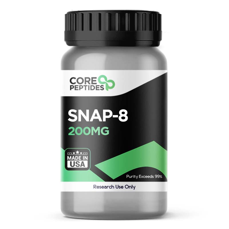
SNAP-8 (200mg)
Synaptosomal-associated protein 8, or the SNAP-8 peptide, is a synthetic octapeptide analog of the N-terminal end of the SNAP-25 peptide. The peptide is made of eight amino acids. It is acetylated at the N-terminus and amidated at the C-terminus, which leads to the following structure: Ac-Glu-Glu-Met-Gln-Arg-Arg-Ala-Asp-NH2. Overview This peptide was developed to compete with the SNAP-25 protein to bind with the vehicle-associated membrane proteins. The SNAP-25 protein, which stands for Synaptosomal-Associated Protein of 25 kDa, is considered a key component in neurotransmitter release. It normally interacts with vehicle-associated membrane proteins to facilitate the fusion of vesicles with the cell membrane, releasing their contents into the synaptic gap. Such a vesicle-associated membrane protein is thought to be synaptic vesicle protein Synaptotagmin 1 (Syt1). Synaptotagmin 1 (Syt1) serves as a calcium sensor and is deemed crucial for regulating neurotransmitter release in response to changes in calcium ion concentrations inside nerve cells. Molecular docking experiments posited that SNAP-8 and other peptides might bind to the C2A–C2B interface, primarily driven by hydrophobic contacts, suggesting a plausible site for inhibitory action on Syt1. The C2A and C2B regions are parts of the protein structure of Synaptotagmin 1 involved in calcium binding and membrane interaction. The binding at this interface by SNAP-8 suggests that the peptide might interfere with Synaptotagmin 1’s ability to respond to calcium signals, potentially disrupting normal neurotransmission. Once the SNAP-8 peptide binds with these proteins, it appears to destabilize the formation of the Soluble N-ethylmaleimide-sensitive factor Attachment Protein Receptor (SNARE) complex. The SNARE complex is instrumental in the docking and fusion of vesicles at the cell membrane. Destabilization of this complex may prevent the release of acetylcholine, reducing localized muscle contractions. Acetylcholine is a neurotransmitter involved in stimulating muscle contractions. Its reduced release may therefore lead to decreased muscle activity and reduced wrinkle depth. Ultimately, all these biological activities induced by SNAP-8 peptide may cause a reduction in lines and wrinkles.(1) Thus, the potential of SNAP-8 to interact at the molecular level with components of the neurotransmitter release mechanism showcases it may act as a modulator of neural and muscular function, specifically in experimental models of skin cell aging. Chemical Makeup(2) Molecular Formula: C41H70N16O6S Molecular Weight: 1075.16 g/mol Other Known Titles: SNAP-8 (Acetyl Glutamyl Heptapeptide-3), Synaptosomal-associated protein 8, Acetyl octapeptide-3 Research and Clinical Studies SNAP-8 Peptide and Skin Wrinkling A study(3) was conducted in 2021 to study the potential of the peptide on expression lines and wrinkles. In this double-blind, randomized clinical trial, 55 research models were observed. The entire cohort exhibited Fitzpatrick skin type I to VI, and were exposed either the peptide serum or a control compound twice a day for 12 weeks. Short-term peptide impact was measured 15 minutes after exposure, and long-term impact was measured at weeks 4, 8, and 12. After completing this study, it was observed that the peptide appeared to cause a notable reduction in the lines within 15 minutes after introduction. For long-term impact, it was suggested by the researchers that the peptide had the potential to induce significant skin improvement after 12 weeks. In another clinical trial conducted in 2013,(4) scientists focused on the Argireline peptide, a synthetic analog of the N-terminal end of the SNAP-25 peptide, which was developed for anti-aging action within the cell by inhibiting the catecholamine release (as opposed to acetylcholine release). Since both peptides are similar in structure, this study's results may reflect parallel actions of the SNAP-8 peptide. For this trial, 60 models were examined, of which 45 were presented with the peptide, and 15 were presented with a control compound. The peptide or the control was introduced every day for four weeks. After the completion of the study, it was reported that the peptide group appeared to have improved skin, with almost a 49% reduction in lines and decreased skin roughness. As per Yuan Wang et al.: "In the subjective evaluation, the total anti-wrinkle efficacy in the argireline group was 48.9%, compared with 0% in the placebo group. In the objective evaluation, the parameters of roughness were all decreased in the argireline group (p < 0.01), while no decrease was obvious in the placebo group (p > 0.05).” SNAP-8 Peptide and Skin Topography A study(5) aimed to evaluate whether the SNAP-8 peptide might be incorporated into an oil and water emulsion form to understand the peptide’s stability. When the 10% peptide oil-in-water emulsion was evaluated over a month-long routine exposure, there appeared to be a ~30% reduction in the depth and appearance of wrinkles. Researchers suggest that the peptide potentially mimics the action of botulinum neurotoxins.(6) SNAP-8 Peptide and SNARE Protein Complex SNAP-8 peptide has shown the potential to reduce muscle contractions by modulating the SNARE protein complex. One study reported that the peptide appeared to reduce the release of glutamate amino acid by almost 43%. Researchers reportedly claimed that the SNAP-8 peptide might reduce wrinkles by an average of 35%, up to a maximum of 62%,(7) a notable improvement in skin texture. SNAP-8 peptide is available for research and laboratory purposes only. Please review and adhere to our Terms and Conditions before ordering. References Lim SH, Sun Y, Thiruvallur Madanagopal T, Rosa V, Kang L. Enhanced Skin Permeation of Anti-wrinkle Peptides via Molecular Modification. Sci Rep. 2018 Jan 25;8(1):1596. doi: 10.1038/s41598-017-18454-z. Erratum in: Sci Rep. 2018 Apr 20;8(1):6500. PMID: 29371611; PMCID: PMC5785486. https://www.ncbi.nlm.nih.gov/pmc/articles/PMC5785486/ National Center for Biotechnology Information (2023). PubChem Compound Summary for CID 86080331, SNAP-8(Acetyl Glutamyl Heptapeptide-3). from https://pubchem.ncbi.nlm.nih.gov/compound/SNAP-8_Acetyl-Glutamyl-Heptapeptide-3 Nguyen TQ, Zahr AS, Kononov T, Ablon G. A Randomized, Double-blind, Placebo-controlled Clinical Study Investigating the Efficacy and Tolerability of a Peptide Serum Targeting Expression Lines. J Clin Aesthet Dermatol. 2021 May;14(5):14-21. Epub 2021 May 1. PMID: 34188744; PMCID: PMC8211334. https://www.ncbi.nlm.nih.gov/pmc/articles/PMC8211334/ Wang Y, Wang M, Xiao S, Pan P, Li P, Huo J. The anti-wrinkle efficacy of argireline, a synthetic hexapeptide, in Chinese subjects: a randomized, placebo-controlled study. Am J Clin Dermatol. 2013 Apr;14(2):147-53. doi: 10.1007/s40257-013-0009-9. PMID: 23417317. https://pubmed.ncbi.nlm.nih.gov/23417317/ Blanes-Mira C, Clemente J, Jodas G, Gil A, Fernández-Ballester G, Ponsati B, Gutierrez L, Pérez-Payá E, Ferrer-Montiel A. A synthetic hexapeptide (Argireline) with anti-wrinkle activity. Int J Cosmet Sci. 2002 Oct;24(5):303-10. doi: 10.1046/j.1467-2494.2002.00153.x. PMID: 18498523. https://pubmed.ncbi.nlm.nih.gov/18498523/ Apland JP, Adler M, Oyler GA. Inhibition of neurotransmitter release by peptides that mimic the N-terminal domain of SNAP-25. J Protein Chem. 2003 Feb;22(2):147-53. doi: 10.1023/a:1023423013741. PMID: 12760419. https://pubmed.ncbi.nlm.nih.gov/12760419/ Errante F, Ledwoń P, Latajka R, Rovero P, Papini AM. Cosmeceutical Peptides in the Framework of Sustainable Wellness Economy. Front Chem. 2020 Oct 30;8:572923. doi: 10.3389/fchem.2020.572923. PMID: 33195061; PMCID: PMC7662462. https://www.ncbi.nlm.nih.gov/pmc/articles/PMC7662462/ Sadowski G, Sadowski J. Safety and Efficacy of a Novel Anti-aging Skin Care Regimen Containing Neutraceuticals and Growth Factors on the Facial Skin of Women: A 12-Week Open-label Study. J Clin Aesthet Dermatol. 2020 Jun;13(6):24-34. Epub 2020 Jun 1. PMID: 32884616; PMCID: PMC7442306. https://www.ncbi.nlm.nih.gov/pmc/articles/PMC7442306 Dr. MarinovDr. Marinov (MD, Ph.D.) is a researcher and chief assistant professor in Preventative Medicine & Public Health. Prior to his professorship, Dr. Marinov practiced preventative, evidence-based medicine with an emphasis on Nutrition and Dietetics. He is widely published in international peer-reviewed scientific journals and specializes in peptide therapy research.
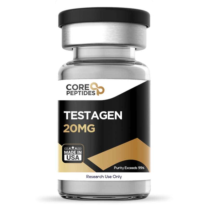
Testagen (20mg)
Testagen, alternatively known as KEDG, is classified among the Khavinson peptides and functions as a short signaling peptide. Short peptides such as Testagen are also suggested to serve as bioregulators which means that they may potentially cross both the cell and nuclear membranes to interact directly with DNA of target tissues.(1) Potentially, it may interact with the function of the anterior pituitary gland cells and interact with endocrine pathways that play a role in the synthesis of testosterone and thyroid-stimulating hormone (TSH).(2) This is likely due to the synthesis of Testagen, which is based on the amino acid composition of extracts from the anterior lobe of the pituitary gland.(3) As a result, it is made of the amino acids lysine, glutamine, asparagine and glycine, forming a tetrapeptide. Chemical Makeup Molecular formula: C17H29N5O9 Molecular weight: 447.2 g/mol Sequence: Lys-Glu-Asp-Gly Reconstitution: Required Other known titles: KEDG, Anterior pituitary peptide (APP) Research and Clinical Studies Testagen and the Hypothalamic-Pituitary-Gonadal Axis Testagen appears to be a bioregulator that may interact with the pituitary gland to upregulate testosterone production. Some trials suggest that the peptide may enhance testosterone production in research models of chronic inflammation and low testosterone as a result. Testagen was suggested to potentially improve uroflowmetry indicators, reduce markers of inflammation, and potentially cause an elevation in the total levels of testosterone. The researchers commented about “a decrease in the level of inflammation in the prostate, an increase in the level of total testosterone.”(4) While the exact mechanisms behind the potential of testagen remain unknown, some researchers have posited that the peptide may potentially have interactions with histones, which might influence epigenetic mechanisms, affecting cellular activities and differentiation. This interaction between short peptides and histones, particularly in the N-terminal histone regions containing seemingly homologous peptide-binding motifs, suggests an intriguing role in the regulation of chromatin structure and gene expression. Testagen has been observed to bind to certain histone regions with a specific conformational structure, this implies a potential impact on the structural dynamics of chromatin. Although core histones did not reveal homologous amino acid sequences, the specific binding indicates that peptides might interact with them through unique spatial conformation rather than sequence homology. This complexation with histones, as well as histone-deoxyribooligonucleotide complexes, suggests a site-specific nature, being influenced by the primary structures of peptides and oligonucleotides involved. Testagen appears to be interacting with histones, and may play a role in the conformation and functional state of chromatin, consequently influencing gene activity and cellular differentiation through epigenetic mechanisms. Understanding the exact pathways and impacts of these interactions would require further exploration and detailed studies into how such peptide-histone complexes influence gene transcription, DNA repair, replication, and other cellular processes influenced by chromatin structure.(5) Testagen and the Hypothalamic–Pituitary–Thyroid Axis In a thorough exploration of Testagen, its impact was conducted on hypophysectomized avian models. The models were apparently used to analyze the synthetic peptide's potential on the functional activity and morphological structure of the thyroid gland. Hypophysectomization, a procedure involving the removal or disabling of the pituitary gland, apparently induced a series of physiological alterations in the avian models, such as growth retardation, reduced excitability, and anorexia, along with changes including decreased body and thyroid gland weight, increased thyroid follicle size, and flattened thyrocytes. Furthermore, a notable decrease was reported in thyroid hormone concentrations, including thyroid-stimulating hormone (TSH), T3 (Triiodothyronine), T4 (Thyroxine), and free thyroxine.(3) After introducing Testagen there appeared to be distinct outcomes noticed among the avian models. An apparent 23% increase in body weight was suggested, complemented by a slight apparent augmentation in the thyroid gland's weight. Morphological adaptations also appeared to take place, such as an apparent reduction in the mean sectional area of follicles and a potential prevention of follicular epithelium flattening and colloid accumulation within the follicular cavity. Noteworthy appeared to be a well-defined interfollicular epithelium, coupled with variations in the nuclear-cytoplasmic ratio and a decrease in the height of thyrocytes, though these were considered less significant than in the control group. Additionally, Testagen exhibited an apparent stimulating impact on the growth of the thyroid capsule, despite the width of the thyroid capsule in these models still appearing to be inferior to that of control models.(3) Diving into the realm of aged avian models a normalization of the thyroid gland weight post-Testagen introduction was apparently observed. The synthetic peptide appeared to exert a moderate stimulating impact on the proliferation of epithelial cells. Consequently, the follicle size in these older avian models may have exceeded that of their younger counterparts. While the epithelium shape may have remained almost constant, a minor elevation was apparently seen in the nuclear-cytoplasmic ratio. Overall, the researchers suggested that the peptide may have better action in younger models and commented that “Restoration of the thyroid functions and morphology was registered to be greater in one-year-old chicken as compared to five-year-old ones.”(3) Testagen peptide is available for research and laboratory purposes only. Please review and adhere to our Terms and Conditions before ordering. References: Fedoreyeva, L. I., Kireev, I. I., Khavinson, V. K.h, & Vanyushin, B. F. (2011). Penetration of short fluorescence-labeled peptides into the nucleus in HeLa cells and in vitro specific interaction of the peptides with deoxyribooligonucleotides and DNA. Biochemistry. Biokhimiia, 76(11), 1210–1219. https://doi.org/10.1134/S0006297911110022 Khavinson, V. K., Popovich, I. G., Linkova, N. S., Mironova, E. S., & Ilina, A. R. (2021). Peptide Regulation of Gene Expression: A Systematic Review. Molecules (Basel, Switzerland), 26(22), 7053. https://doi.org/10.3390/molecules26227053 Kuznik, B. I., Pateiuk, A. V., Rusaeva, N. S., Baranchugova, L. M., & Obydenko, V. I. (2011). Advances in gerontology = Uspekhi gerontologii, 24(1), 93–98. https://pubmed.ncbi.nlm.nih.gov/21809626/ Rossikhin, V. V., Hoshchenko, Y. O., & Osipov, P. G. (2011). EFFICACY OF TESTOSTERONE SYNTHESIS INDUCTOR APPLICATION" TESTAGEN" IN ANDROGENIC DEFICIENCY IN PATIENTS WITH CHRONIC ABACTERIAL PROSTATITIS. Problems of Endocrine Pathology, 36(2), 17-22. DOI: 10.21856/j-PEP.2011.2.03 Fedoreyeva, L. I., Smirnova, T. A., Kolomijtseva, G. Y., Khavinson, V. K., & Vanyushin, B. F. (2013). Interaction of short peptides with FITC-labeled wheat histones and their complexes with deoxyribooligonucleotides. Biochemistry (Moscow), 78, 166-175. https://doi.org/10.1134/S0006297913020053 { "@context": "https:\/\/schema.org", "@type": "Product", "name": "Testagen (20mg)", "description": "Testagen for sale online (20mg). Peptides for sale at 99% purity with top customer service. Get research study results and information.", "image": "https://www.painandanxietymeds.shop/wp-content/uploads/2023/10/Testagen-20mg-300x300.jpg", "offers": [ { "@type": "Offer", "priceCurrency": "USD", "price": "63", "availability": "https:\/\/schema.org\/InStock", "itemCondition": "https:\/\/schema.org\/NewCondition", "seller": { "@type": "Organization", "name": "painandanxietymeds.shop" }, "url": "https:\/\/www.painandanxietymeds.shop\/testagen-20mg/", "hasMerchantReturnPolicy": { "@type": "MerchantReturnPolicy", "applicableCountry": "US", "returnPolicyCategory": "https:\/\/schema.org\/MerchantReturnNotPermitted" }, "shippingDetails": { "@type": "OfferShippingDetails", "shippingRate": { "@type": "MonetaryAmount", "minValue": 0, "maxValue": 9.25, "currency": "USD" }, "shippingDestination": { "@type": "DefinedRegion", "addressCountry": "US" }, "deliveryTime": { "@type": "ShippingDeliveryTime", "handlingTime": { "@type": "QuantitativeValue", "minValue": 1, "maxValue": 2, "unitCode": "d" }, "transitTime": { "@type": "QuantitativeValue", "minValue": 1, "maxValue": 5, "unitCode": "d" } } }, "priceValidUntil": "2027-12-09T15:11:59+00:00" } ], "url": "https:\/\/www.painandanxietymeds.shop\/testagen-20mg/", "aggregateRating": { "@type": "AggregateRating", "ratingValue": 96, "bestRating": 100, "reviewCount": 118 }, "review": [] } Dr. MarinovDr. Marinov (MD, Ph.D.) is a researcher and chief assistant professor in Preventative Medicine & Public Health. Prior to his professorship, Dr. Marinov practiced preventative, evidence-based medicine with an emphasis on Nutrition and Dietetics. He is widely published in international peer-reviewed scientific journals and specializes in peptide therapy research.
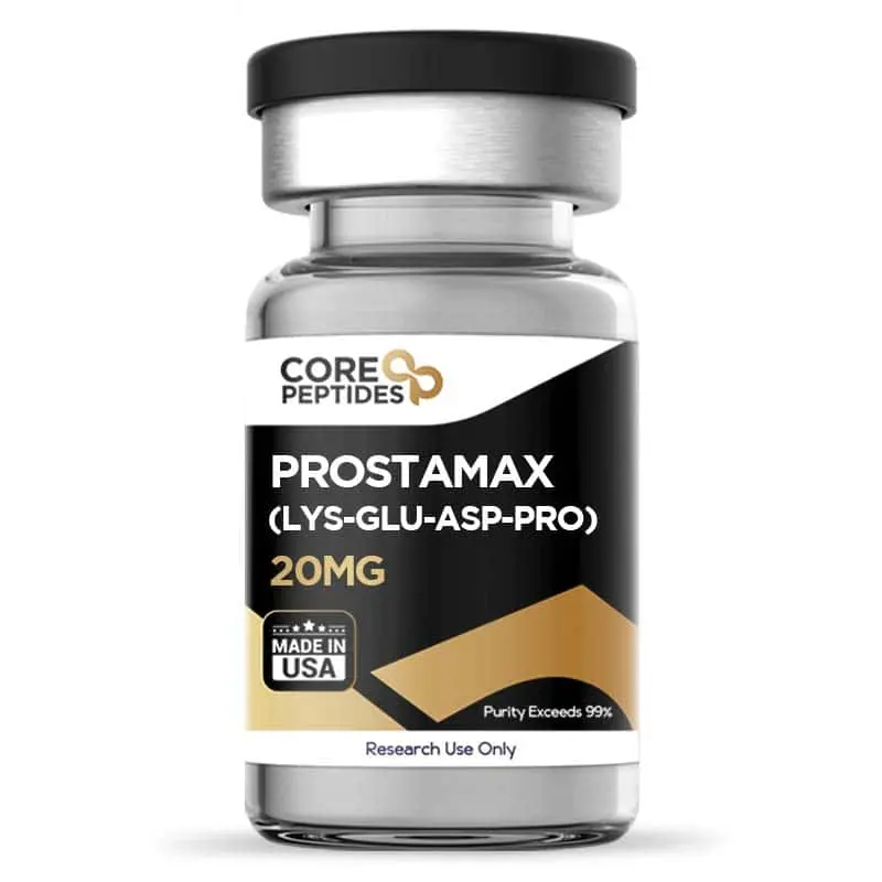
Prostamax (20mg)
Prostamax, aka KEDP, is a synthetic tetrapeptide (Lys-Glu-Asp-Pro) classified among the Khavinson peptides, which researchers have suggested may exhibit primary repair potential on various tissues, notably in prostate cell cultures. It appears to influence the structural dynamics of chromatin, potentially activating previously repressed genes and altering chromosomal dynamics. This may include increasing in the frequency of sister chromatid exchanges and Ag-positive nucleolus organizer regions, indicating enhanced chromosomal exchange activities and changes in ribosomal RNA gene activity as well as reducing the frequency of large segments of C-pericentromeric heterochromatin, suggesting a decondensation action on the chromatin. In prostatic gland tissue cultures, Prostamax may have a role in stimulating reparation processes, reducing inflammation and preventing sclerotic and atrophic processes. Chemical Makeup Molecular formula: C20H33N5O9 Molecular weight: 487.5 g/mol Sequence: Lys-Glu-Asp-Pro Other known titles: KEDP, SCHEMBL6660498 Research and Clinical Studies The content presented here integrates recent findings from preliminary studies about Prostamax's potential, as evidenced by diverse experimental methods. Prostamax Peptide and Gene Expression Related to Cellular Aging One study highlights the potential impact of Prostamax on the chromatin structure in lymphocytes.(1) Chromatin, the complex of DNA and proteins found in cells, has been observed to undergo two stages of denaturation at specific temperatures and energy states, denoted as T(d)VII and T(d)VIII. Prostamax appears to induce notable changes in this chromatin structure. Specifically, it appears to cause a redistribution of heat among two endotherms, T(d)III and T(d)IV, and leads to a decrease in their temperatures by 2.9 and 1.0 degrees Celsius, respectively. This redistribution and temperature shift might be linked to a relaxation in the chromatin structure, particularly the 30-nm-thick fiber relaxing into a 10-nm filament. Furthermore, there's a suggestion that Prostamax may lead to minor structural alterations in the nucleosomal organization within the chromatin, indicated by a slight decrease in the temperatures of T(d)VIII and T(d)VII in lymphocytes introduced to Prostamax compared to control cells. These changes are possibly due to adjustments in the structural organization of both the 10-nm filament and the 30-nm fiber. Another trial further investigates the impact of Prostamax on several chromosomal characteristics in aged cells, revealing that Prostamax possibly influences these characteristics.(2) More specifically, the study suggests that Prostamax might have a modifying action on chromatin, particularly in the context of aging cells. This could potentially lead to the activation of previously repressed genes and changes in chromosomal dynamics. For example, Prostamax apparently increases the frequency of sister chromatid exchanges (SCE). In cells introduced to Prostamax, the frequency of SCE rose to an average of 12.0 exchanges per cell, compared to 5.9 exchanges in control cells. This suggests that Prostamax may have a role in enhancing chromosomal exchange activities. Furthermore, the study posits that Prostamax may potentially increase the frequency of Ag-positive nucleolus organizer regions (NORs). In Prostamax-exposed cells, the average was 2.5 Ag-positive NORs per cell, compared to just 0.95 in intact cells. This increase might indicate changes in ribosomal RNA gene activity or chromatin structure modifications. Prostamax also seemingly reduces the frequency of large segments of C-pericentromeric heterochromatin, particularly in chromosomes 1 and 9. This change might imply a decondensation and deheterochromatinization action on the chromatin, possibly leading to the release of genes previously repressed by heterochromatinization. Ultimately, the authors concluded that “basis for the protective action of Prostamax is its modifying effect on chromatin.” Another trial also reports that Prostamax may exhibit a potential to affect genetic processes in aging cells by modulating chromatin structure and possibly reactivating certain genes.(3) The trial focused on the potential role of Prostamax in the activation of ribosome genes, decondensation of chromatin, and altering the structure of heterochromatin. Prostamax, along with other peptides like Epitalon and Livagen, possibly led to the decondensation of chromosome 1 pericentromeric structural chromatin. This suggests that Prostamax may have a role in modulating the structure of chromatin in aging cells. The decondensation of chromatin is significant as it potentially indicates an activation of previously inactivated genes due to age-related chromatin condensation. The research posited that the peptides, including Prostamax, potentially "release" genes repressed as a result of the age-specific condensation of cellular euchromatin regions, referred to as deheterochromatinization of facultative chromatin. This implies that Prostamax may contribute to the reactivation of certain genetic activities that diminish with age. Furthermore, Prostamax possibly induced changes specifically in chromosome 1, suggesting a selective action on certain chromosomal regions. However, the exact mechanisms through which Prostamax exerts these actions, and the full scope of its potential impact on aging and age-related genetic processes, remain somewhat unclear and require further investigation. Prostamax Peptide and the Prostate Gland Prostamax has been the subject of research exploring its potential action on prostatic gland tissue cultures. One investigation, utilizing organotypic tissue cultures, incorporated explants from both young and aged murine models as the basis for the study.(4) The focus was to observe any possible stimulating action of Prostamax on these tissues. In the course of this research, Prostamax was introduced at a specific concentration, which was posited to potentially elicit a stimulating action on the prostatic gland tissue cultures. This action was observed when these cultures were compared to control explants, suggesting that Prostamax might have a role in stimulating reparative processes in prostatic tissues, especially during the aging process in these murine models. The authors suggested that peptide such as Prostamax may have potential “for the stimulation of reparative processes in the appropriate tissues while aging.” Another study explored the potential of Prostamax in models of chronic aseptic prostatitis.(5) Prostamax, through experimental studies, apparently indicated a capacity to reduce the intensity of chronic inflammation, such as swelling, hyperemia of vessels, and lymphoid infiltration in murine models. The experimental setup involved murine models that underwent a procedure designed to induce chronic aseptic inflammation in the prostate gland. Post-procedure, the models were exposed to Prostamax and its impact was then evaluated in contrast to active controls. The primary goal was to gauge the potential of Prostamax in lessening the impact of chronic prostatitis and its associated complications, and to compare its potential to active controls. The authors of this study suggested that Prostamax might have the capability to diminish inflammation-related symptoms such as swelling, vessel hyperemia, and cellular infiltration commonly seen in murine models of chronic prostatitis. Notably, it also seemed to have a role in possibly curtailing the development of sclerotic and atrophic processes in the prostate gland models. These processes are typically seen as complications in such models and their potential mitigation by Prostamax is a point of interest. Prostamax's potential in preventing these processes appeared more pronounced compared to the comparator agents, suggesting its superiority in this regard. Additionally, Prostamax was observed to possibly enhance sexual activity in the animal models, a finding that may indicate broader research potential. Prostamax peptide is available for research and laboratory purposes only. Please review and adhere to our Terms and Conditions before ordering. References: Meskhi T, Khachidze D, Barbakadze Sh, Madzhagaladze G, Gorgoshidze M, Monaselidze D, Lezhava T, Tadumadze N. Vliianie peptidnogo bioreguliatora prostamaksa na geterokhromatin limfotsitov cheloveka in situ [The influence of the peptide bioregulator prostamax on heterochromatin of human lymphocytes in situ]. Biofizika. 2004 Nov-Dec;49(6):1091-3. Russian. PMID: 15612551. Dzhokhadze TA, Buadze TZh, Gaĭozishvili MN, Baratashvili NA, Lezhava TA. [Deheterochromatinization of the chromatin in old age induced by oligopeptide bioregulator (Lys-Glu-Asp-Pro)]. Georgian Med News. 2012 Nov;(212):76-82. Russian. PMID: 23221144. Khavinson VKh, Lezhava TA, Malinin VV. Effects of short peptides on lymphocyte chromatin in senile subjects. Bull Exp Biol Med. 2004 Jan;137(1):78-81. doi: 10.1023/b:bebm.0000024393.40560.05. PMID: 15085253. Zakutskiĭ AN, Chalisova NI, Ryzhak GA, Aniskina AI, Filippov SV, Zeziulin PN. [The tissue-specific effect of synthetic peptides-biologic regulators in organotypic tissues culture in young and old rats]. Adv Gerontol. 2006;19:93-6. Russian. PMID: 17152728. Borovskaya, T. G., Pakhomova, A. V., Vychuzhanina, A. V., Poluektova, M. E., Fomina, T. I., Ermolaeva, L. A., ... & Neplochov, E. A. (2013). Experimental studying of the drug efficiency Prostamax in the therapy of chronic aseptic prostatitis and its complications. Modern Research in Inflammation, 2013. Dr. MarinovDr. Marinov (MD, Ph.D.) is a researcher and chief assistant professor in Preventative Medicine & Public Health. Prior to his professorship, Dr. Marinov practiced preventative, evidence-based medicine with an emphasis on Nutrition and Dietetics. He is widely published in international peer-reviewed scientific journals and specializes in peptide therapy research.
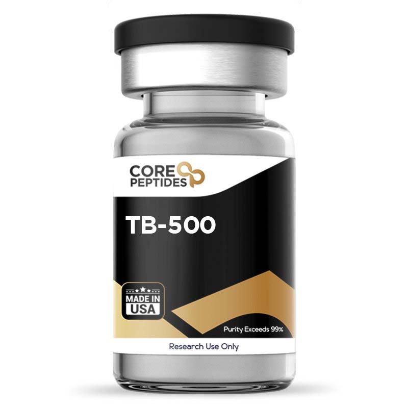
TB-500 (Thymosin Beta 4) (5mg / 10mg)
TB-500 peptide, also known as synthetic Thymosin Beta 4 or Tβ4 is the synthetic version of the naturally occurring protein Thymosin beta 4. The latter is found naturally within the cells of the thymus organ and encoded by the TMSB4X gene. Researchers have suggested TB-500 peptide hosts similar potential as Thymosin beta 4. These potential mechanisms of action may include inducing angiogenesis, elevating wound healing, or possibly elevated metastatic potential of tumor cells and hair growth. Besides being highly soluble in water and light in weight, TB-500 is a 43 amino acid-containing peptide found in abundance in the wound fluid comprising multiple blood platelets. This peptide may exhibit possible anti-inflammatory potential and may support neurological healing, as well as potentially supporting healing processes in the spinal cord, heart, and epidermis.(1) Overview TB-500 peptide, also known as thymosin β(4), includes a distinct peptide segment (17)LKKTETQ(23), which acts as the active site and which researchers consider potentially impactful in actin binding, cell migration, and wound healing.(2) The amino acid sequence of TB-500 is: Ac-Ser-Asp-Lys-Pro-Asp-Met-Ala-Glu-Ile-Glu-Lys-Phe-Asp-Lys-Ser-Lys-Leu-Lys-Lys-Thr-Glu-Thr-Gln-Glu-Lys-Asn-Pro-Leu-Pro-Ser-Lys-Glu-Thr-Ile-Glu-Gln-Glu-Lys-Gln-Ala-Gly-Glu-Ser-OH. Actins are essential proteins that form a key component of the cytoskeleton within cells, serving not only to maintain cellular structure but also to facilitate various cellular functions, including movement. Actin is suggested to be critical in supporting these cellular structures and processes. Thymosin beta-4 and, thus, TB-500 are believed to interact with actin, potentially by binding to globular actin (G-actin), a precursor to filamentous actin (F-actin). This interaction is thought to hinder the transformation of G-actin into F-actin, a process known as actin sequestration, and is likely to increase the availability of G-actin. The inhibition of F-actin formation by thymosin beta-4 may conceivably modify the structure of the cellular cytoskeleton, impacting cellular abilities for movement and morphological changes. Such changes are tentatively linked to various physiological and pathological states where cell motility is essential, including wound healing, tissue regeneration, and the progression of cancer through metastasis.(3) Furthermore, Thymosin beta-4 has been detected not only within cells but also extracellularly, such as in blood plasma and wound exudates. Preliminary studies involving vascular cells suggest that Thymosin beta-4 is extracellular, it might influence cellular functions like motility and the formation of new blood vessels (angiogenesis).(11,12) It is postulated that Thymosin beta-4 might exert this potential through its interactions with ATP synthase enzymes located on the cell surface, which are critical for cellular energy production. These findings indicate a broader scope of action for thymosin beta-4, impacting both intra- and extracellular processes. Chemical Makeup Molecular Formula: C212H350N56O78S Molecular Weight: 4963 g/mol Other Known Titles: Thymosin Beta 4 Research and Clinical Studies TB-500 Peptide and Inflammation Tβ4, and thus TB-500, is thought to potentially increase the levels of microRNA-146a (miR-146a), which might function as a suppressive regulator of specific cellular signaling pathways, particularly those associated with the functions of inflammation-related cytokines, including L-1 receptor-linked kinase 1 (IRAK1) and tumor necrosis factor receptor-associated factor 6 (TRAF6). The researchers of a study investigating the potential of the peptide on these factors propose this as a possible mechanism of action for TB-500. More specifically, the authors observed that "transfection of anti-miR-146a nucleotides reversed the inhibitory effect of Tβ4 on IRAK1 and TRAF6," thus suggesting this as a potential mechanism. Consequently, it is suggested that TB-500 may contribute to anti-inflammatory potential via these mechanisms.(4) TB-500 Peptide and Acute Wounds In 1999, a research study was conducted on wounded murine test models, who were introduced to TB-500 as a form of synthetic Thymosin Beta 4.(5) Four days after the presentation, it was reported by the researchers that the TB-500 peptide rats exhibited an apparent 41% increase in re-epithelialization than control murine models presented with saline. Seven days after the study, the TB-500 wounds were reported to be contracted by at least 11% more than the saline wounds. Upon analysis, it was concluded that TB-500 may possibly induce angiogenesis and collagen deposition, increasing the wound healing rate.The authors commented that their observations “suggest that Tβ4 is a potent wound healing factor with multiple activities...” TB-500 Peptide and Chronic Wounds Research studies were carried out on normal rats and mice, diabetic mice, aged mice, and steroid-influenced rats. All these animals were served full-thickness punch wounds and introduced to the TB-500 peptide. It was reported that the TB-500 appeared to accelerate the wound-healing process in all test models, regardless of the stated pre-existing conditions. Furthermore, phase 2 clinical trials were conducted on models of stasis and pressure ulcers. It was reported that TB-500 might accelerate the healing process by as much as one month.(6) TB-500 Peptide and Heart Cells Pulmonary hypertension is considered by scientists to be a progressive cardiac disease where the pulmonary arteries restrict the blood ejection by the right ventricle. This may result in increased pulmonary vascular resistance and pressure, potentially leading to ventricular failure of the heart. It was reported by researchers that TB-500 might be action specific on the Notch3-Col 3A-CTGF gene axis in MCT-influenced mice, which appeared to result in the case of the test study in decreasing the right ventricular heart cell hypertrophy by a significant amount.(7) Based on Tβ4 research, TB-500 might also influence the regeneration of cardiac cells. Preliminary research indicates that TB-500 may enhance the resilience of myocardial cells under hypoxic conditions and may also promote angiogenesis, which could facilitate the repair of cardiac cells. There is a suggestion from researchers that cardiac fibroblasts could potentially differentiate into cells akin to cardiomyocytes.(8) Furthermore, it has been proposed that the combination of TB-500 with cardiac reprogramming methods could synergistically mitigate damage to cardiac cells and support their regeneration by activating intrinsic cells in the heart area. Further experimentation employing mouse models, in which coronary arteries were ligated, suggested that TB-500 could potentially increase the activity of integrin-linked kinase (ILK) and protein kinase B (Akt) in cardiac tissue. This observation indicates a possible enhancement in the early survival of cardiomyocytes and an apparent improvement in cardiac function.(9) Further, the research indicates that TB-500 may facilitate the migration of myocardial and endothelial cells in the fetal heart, and this function appears to be preserved in adult cardiomyocytes. TB-500 and Hair Follicle Growth In 2003, studies were carried out on mice to examine the potential of TB-500 in hair growth. Under the influence of the TB-500 peptide, it was reported by the researchers that, via histological examination of the mouse skin cells, the peptide appeared to increase the number of hair shafts and hair follicles, thereby inducing hair growth. Upon real-time PCR and western blotting techniques, changes in the expression of m-RNA cells were observed between the TB500 and control mice. The m-RNA and protein levels were reported elevated in TB-500 mice, which might have significantly induced hair growth.(10) TB-500 and Blood Vessel Formation It is hypothesized that TB-500 might influence angiogenesis via several molecular interactions. This is based on studies involving TB-500 overexpression lentiviral vector in transfecting umbilical vein endothelial cells (HUVEC) and murine critical limb ischemia (CLI) models.(13) Researchers have also employed inhibitors such as DAPT, targeting the Notch pathway, and BMS, affecting the NF-κB pathway, in both HUVEC and murine CLI experiments to probe the intricate biological processes involved. The potential of TB-500 on angiogenesis and cellular migration were evaluated using MTT assays to measure cell viability, alongside tube formation and wound healing assays to assess angiogenic and migratory capabilities, respectively. Additionally, a variety of molecular methodologies were utilized, including Western blotting, reverse transcription, quantitative PCR, immunofluorescence, and immunohistochemistry. These techniques were instrumental in investigating the expression levels of angiogenesis-associated markers and elements related to the Notch/NF-κB pathways. Preliminary findings indicate that TB-500 might enhance not only the viability, angiogenesis, and migration of HUVEC but could also elevate the expression of angiopoietin-2 (Ang2), TEK receptor tyrosine kinase 2 (tie2), vascular endothelial growth factor A (VEGFA), NOTCH1 intracellular domain (N1ICD), Notch receptor 3 (Notch3), NF-κB, and phosphorylated (p)-p65 in these cells. In the muscle tissues of murine CLI models, similar increases in the expression of CD31, α-smooth muscle actin (α-SMA), Ang2, tie2, VEGFA, N1ICD, and p-p65 were observed, suggesting a regulatory potential of TB-500 on these molecular targets. Interestingly, the application of DAPT and BMS in these studies seemed to counteract the actions of TB-500, potentially indicating that the mechanisms of action of TB-500 in promoting angiogenesis might be mediated through its interactions with the Notch and NF-κB pathways. Moreover, the apparent reversal of the actions of DAPT and BMS by TB-500 could underscore its role in modulating these pathways, supporting the proposition of its regulatory functions in angiogenesis. Researchers have noted that these observations might imply a role for Tβ4 in promoting angiogenesis through regulation of these critical pathways. TB-500 and Corneal Tissues Studies have posited that TB-500 may modulate cytokine production and thus accelerate healing in corneal wound models.(14) Following injury, there is some indication that TB-500 could promote increased expression of IL-1β and IL-6 mRNA in the corneas of murine models. Moreover, TB-500 experimentation after alkali injury might lead to a decrease in the expression of chemoattractants such as MIP-2 and KC for polymorphonuclear neutrophils (PMNs) in mouse corneas, potentially resulting in diminished PMN infiltration. Concerning the inflammatory signaling pathways in the cornea, it is speculated that TB-500 may influence NFκB pathways, possibly exerting anti-inflammatory actions. TB-500 is also theorized to possess anti-apoptotic attributes. An overexpression of TB-500 in cellular models is observed to potentially increase growth rates, diminish basal apoptosis, and confer resistance to factors that induce cell death. In corneal epithelial cells, TB-500 could potentially inhibit apoptosis by blocking caspases and curtailing the release of the pro-apoptotic protein bcl-2 from mitochondria. The mechanism of TB-500’s anti-apoptotic action might include reducing the initiation signals of early cell death and activating the survival kinase Akt via complex interactions with PINCH and integrin-linked kinase. It is conceivable that TB-500’s anti-apoptotic influence operates through several molecular pathways. Nonetheless, it is crucial to acknowledge that these mechanisms remain conjectural and warrant further empirical investigation to be substantiated. TB-500 peptide is available for research and laboratory purposes only. Please review and adhere to our Terms and Conditions before ordering. References: Kleinman HK, Sosne G. Thymosin β4 Promotes Dermal Healing. Vitam Horm. 2016;102:251-75. doi: 10.1016/bs.vh.2016.04.005. Epub 2016 May 24. Ho EN, Kwok WH, Lau MY, Wong AS, Wan TS, Lam KK, Schiff PJ, Stewart BD. Doping control analysis of TB-500, a synthetic version of an active region of thymosin β₄, in equine urine and plasma by liquid chromatography-mass spectrometry. J Chromatogr A. 2012 Nov 23;1265:57-69. doi: 10.1016/j.chroma.2012.09.043. Epub 2012 Sep 23. Gurtner GC, Werner S, Barrandon Y, Longaker MT. Wound repair and regeneration. Nature. 2008 May 15;453(7193):314-21. doi: 10.1038/nature07039. PMID: 18480812. Santra, M., Zhang, Z. G., Yang, J., Santra, S., Santra, S., Chopp, M., & Morris, D. C. (2014). Thymosin β4 up-regulation of microRNA-146a promotes oligodendrocyte differentiation and suppression of the Toll-like proinflammatory pathway. The Journal of biological chemistry, 289(28), 19508–19518. https://doi.org/10.1074/jbc.M113.529966 Katherine M. Malinda et.al, Thymosin β4 Accelerates Wound Healing, Journal of Investigative Dermatology, Volume 113, Issue 3, 1999, Pages 364-368, ISSN 0022-202X. Treadwell T, Kleinman HK, Crockford D, Hardy MA, Guarnera GT, Goldstein AL. The regenerative peptide thymosin β4 accelerates the rate of dermal healing in preclinical animal models and in patients. Ann N Y Acad Sci. 2012 Oct. Wei C, Kim IK, Li L, Wu L, Gupta S. Thymosin Beta 4 protects mice from monocrotaline-induced pulmonary hypertension and right ventricular hypertrophy. PLoS One. 2014 Nov 20;9(11):e110598. Srivastava, D., Ieda, M., Fu, J., & Qian, L. (2012). Cardiac repair with thymosin β4 and cardiac reprogramming factors. Annals of the New York Academy of Sciences, 1270, 66–72. https://doi.org/10.1111/j.1749-6632.2012.06696.x Bock-Marquette, I., Saxena, A., White, M. D., Dimaio, J. M., & Srivastava, D. (2004). Thymosin beta4 activates integrin-linked kinase and promotes cardiac cell migration, survival and cardiac repair. Nature, 432(7016), 466–472. https://doi.org/10.1038/nature03000 Gao, Xy., Hou, F., Zhang, Zp. et al. Role of thymosin beta 4 in hair growth. Mol Genet Genomics 291, 1639–1646 (2016). Huff, T., Müller, C. S., Otto, A. M., Netzker, R., & Hannappel, E. (2001). beta-Thymosins, small acidic peptides with multiple functions. The international journal of biochemistry & cell biology, 33(3), 205–220. https://doi.org/10.1016/s1357-2725(00)00087-x Freeman, K. W., Bowman, B. R., & Zetter, B. R. (2011). Regenerative protein thymosin beta-4 is a novel regulator of purinergic signaling. FASEB journal : official publication of the Federation of American Societies for Experimental Biology, 25(3), 907–915. https://doi.org/10.1096/fj.10-169417 Lv, S., Cai, H., Xu, Y., Dai, J., Rong, X., & Zheng, L. (2020). Thymosin‑β 4 induces angiogenesis in critical limb ischemia mice via regulating Notch/NF‑κB pathway. International journal of molecular medicine, 46(4), 1347–1358. https://doi.org/10.3892/ijmm.2020.4701 Sosne, G., Qiu, P., & Kurpakus-Wheater, M. (2007). Thymosin beta 4: A novel corneal wound healing and anti-inflammatory agent. Clinical ophthalmology (Auckland, N.Z.), 1(3), 201–207. Dr. MarinovDr. Marinov (MD, Ph.D.) is a researcher and chief assistant professor in Preventative Medicine & Public Health. Prior to his professorship, Dr. Marinov practiced preventative, evidence-based medicine with an emphasis on Nutrition and Dietetics. He is widely published in international peer-reviewed scientific journals and specializes in peptide therapy research.
Pinealon (20mg)
Pinealon is a short synthetic peptide composed of three amino acids Glu-Asp-Arg, also known as the EDR peptide.(1)(2) Pinealon is suggested to impact the central nervous system, which may lead to behavior modification while also possibly protecting neurons and various cell types from oxidative stress. The primary research focus on this peptide is evaluating its potential to penetrate the blood-brain barrier, cellular membrane, and nuclear membrane. However, researchers have also suggested that it may also interact directly with DNA molecules. Pinealon tripeptide is isolated from another chemical compound, Cortexin.(2) Cortexin is a polypeptide with a particularly low molecular weight. Due to this low weigh, Cortexin has been suggested by researchers to cross the blood-brain barrier to produce potential stimulatory action on the neurotransmitters in the brain.(3) Overview The neuroprotective and anti-apoptotic potential of the Pinealon peptide has been posited by researchers to be exerted via the MAPK / ERK signaling pathway.(2) The ERK signaling cascade may play a role in the phosphorylation of the substrates, which may lead to elevated neuron plasticity and increased cellular stress. In turn, this action may lead to cellular apoptosis. Similarly, MAPK signaling has been associated with certain metabolic disorders and inflammatory reactions. Researchers suggest that Pinealon may potentially reduce the synthesis of reaction oxygen species (ROS) on the cellular level. ROS is considered a messenger to MAPK and ERK signaling pathways, and the reduced synthesis may lead to a reduction in signaling reactions, preventing cellular stress and cell death.(2) Studies on Pinealon have suggested a concentration-dependent action. At lower concentrations, the peptide may potentially restrict the synthesis and subsequent accumulation of ROS and cell death. In comparison, the peptide might lead to a modulation of the cell cycle at higher concentrations. Researchers reporting this apparent correlation in peptide presence have suggested that it may exert antioxidant potential at lower concentrations while possibly interacting with the cell genome and altering the cell cycle.(4) Chemical Makeup Molecular Formula: C15H26N6O8 Molecular Weight: 418.4 g/mol Other Known Titles: EDR, Glu-Asp-Arg Research and Clinical Studies Pinealon Peptide and Cell Aging The main purpose of this clinical study was to analyze the cellular and metabolic aspects of synthetic tripeptides, including Pinealon and a similar peptide called Vesugen. As part of this study,(5) research models of poly-morbidity and organic brain syndrome were observed. Both peptides exhibited apparent anabolic potential; researchers reported improvement in the functioning of the central nervous system and other vital organs compared to control models. The peptides did not appear to affect the degree of chromatin condensation, which the researchers indicated may suggest that the peptides do not act on cellular levels. Another separate study(6) has suggested that the Pinealon peptide may potentially act on muscle cells, modulating the levels of irisin. Scientists consider irisin to be central to muscle cell protection and mainly secreted during physical strain. Irisin may lead to the burning of excessive fat cell stores and is assumed by researchers to induce an elongation of telomeres (DNA caps) as well. By potentially increasing irisin levels, Pinealon may potentially exert protection of DNA telomeres and counteract cell aging action to some degree. Pinealon Peptide and Prenatal Hyperhomocysteinemia Scientists characterize hyperhomocysteinemia by the excessive concentration of homocysteine (HC) amino acid in the blood, indicating extreme vitamin deficiency, which might lead to an increased risk of neurological deterioration.(7) The main goal of this study(8) was to determine the potential of Pinealon on experimentally induced hyperhomocysteinemic pregnant female murine models. The female rats were given methionine from their second trimester onwards, which appeared to lead to increased concentration of homocysteine (HC) levels. The rat offspring from the control and the experimental models were then observed for this study. Upon analysis, it was suggested by the researchers that the peptide did not appear to reduce or inhibit the development of homocysteine in the offspring, however, there appeared to be some action seen in the experimental models, as they appeared to exhibit increased cognitive processing. The researchers suggested that the peptide did not appear to induce the metabolism of homocysteine but may have led to reduced toxic action of the compound. Pinealon Peptide and Serotonin Expression Studies(9) were conducted on the isolated brain cell cultures where the Pinealon peptide was presented. Upon analysis, it was suggested that the peptide might stimulate levels of serotonin expression in the cell cultures. Preliminary results seemed to indicate that after exposure to Pinealon, there was a notable increase in serotonin synthesis compared to the control groups. This increase was quantitatively assessed to be 1.9 times greater in younger cell cultures. The molecular dynamics behind this observed action might involve Pinealon's interaction with the DNA. It is speculated that this augmentation in serotonin production may be facilitated by Pinealon's potential to bind specifically to a DNA sequence within the promoter region of the gene encoding 5-tryptophan hydroxylase, an enzyme considered to be critical for serotonin synthesis. More specifically, the peptide is hypothesized to bind to a particular nucleotide sequence in the promoter region of the 5-tryptophan hydroxylase gene, potentially enhancing the transcriptional activity of the gene and thus increasing 5-tryptophan hydroxylase enzyme production. Additionally, molecular docking simulations were employed to formulate hypotheses about the peptide’s binding orientation and stability with the DNA. These simulations, which consider factors like hydrophobic interactions, electrostatic forces, and hydrogen bonding, indicated a lower (negative) binding energy for Pinealon compared to another peptide, Lys-Glu-Asp. Such a decrease in binding energy suggests a more stable potential interaction with DNA, which might lead to elevated levels of 5-tryptophan hydroxylase enzyme synthesis and, subsequently, increased serotonin production. Scientists consider serotonin a key mood-stabilizing hormone, and further studies are being conducted to observe the potential of Pinealon on serotonin synthesis. Another study suggests that Pinealon may potentially influence serotonin levels in the cerebral cortex of murine models under conditions of mild hypothermia. The possible elevation of serotonin levels postulated in the research might indicate that Pinealon could be involved in modulating these aspects of brain function, particularly in response to stress induced by mild hypothermia.(10) The same study also suggests that such models may exert an accumulation of adrenergic mediators when exposed to Pinealon and acute hypobaric hypoxia. Adrenergic mediators are chemicals that transmit nerve impulses and may potentially influence various brain functions, particularly under stress. The increase in these mediators might indicate a response mechanism of Pinealon that may be associated with neuroprotective activities. Pinealon Peptide and Anti-apoptotic Potential Studies(11) have suggested that Pinealon peptide may impact the levels of the caspase 3 enzyme. Scientists consider the caspase 3 enzyme an initiator of cell apoptosis (controlled cell death). When Pinealon peptide was presented in experimental murine models of ischemic stroke, the peptide appeared to modulate the levels of this enzyme, thereby shutting down the pathway of cellular death. This, in turn, reportedly reduced hypoxia action during the stroke. The research posits that Pinealon may play a role in reducing neuroinflammation and potentially restoring neurogenic processes to their reference levels, presumably those observed under normoxic (normal oxygen) conditions. By potentially fostering an environment conducive to neurogenesis during hypoxic stress, researchers also posit that Pinealon may contribute to the brain's resilience against oxygen deprivation. In addition to its apparent role in modulating the activity of caspase-3, Pinealon was also suggested to influence levels of interleukin-6 and tumor necrosis factor. Interleukin-6 and tumor necrosis factors are cytokines commonly linked with inflammation and cellular stress responses. The implication is that Pinealon might help bring these inflammatory markers back to baseline levels, thus potentially mitigating the inflammatory response and apoptosis triggered by hypoxic conditions. The action of suppressing the caspase 3 enzyme was also observed in skin cells. By reducing cellular apoptosis, the peptide may potentially stimulate cell proliferation, possibly leading to an increased regenerative process.(12) Pinealon peptide is available for research and laboratory purposes only. Please review and adhere to our Terms and Conditions before ordering. References: Khavinson, V., Linkova, N., Kozhevnikova, E., & Trofimova, S. (2020). EDR Peptide: Possible Mechanism of Gene Expression and Protein Synthesis Regulation Involved in the Pathogenesis of Alzheimer's Disease. Molecules (Basel, Switzerland), 26(1), 159. https://www.ncbi.nlm.nih.gov/pmc/articles/PMC7795577/ National Center for Biotechnology Information. PubChem Compound Summary for CID 10273502, Glu-Asp-Arg. https://pubchem.ncbi.nlm.nih.gov/compound/Glu-Asp-Arg Eroğlu, O., Karlıdağ, T., Kuloğlu, T., Keleş, E., Kaygusuz, İ., & Yalçın, Ş. (2018). The Protective Effect of Cortexin on Cisplatin-Induced Ototoxicity. The journal of international advanced otology, 14(1), 27–33. https://www.ncbi.nlm.nih.gov/pmc/articles/PMC6354512/ Khavinson V, Ribakova Y, Kulebiakin K, Vladychenskaya E, Kozina L, Arutjunyan A, Boldyrev A. Pinealon increases cell viability by suppression of free radical levels and activating proliferative processes. Rejuvenation Res. 2011 Oct;14(5):535-41. https://pubmed.ncbi.nlm.nih.gov/21978084/ Meshchaninov VN, Tkachenko EL, Zharkov SV, Gavrilov IV, Katyreva Iue. EFFECT OF SYNTHETIC PEPTIDES ON AGING OF PATIENTS WITH CHRONIC POLYMORBIDITY AND ORGANIC BRAIN SYNDROME OF THE CENTRAL NERVOUS SYSTEM IN REMISSION. Adv Gerontol. 2015;28(1):62-7. Russian. PMID: 26390612. https://pubmed.ncbi.nlm.nih.gov/26390612/ Khavinson VKh, Kuznik BI, Tarnovskaya SI, Lin'kova NS. Short Peptides and Telomere Length Regulator Hormone Irisin. Bull Exp Biol Med. 2016 Jan;160(3):347-9. doi: 10.1007/s10517-016-3167-y. Epub 2016 Jan 8. PMID: 26742748. https://pubmed.ncbi.nlm.nih.gov/26742748/ Homocysteine. https://my.clevelandclinic.org/health/articles/21527-homocysteine Arutjunyan, A., Kozina, L., Stvolinskiy, S., Bulygina, Y., Mashkina, A., & Khavinson, V. (2012). Pinealon protects the rat offspring from prenatal hyperhomocysteinemia. International journal of clinical and experimental medicine, 5(2), 179–185. https://www.ncbi.nlm.nih.gov/pmc/articles/PMC3342713/ Khavinson, V.K., Lin’kova, N.S., Tarnovskaya, S.I. et al. Short Peptides Stimulate Serotonin Expression in Cells of Brain Cortex. Bull Exp Biol Med 157, 77–80 (2014). https://link.springer.com/article/10.1007/s10517-014-2496-y#citeas Mendzheritsky AM, Karantysh GV, Ryzhak GA, Prokofiev VN. [Pinealon and Cortexin influence on behavior and neurochemical processes in 18-month aged rats within hypoxia and hypothermia]. Adv Gerontol. 2015;28(3):532-539. Russian. PMID: 28509493. Mendzheritskiĭ AM, Karantysh GV, Ryzhak GA, Dem'ianenko SV. [Regulation of content of cytokines in blood serum and of caspase-3 activity in brains of old rats in model of sharp hypoxic hypoxia with Cortexin and Pinealon]. Adv Gerontol. 2014;27(1):94-7. Russian. PMID: 25051764. https://pubmed.ncbi.nlm.nih.gov/25051764/ Voicekhovskaya MA, Chalisova NI, Kontsevaya EA, Ryzhak GA. Effect of bioregulatory tripeptides on the culture of skin cells from young and old rats. Bull Exp Biol Med. 2012 Jan;152(3):357-9. doi: 10.1007/s10517-012-1527-9. PMID: 22803085. https://pubmed.ncbi.nlm.nih.gov/22803085/ Dr. MarinovDr. Marinov (MD, Ph.D.) is a researcher and chief assistant professor in Preventative Medicine & Public Health. Prior to his professorship, Dr. Marinov practiced preventative, evidence-based medicine with an emphasis on Nutrition and Dietetics. He is widely published in international peer-reviewed scientific journals and specializes in peptide therapy research.
Selank (10mg)
Selank is a short, synthetic heptapeptide composed of seven amino acids. Selank was developed to mimic the naturally occurring peptide Tuftsin,(5) a short fragment from the immunoglobulin G (IgG), a natural tetrapeptide involved in certain biological functions that regulate the immune system. After the discovery and initial isolation of Tuftsin, researchers suggested that the peptide might be involved in the functions of phagocytic cells, including phagocytosis, motility, and immunological cell functions.(2) Since then, various synthetic analogs of Tuftsin have been synthesized in laboratories by conventional and polymeric reagent methods.(4) Overview Selank is a peptide chain composed of two fragments – one is Tuftsin at the N-terminus, and the other is a tripeptide Pro-Gly-Pro (PGP) at the C-terminal end of the molecule. The inclusion of a Pro-Gly-Pro (PGP) sequence in the peptide Selank might enhance its potential to penetrate various biological barriers, including the blood-brain barrier (BBB). The BBB is a highly selective and semi-permeable membrane that delineates the circulating blood from brain tissues and extracellular fluid within the central nervous system. It is deemed to play a pivotal role in controlling the entry of molecules. Integrating the PGP sequence might modify the peptide's hydrophilicity or lipophilicity, which might increase its compatibility with the lipid-rich milieu of the BBB. Moreover, the PGP motif might interact with certain transport systems or receptors on the BBB membrane, potentially facilitating receptor-mediated endocytosis or active transport. These mechanisms may permit Selank to circumvent the tight junctions that typically impede the transit of larger molecules through the BBB. Additionally, the presence of the PGP sequence might alter the tertiary structure of Selank, potentially rendering it more amenable to traversing the BBB. This alteration might arise from changes in the peptide's spatial configuration, which may influence its interaction with the cellular components of the BBB.(6) Chemical Makeup Molecular Formula: C33H57N11O9 Molecular Weight: 751.88 g/mol Other Known Titles: TP-7, Selanc Research and Clinical Studies Selank Peptide and BDNF Levels There is a possibility that Selank may influence the expression of brain-derived neurotrophic factor (BDNF), deemed a crucial protein in the brain that supports neuronal survival and growth.(1) Research indicates that Selank may markedly increase the levels of BDNF mRNA in the hippocampus, an integral region of the brain involved in memory and emotional responses. The potential of Selank to boost BDNF expression, particularly under conditions where stress and glucocorticoids suppress BDNF levels, suggests its relevance in research studies within the context of neuroplasticity decline. This protein's perceived role in synaptic function and neuronal adaptation underscores the significance of such investigations. Selank Peptide and Serotonin Signaling Selank may potentially influence serotonin signaling mechanisms. Serotonin signaling is theorized to play a crucial role in the management of mood and anxiety within the brain. Research utilizing murine models where serotonin synthesis was inhibited has indicated that Selank might be able to alter serotonin levels in cases where the serotonergic system is impaired. It has been proposed by researchers that Selank may potentially boost serotonin metabolism in the brainstem, indicating its potential action on the serotonin system. More specifically, the peptide is thought to facilitate an increase in the metabolic processing of serotonin in brain regions considered essential for mood and anxiety regulation. Additionally, the hypothesis that Selank may enhance serotonin metabolism suggests a potential pathway by which Selank might ameliorate issues stemming from diminished serotonin activity. Selank Peptide and GABA Signaling Studies have suggested that Selank may act on the gamma aminobutyric acid (GABA) receptors. GABA is considered an inhibitory neurotransmitter within this context, playing a role in diminishing neuronal excitability, fostering relaxation, and mitigating anxiety observations, as observed in animal research models. In one study,(7) the expression of 84 genes involved in neurotransmission was studied in murine models. The experimental murine models were exposed to either Selank or GABA, and gene expression was studied after one and three hours via a PCR method. All the gene expressions studied for Selank and GABA appeared positively correlated. The results suggested that Selank had the potential to induce several alterations in the neurotransmission process, suggesting by proxy that Selank may exert possible action via modulating the GABAergic system. Moreover, the literature indicates that the influence of Selank might not be confined to mere direct actions on the transcriptional activity of genes associated with GABA receptors. It might also involve allosteric modulation of the GABAergic system. This is inferred from observed variations in gene expression following exposure to Selank compared to GABA, where Selank has been speculated to distinctly affect the expression of specific genes. Such differential gene expression alludes to a more complex interaction of Selank with the GABAergic system, possibly diverging from the direct receptor activation typically seen with GABA. Selank has also been proposed to instigate enduring modifications within neurotransmitter systems, a characteristic that might account for its extended anxiolytic actions observed in experimental frameworks. These alterations suggest a broader and potentially long-term impact on neurotransmitter dynamics beyond potential receptor interaction, emphasizing the complex potential of Selank on neural regulation. Selank Peptide and Genome Expression Studies have been conducted to understand the potential of Selank peptide on genome expression and its involvement in the inflammatory process. These studies(8) were conducted on male murine models weighing 250 grams. These murine models were separated into three groups – a control group, an experimental group with a single exposure to Selank, and an experimental group under routine exposure to Selank. After the study, the RNA was isolated from the rat spleen and hippocampus and studied via PCR method. Based on the results, it was suggested by the researchers that Selank might have the potential to impact gene expression, exhibited more definitively in the spleen and the hippocampus. One of the gene expressions reported during the study was the change in CX3CR1, which was involved in the inflammatory process. This suggested that Selank might regulate the inflammatory process through the mechanism of gene expression, mainly the CX3CR1 alteration. Selank Peptide and Enkephalin Signaling In a clinical study,(9) 62 research models of Generalized Anxiety Disorder (GAD) were examined. These models were divided into two groups – 48% were exposed to Selank, whereas 52% were exposed to a generic benzodiazepine compound. Following the study, the psychometric levels of all models were analyzed. Results suggested that the impact of Selank appeared to be similar to those of the generic compound. Researchers also reported that enkephalin levels of tau leu-enkephalin were apparently reduced in the Selank-exposed group before the experiment, and the addition of Selank may have reversed this observation. Selank is hypothesized to exert suppressive actions on enzymes that break down enkephalins. Enkephalins, which are endogenous ligands for opioid receptors, have been implicated in the regulation of pain, mood, and stress responses. Therefore, the potential inhibition of these degrading enzymes by Selank might result in increased levels of enkephalins, potentially augmenting their physiological actions. This could lead to a noticeable elevation in tau(1/2) leu-enkephalin concentrations during experiments involving anxiety models where Selank is evaluated. Selank Peptide and Memory This study(11) was conducted on murine models that were enrolled in a "training" session of four days for learning conditioned avoidance response (CAR). CAR is a learned response in delaying or preventing avoidance behavior. Selank exposure was begun 15 minutes before the training session on all four days. Upon monitoring the behavior of the murine models, researchers suggested that the learning abilities of the murine models appeared to improve with each exposure of the peptide, as the number of errors reduced and the number of correct solutions increased. This potential impact might be mediated through multiple interconnected pathways, including the modulation of neuropeptide systems within the brain. These neuropeptides are deemed critical as they are believed to play significant roles in various cognitive functions, potentially boosting processes related to learning and memory. Additionally, the peptide Selank might alter neural circuits involved in memory consolidation. This alteration may enhance synaptic stability and efficiency in learning processes. Moreover, Selank might indirectly boost cognitive performance by mitigating anxiety-related factors that frequently impede learning efficiency. This suggests that Selank may influence the emotional aspects of cognition, enhancing the overall cognitive process. Furthermore, Selank may promote neural plasticity or the capacity of neurons to adapt, particularly in cognitive circuits that are not performing optimally, thus potentially elevating their function. This enhancement of neural adaptability may be vital for the maintenance and improvement of cognitive abilities, suggesting that Selank might be a valuable compound for further research in neurocognitive fields. Selank Peptide and Immunomodulation This clinical study(12) was conducted on research models of GAD with neurasthenia, with a control group and experimental group exposed Selank peptide for 14 days. After the study, the peripheral blood samples were collected and analyzed. The analytical results suggested peak elevation in the levels of IL-6 cytokine and changes in the Th1 and Th2 cytokine ratio, all of which are believed to regulate the immune system. Selank Peptide and Withdrawal In this study,(13) murine models were infused with 10% ethanol as their sole fluid source for 24 weeks. Consequently, these murine models were experimentally induced with alcohol withdrawal symptoms once the ethanol drip was removed. The murine models were then exposed to Selank, to study its potential impact on the withdrawal symptoms. After 48 hours, researchers reported that the alcohol withdrawal symptoms appeared to have reduced, based on the results of their social interaction and maze tests. Selank Peptide and Cardiovascular Activity In this study,(14) Selank was presented to feline models. The main goal of this study was to examine the cardiovascular and respiratory potential of the peptide in this specific model. Upon exposure to the peptide, it was reported by the researchers that there appeared to be a 32% decrease in arterial blood pressure within 3 minutes of peptide presence. Moreover, the peptide possibly induced a 24% increase in cerebral blood flow within the first 10 minutes, slowly decreasing to optimal levels. Selank was not reported to have induced any action on the respiratory system or the heart rate. Selank Peptide and Weight, Cholesterol In this study,(15) murine models were first exposed to a high-fat diet for 6 consecutive weeks until the weights of murine models were measured between 280g to 300g. Later, these murine models were divided into two groups, a control group given sodium chloride, and an experimental group exposed to Selank peptide. Additionally, a group of control murine models were used for this study, which were not presented with either of the agents but were simply monitored for the purpose of the study. Upon analysis, it was suggested by the researchers that the Selank group exhibited apparently decreased levels of cholesterol and fat anywhere between 25% and 58%. Based on the findings, Selank may also reduce specific forms such as low-density lipoprotein (LDL), very-low-density lipoprotein (VLDL) cholesterol, and triglycerides. This indicates that Selank might play either a direct or an indirect role in influencing the mechanisms of lipid metabolism and could potentially display actions that lower cholesterol (hypocholesterolemic) and lipid levels (hypolipidemic). Moreover, the investigation noted discernible enhancements in parameters related to hemostasis, including elevated total fibrinolytic activity and decreased platelet aggregation, which may suggest improvements in conditions that favor clot formation. Additionally, the study points to a possible modulatory action of Selank on glucose homeostasis, which involves maintaining stable blood glucose levels. The fat metabolism rate of the Selank group also was reportedly improved and was eventually measured at the same rate as those in control models. Upon weight determination of the murine models, it was noted that the control group exhibited an average weight gain of 40g throughout the study, whereas the experiment group maintained the same weight throughout the study, with gradual weight reduction upon peptide presentation. Selank peptide is available for research and laboratory purposes only. Please review and adhere to our Terms and Conditions before ordering. References: Inozemtseva, L. S., Karpenko, E. A., Dolotov, O. V., Levitskaya, N. G., Kamensky, A. A., Andreeva, L. A., & Grivennikov, I. A. (2008). Intranasal administration of the peptide Selank regulates BDNF expression in the rat hippocampus in vivo. Doklady biological sciences : proceedings of the Academy of Sciences of the USSR, Biological sciences sections, 421, 241–243. https://doi.org/10.1134/s0012496608040066 Najjar VA. Tuftsin, a natural activator of phagocyte cells: an overview. Ann N Y Acad Sci. 1983;419:1-11. doi: 10.1111/j.1749-6632.1983.tb37086.x. https://pubmed.ncbi.nlm.nih.gov/6370072/ Semenova, T. P., kozlovskiĭ, I. I., Zakharova, N. M., & Kozlovskaia, M. M. (2009). Eksperimental'naia i klinicheskaia farmakologiia, 72(4), 6–8. Fridkin M, Stabinsky Y, Zakuth V, Spirer Z. Tuftsin and some analogs: synthesis and interaction with human polymorphonuclear leukocytes. Biochim Biophys Acta. 1977 Jan 24;496(1):203-11. https://pubmed.ncbi.nlm.nih.gov/576412/ Kozlovskaya MM, Kozlovskii II, Val'dman EA, Seredenin SB. Selank and short peptides of the tuftsin family in the regulation of adaptive behavior in stress. Neurosci Behav Physiol. 2003 Nov;33(9):853-60. https://pubmed.ncbi.nlm.nih.gov/14969422/ Elena Filatova et al., GABA, Selank, and Olanzapine Affect the Expression of Genes Involved in GABAergic Neurotransmission in IMR-32 Cells. https://doi.org/10.3389/fphar.2017.00089 Volkova, A., Shadrina, M., Kolomin, T., Andreeva, L., Limborska, S., Myasoedov, N., & Slominsky, P. (2016). Selank Administration Affects the Expression of Some Genes Involved in GABAergic Neurotransmission. Frontiers in pharmacology, 7, 31. https://www.ncbi.nlm.nih.gov/pmc/articles/PMC4757669/ T.A Kolomin et al., Transcriptomic Response of Rat Hippocampus and Spleen Cells to Single and Chronic Administration of the Peptide Selank. June 2, 2009. DOI: 10.1134/S1607672910010023 Zozulia AA, Neznamov GG, Siuniakov TS, Kost NV, Gabaeva MV, Sokolov OIu, Serebriakova EV, Siranchieva OA, Andriushenko AV, Telesheva ES, Siuniakov SA, Smulevich AB, Miasoedov NF, Seredenin SB. Efficacy and possible mechanisms of action of a new peptide anxiolytic selank in the therapy of generalized anxiety disorders and neurasthenia. Zh Nevrol Psikhiatr Im S S Korsakova. 2008;108(4):38-48. Russian. https://pubmed.ncbi.nlm.nih.gov/18454096/ Medvedev VE, Tereshchenko ON, Israelian AIu, Chobanu IK, Kost NV, Sokolov OIu, Miasoedov NF. A comparison of the anxiolytic effect and tolerability of selank and phenazepam in the treatment of anxiety disorders. Zh Nevrol Psikhiatr Im S S Korsakova. 2014;114(7):17-22. Russian. https://pubmed.ncbi.nlm.nih.gov/25176261/ Kozlovskii II, Danchev ND. The optimizing action of the synthetic peptide Selank on a conditioned active avoidance reflex in rats. Neurosci Behav Physiol. 2003 Sep;33(7):639-43. https://pubmed.ncbi.nlm.nih.gov/14552529/ Uchakina ON, Uchakin PN, Miasoedov NF, Andreeva LA, Shcherbenko VE, Mezentseva MV, Gabaeva MV, Sokolov OIu, Zozulia AA, Ershov FI. Immunomodulatory effects of selank in patients with anxiety-asthenic disorders. Zh Nevrol Psikhiatr Im S S Korsakova. 2008;108(5):71-5. Russian. https://pubmed.ncbi.nlm.nih.gov/18577961/ Kolik LG, Nadorova AV, Kozlovskaya MM. Efficacy of peptide anxiolytic selank during modeling of withdrawal syndrome in rats with stable alcoholic motivation. Bull Exp Biol Med. 2014 May;157(1):52-5. https://pubmed.ncbi.nlm.nih.gov/24913576/ Gan'shina TS, Kozlovskiĭ II. [Effects of the new peptide anxiolytic drug selank on the cardiovascular system functioning and respiration in cats]. Eksp Klin Farmakol. 2005 Jul-Aug;68(4):33-5. Russian. https://pubmed.ncbi.nlm.nih.gov/16193654/ N.F. Mjasoedov et al, The Influence of Selank on the Parameters of the Hemostasis System, Lipid Profile, and Blood Sugar Level in the Course of Experimental Metabolic Syndrome. April 14, 2014. Dr. MarinovDr. Marinov (MD, Ph.D.) is a researcher and chief assistant professor in Preventative Medicine & Public Health. Prior to his professorship, Dr. Marinov practiced preventative, evidence-based medicine with an emphasis on Nutrition and Dietetics. He is widely published in international peer-reviewed scientific journals and specializes in peptide therapy research.
Livagen (20mg)
Livagen is a short peptide containing four amino acids: lysine, alanine, aspartic acid, and glutamic acid. Considered to be a bioregulator, this peptide may act on the DNA structure and impact functionality. The primary potential of Livagen is identified in how it acts on chromatin, DNA, and genes. Scientists have formalized that DNA is a double helix structure surrounded by proteins called "histones." These histones bind together, forming chromatin, and several chromatins then condense together, forming the chromosomes. These chromosomes appear crucial to forming genetic material specific to each organism.(1) Livagen has been considered by researchers for its potential to 'de-condense' these chromatin materials. As a result, some genes that had become non-functional might become active, possibly improving cell activity and production.(2) Researchers posit that Livagen may increase energy levels and skin elasticity and possibly induce improvements in immune system functionality via this mechanism. Researchers have suggested that the lipid peptide acts on the lymphocytes, possibly reactivating the ribosomes by 'unpacking' the chromatin and modifying gene expression. These lymphocytes are white blood cells that are deemed essential in attacking foreign impurities entering the system and enhancing immunity. In this manner, Livagen may exhibit potential to improve the immune system by coordinating cellular responses and controlling inflammatory responses.(3) T. Lezhava et al., who conducted a clinical research study stated that “These results indicate that peptide bioregulators Epitalon, Livagen, and Vilon cause activation (deheterochromatinization) of chromatin in lymphocytes of [aged models].” Chemical Makeup Molecular Formula: C18H31N5O9 Molecular Weight: 461.5 g/mol Other Known Titles: KEDA Research and Clinical Studies Livagen and Gene Regulation Cardiac disorders such as hypertrophic cardiomyopathy (HCM), atherosclerosis, and cardiac injury are considered to be characterized by dysregulation in gene expression and chromatin structure. A bioregulatory peptide such as Livagen has been suggested by researchers to enact potential mitigation of such gene regulation dysfunctions. Studies(4,5) have suggested that Livagen peptides may help reduce specific long-term impacts of such conditions when applied to cell culture models of HCM. A publication related to the same study(6) also reported on the potential of the peptide’s exposure in the presence of cobalt ions to evaluate their combined potential on chromatin structures in experimental models of hypertrophic cardiomyopathy. The study suggested that this combination possibly induces the de-condensation of chromatin. The results of this study are stated to be critical “because it provides new information about the protective effect of Livagen and Livagen + Cobalt ions on the lymphocytes” in hypertrophic cardiomyopathy models.(6) Livagen, either alone or in combination with cobalt ions, appears to modify the activity of nucleolar organizer regions (NORs) and the associative behavior of acrocentric chromosomes. The study posits that the combined application of Livagen and cobalt ions increases frequency, scoring 2 NORs, which are integral to ribosomal RNA synthesis. Such an action on NORs may indicate enhanced or altered protein synthesis capabilities in these cells, which might be crucial in understanding how cellular function is modified in the context of cardiac hypertrophy seen in HCM. Additionally, an increase in the association of acrocentric chromosomes was noted, which is speculated to be more pronounced with the combined exposure of Livagen and cobalt ions compared to each agent alone. This phenomenon might reflect changes in the chromatin state, specifically the decondensation of heterochromatin. The study suggests that this chromatin remodeling might facilitate the exposure and possible transcriptional activation of previously inactivated genes within these regions. Livagen and Nociception Enkephalins are considered to be naturally occurring neurotransmitters that may bind with mu- and delta- receptors and reduce pain signal transmission. Bioregulatory peptides such as Livagen may possibly act on enzymes and prevent the degradation of these neurotransmitters, possibly increasing enkephalin levels. The activity of these degrading enzymes was assessed in vitro, based on the rate of hydrolysis of 3H-Leu-enkephalin, a labeled enkephalin peptide.(7) The study also explored whether Livagen might interact directly with mu- and delta- receptors in the brain, using a radioreceptor method with the tracer [3H][D-Ala2, D-Leu5]-enkephalin. This component of the study posited that there appeared to be no observable interaction between Livagen and the mu- or delta-opioid receptors of the membrane fraction from murine models' brains. This outcome suggests that while Livagen may influence opioid peptide levels by inhibiting their degradation, it does not appear to directly bind or affect opioid receptors in the brain. Livagen and Antioxidant Systems A study explored the potential hepatoprotective and immunoprotective properties of the Livagen, particularly in liver fibroid induration and both acute and chronic hepatitis models. The findings suggest that Livagen may help normalize immune response and antioxidant status, which might restore liver function during episodes of hepatitis, particularly in cases where liver function typically declines. Livagen potentially offers positive actions in mitigating liver fibroid induration and combating various forms of hepatitis, though the exact mechanisms of action remain unclear.(8) Livagen and DNA Repair DNA alteration over time is considered to lead to increased chromatin condensation and decreased cellular repair.(9) Researchers have suggested the Livagen peptide to inhibit and possibly reverse these alterations, improving cellular functions and 'reversing' cell aging.(3) Thus, specific chromosomal abnormalities may also be addressed through Livagen based on the exact mechanism. However, no direct studies have been conducted. Livagen peptide is available for research and laboratory purposes only. Please review and adhere to our Terms and Conditions before ordering. References: National Human Genome Research Institute. Chromatin. May 10, 2022. https://www.genome.gov/genetics-glossary/Chromatin Khavinson VKh, Lezhava TA, Monaselidze JG, Dzhokhadze TA, Dvalishvili NA, Bablishvili NK, Ryadnova IY. Effects of Livagen peptide on chromatin activation in lymphocytes from old people. Bull Exp Biol Med. 2002 Oct;134(4):389-92. https://pubmed.ncbi.nlm.nih.gov/12533768/ Lezhava T, Monaselidze J, Kadotani T, Dvalishvili N, Buadze T. Anti-aging peptide bioregulators induce reactivation of chromatin. Georgian Med News. 2006 Apr;(133):111-5. PMID: 16705247. https://pubmed.ncbi.nlm.nih.gov/16705247/ Dzhokhadze Ta et al., Functional regulation of genome with peptide bioregulators by hypertrophic cardiomyopathy (by patients and relatives), December 1, 2013. https://www.semanticscholar.org/paper/Functional-regulation-of-genome-with-peptide-by-(by-Ta-TZh/ddda519986d5793a0aedc2293f00f0e5fa540b4d Lezhava T et al., Activation of pericentromeric and telomeric heterochromatin in cultured lymphocytes from old individuals, 01 Apr 2007. https://europepmc.org/article/MED/17460203 [Effect of peptide bioregulator and cobalt ions on the activity of NORs and associations of acrocentric chromosomes in lymphocytes of patients with hypertrophic cardiomyopathy and their relatives]. Georgian Med News. 2014 Sep;(234):134-7. Russian. https://pubmed.ncbi.nlm.nih.gov/25341254/ Kost NV, Sokolov OIu, Gabaeva MV, Zolotarev IuA, Malinin VV, Khavinson VKh. Vliianie novykh peptidnykh bioreguliatorov livagena i épitalona na énkefalindegradiruiushchie fermenty syvorotki krovi cheloveka [Effect of new peptide bioregulators livagen and epitalon on enkephalin-degrading enzymes in human serum]. Izv Akad Nauk Ser Biol. 2003 Jul-Aug;(4):427-9. https://pubmed.ncbi.nlm.nih.gov/12942748/ Kuznik BI, Khasanova NB, Ryzhak GA, Mezsheriakova IE, Khavinson VK. [The influence of polypeptide liver complex and tetrapeptide KEDA on organism physiological function in norm and age-related pathology.]. Adv Gerontol. 2020;33(1):159-164. Russian. PMID: 32362099. Lezhava TA. Funktsional'nye osobennosti khromosom cheloveka i starenie [Human chromosome functional characteristics and aging]. Adv Gerontol. 2001;8:34-43. Russian. PMID: 11582753. https://pubmed.ncbi.nlm.nih.gov/11582753/ Dr. MarinovDr. Marinov (MD, Ph.D.) is a researcher and chief assistant professor in Preventative Medicine & Public Health. Prior to his professorship, Dr. Marinov practiced preventative, evidence-based medicine with an emphasis on Nutrition and Dietetics. He is widely published in international peer-reviewed scientific journals and specializes in peptide therapy research.
Ovagen (20mg)
Ovagen, also known by its sequence Glu–Asp–Leu (EDL), is classified among the short regulatory peptides that are proposed to act as genome-level bioregulators.(1) Within this conceptual framework, these compounds are often referred to as cytomedines, aka peptide messengers that may operate as independent signaling molecules, helping cells adapt their functional programs to internal and external cues. Within this broader family, Ovagen has been described by experts as a tripeptide that may form complexes with d(ATATATATAT )sequences located in the minor groove of DNA. It is proposed that this peptide may bind preferentially to AT-rich stretches of mammalian DNA in the minor groove, where many regulatory interactions usually occur. Through these types of interactions, Ovagen is suggested to interact with the expression of genes encoding cellular aging markers and other regulators of cellular stress responses. In vitro data from renal and hepatic models further suggest that Ovagen may display protective potential, possibly by modulating gene expression programs linked to proliferation, redox balance, and cellular resilience. Chemical Makeup Other Known Titles: EDL; H-Glu-Asp-Leu-OH Molecular Weight: 375.37 g/mol Molecular Formula: C15H25N3O8 Research and Clinical Studies Ovagen Research in Cellular Aging Further research by Khavinson et al. describes Ovagen as having possible geroprotective actions on aging kidney (renal) cell cultures.(2) When added to both young and aged renal cells, Ovagen apparently increases cell proliferation while at the same time reducing the expression of several cellular aging-associated markers. Ovagen associates with specific AT-rich DNA regions, possibly modulating the transcription of genes related to p16, p21, p53, and SIRT6, and thereby may shift the balance of renal cells away from senescence and toward renewed proliferative capacity under in-vitro conditions. These proteins are described in the paper as markers of cellular aging, so their downregulation in the presence of Ovagen may indicate a partial shift away from a senescent-like state toward a more proliferative phenotype. In parallel, Ovagen is reported to increase the expression of SIRT6 in renal cell cultures. Because the authors state that “the reduction of SIRT-6 synthesis in cells is one of the causes of cell senescence”, and the observed upregulation of SIRT6 in response to Ovagen is posited as a key element of its potential geroprotective activity at the cellular level. Taken together, within the limits of this single cell-culture study, Ovagen potential is described as a combination of better-supported proliferation, reduced expression of classical cellular aging markers, and increased expression of SIRT6. Ovagen Research in Kidney Cells Further research by Zamorskii et al in kidney cells suggests that Ovagen may induce a reduction in tubular water reabsorption by around 2.9% and a 1.6-fold rise in sodium excretion, while absolute and relative sodium reabsorption and proximal sodium transport remained stable.(3) The authors also noted that “EDL increased distal sodium transport by 1.2–1.3 times.” At the same time, the experiment by Zamorskii et al. reveals that the distal sodium transport apparently increased, and the correlations that describe glomerulo-tubular and tubular–tubular balance were preserved. Researchers such as these have suggested that Ovagen may shift distal tubular handling of sodium and water without disrupting intrinsic intrarenal autoregulation. Histological assessment after Ovagen exposure revealed no negative consequences in aged renal cells studied in laboratory settings. The study also suggests that Ovagen may relevantly support the prooxidant–antioxidant balance in aged kidney cells. Ovagen was associated with reduced lipid peroxidation and a decrease in oxidatively modified proteins. At the same time, catalase and glutathione peroxidase activities both increased, with the rise in glutathione peroxidase activity being especially pronounced compared to control laboratory models. This pattern is posited as a potential attenuation of oxidative stress in renal cells. Thus, Ovagen may modestly support water and sodium excretion via tubular mechanisms, adjust distal sodium handling, and possibly dampen oxidative damage in kidney cells, all while apparently preserving normal intrarenal regulatory relationships and baseline histoarchitecture. Ovagen Research in Liver Cells Further experimental work, largely reported in the patent by Khavinson et al. and earlier liver-cell investigations, describes Ovagen as a tripeptide with potential hepatoregenerative and hepatoprotective actions under laboratory conditions.(4) In liver cell systems, Ovagen was observed to possibly prolong the survival of liver cells while at the same time stimulating the growth of neoplastic liver tissue, which the authors interpret as an indication of its capacity to modulate hepatic cell proliferation. In parallel, murine models of liver regeneration after partial hepatectomy and experimental cirrhosis suggest that Ovagen apparently increases the proportion of dividing cells in regenerating liver tissue, may support biochemical markers associated with liver injury, and potentially increases hepatic glycogen content. Ovagen’s potential mechanisms behind these actions may involve interacting with genomic targets and thereby modulating the expression of genes that control proliferation, stress responses, and metabolic pathways in hepatic cells. Ovagen peptide is available for research and laboratory purposes only. Please review our Terms and Conditions before ordering. References: Khavinson, Vladimir Khatskelevich, et al. "Peptide regulation of gene expression: A systematic review." Molecules 26.22 (2021): 7053. Khavinson VKh, Tarnovskaia SI, Lin'kova NS, Poliakova VO, Durnova AO, Nichik TE, Kvetnoĭ IM, D'iakonov MM, Iakutseni PP. [Tripeptides slow down the aging process in renal cell culture]. Adv Gerontol. 2014;27(4):651-6. Russian. PMID: 25946838. Zamorskii, I. I.; Shchudrova, T. S.; Zeleniuk, V. G.; Linkova, N. S.; Nichik, T. E.; Khavinson, V. Kh. (2019). The Influence of Peptides on the Morphofunctional State of Kidneys in Old Rats. Advances in Gerontology, 9(1), 75–80. doi:10.1134/S207905701901017X Khavinson, Vladimir Khatskelevich, et al. "Пептид, стимулирующий регенерацию ткани печени, фармацевтическая композиция на его основе и способ ее применения." in Russian (2007). Dr. MarinovDr. Marinov (MD, Ph.D.) is a researcher and chief assistant professor in Preventative Medicine & Public Health. Prior to his professorship, Dr. Marinov practiced preventative, evidence-based medicine with an emphasis on Nutrition and Dietetics. He is widely published in international peer-reviewed scientific journals and specializes in peptide therapy research.
BPC-157 (5mg / 10mg)
The BPC-157 peptide, also known as Pentadecapeptide BPC 157 or Body Protection Compound 157, is a synthetic compound that has been suggested in various studies to assist with healing joint, tendon, and muscle tissue, as well as nerve tissue. BPC-157 is a peptide composed of 15 amino acids with potential protective properties. As the name suggests, Body Protection Compound (BPC) is an amino acid fragment isolated from gastric juice.(1) BPC-157 is also commonly known as pentadecapeptide due to the 15 amino acids it is comprised of.(1) Overview BPC-157 has been steadily researched for its potential in wound healing. Presentation of BPC-157 may stimulate the growth hormone (GH) receptors, thereby inducing similar GH potential. BPC-157 peptide appears to bind with growth hormone receptors, possibly stimulating cell proliferation. This may lead to the development of new tissue composed of collagen and the development of a network of blood vessels in a process also called ‘angiogenesis.’ Consequently, the wound is ‘rebuilt’ and healed faster than the usual rate.(1) BPC-157 has also been studied in correlation to gastrointestinal function. Serotonin, an enteric neurotransmitter, is localized in the GI tract and GI mucosa. Altered serotonin levels may inhibit gastric acid secretion, affecting gut mucosal function and influencing gastric blood flow.(2) BPC-157 appears to have a particular antidepressant activity, which may counteract serotonin-induced action. The peptide may counteract the 5-HT2A receptors, restricting the serotonin binding with these receptors and thereby inhibiting its action.(3) The peptide has been researched for its potential action across diverse functions, including tissue repair, pain perception, gastrointestinal regulation, and tendon, ligament, muscle, and bone cell reparations. Multiple studies have since been conducted to understand the full action of the peptide, especially in the area of healing gastrointestinal ulceration, which is elaborated on below. Studies have suggested the peptide may increase the build-up of the blood vessels and induce anti-inflammation potential via improving functional recovery.(4) Chemical Makeup Molecular Formula: C62H98N16O22 Molecular Weight: 1419.55 g/mol Other Known Titles: Body Protection Compound-157 Research and Clinical Studies BPC-157 Peptide and Wound Healing In a study, three experimental murine models were used – first with skin tissue wounds, second with colon tissue anastomosis, and third with synthetic sponge implantation. A portion of the murine models were presented with a placebo, whereas others were presented with the BPC 157 peptide. After the study, all models were histologically examined. The researchers reported that the BPC-157 murine models appeared to exhibit higher numbers of collagen, reticulin, and blood vessel development than the ones in the control group.(5) In a particular study, researchers explored the theory that the peptide BPC-157 might potentially hasten wound healing compared to a control group. This hypothesis was rooted in observing possible improvements in several key areas of wound healing. These included the formation of new granulation tissue, which is critical in the healing process, along with reepithelialization. In this process, new epithelial cells form to replace those damaged by the wound. Additionally, there was an observation of potential improvements in dermal remodeling, a phase where the skin regains strength and elasticity, and collagen deposition, crucial for tissue repair.(6) The study also suggested that BPC-157 might have enhanced the expression of vascular endothelial growth factor (VEGF) in the injured skin tissues. VEGF is a significant protein that promotes blood vessel growth, vital to healing damaged tissues. The researchers further speculated that the peptide could have influenced umbilical vein endothelial cell proliferation (HUVECs). These cells line the blood vessels and are considered to be integral to forming new blood vessels during wound healing.(6) Additionally, there was a conjecture about a noticeable increase in the migration of HUVECs. This observation was based on results from wound healing assays, tests designed to measure various aspects of wound healing. The presence of BPC-157 might have led to an increased expression of VEGF-a, a variant of VEGF, and consequently accelerated the formation of vascular tubes in a laboratory setting. Moreover, the study hinted at the possibility that BPC-157 might influence the activity of specific proteins and enzymes involved in cellular signaling pathways. Specifically, it seemed that BPC-157 could regulate the phosphorylation level of extracellular signal-regulated kinases 1 and 2 (ERK1/2). Phosphorylation is a process that activates or deactivates many protein enzymes and is a crucial step in sending signals within cells. The affected enzymes, ERK1/2, along with their downstream targets, including c-Fos, c-Jun, and Egr-1, are believed to play significant roles in cell growth, migration, and angiogenesis, which is the development of new blood vessels.(6) BPC-157 Peptide and Tendon Healing An experiment was conducted in the cultured tendon fibroblasts derived from the tendons of murine models. The cultures were divided into two groups; one was the control, whereas the other was presented with the peptide. Following the study, the following was reported:(1) The peptide appeared to promote the outgrowth of tendon fibroblasts and tissue healing; Even under H2O2 stress, BPC-157 appeared to stimulate apparent cell survival under stress; The peptide appeared to promote migration of the tendon fibroblasts; BPC-157 reportedly induced increased levels of phosphorylation of both PAK and paxillin, while the total protein level remained unchanged. Upon analysis, it was suggested that the peptide may impact tendon healing, tendon outgrowth, and cell survival via the F-actin formation and activation of the FAK and paxillin pathways.(1) F-actin formation is considered a key component in the cell's cytoskeleton, providing structure and aiding in cell movement. If BPC-157 enhances F-actin formation, this might indicate an improvement in the cytoskeletal organization and cell motility of tendon fibroblasts, which are essential for the repair and regeneration of tendon tissues. Further into the study, researchers utilized Western blotting, a laboratory method to detect specific proteins in a sample. Through this analysis, they suggested that BPC-157 might activate focal adhesion kinase (FAK) and paxillin, two proteins that play a significant role in cellular processes. The tentative finding was that the phosphorylation levels of FAK and paxillin appeared to increase in the presence of BPC-157. Interestingly, the total amounts of these proteins appeared to have remained unchanged, leading to the speculation that BPC-157's role might be more about activating existing molecules rather than increasing their production. This led to a further hypothesis that BPC-157 might activate the FAK-paxillin pathway. This pathway is considered to promote cell migration and adhesion, especially in tendon fibroblasts. The activation of this pathway could imply that BPC-157 plays a role in enhancing the movement and adherence of these cells, which are key processes in tendon healing and regeneration. BPC-157 Peptide and Gastrointestinal Healing A study was conducted to scrutinize the action of BPC-157 peptide against similar angiogenic growth factors such as EGF, FGF, and VEGF. The primary assumptions were that BPC-157 is highly stable, biocompatible, and sufficient to exert action when presented by itself. While the study reported improved healing, only BPC-157 appeared to have exhibited consistent results in all wound types (i.e., chronic and acute) on the esophagus, stomach, duodenum, and lower GI tract. This study suggested the extent of the angiogenic potential of the peptide is apparently very high as it appeared to extend not only on local wounds and ligaments but also on GI wounds and bone healing.(7) BPC-157 Peptide and Tissue Damage A study was conducted to understand the extent of the angiogenic potential of the peptide beyond local wounds, ligaments, and GI tract wounds and to study its action on multiple gastrointestinal lesions on the pancreas, liver injuries, heart damage, endothelium damage, and blood pressure. Following the results, scientists suggested that the BPC-157 peptide may induce a network of activities via peptidergic defense systems. There is also a possibility that BPC-157 may play a role in addressing both acute and chronic inflammation, aiding in wound healing, and assisting in the healing of fractures, including cases of pseudoarthrosis. This broad spectrum of potential suggests that BPC-157 could be part of the organism's unique peptidergic defense system.(8) There are several neurotransmitters and functions considered by scientists to be important, such as dopamine, nitrous oxide, prostaglandin, and other neuron systems. Any over-activity or inhibition of these systems may lead to lesions in different organs. BPC-157, through its defense system, appears to counteract these systems and possibly reverse their over-activation and inhibition. The researchers commented that these might include important systems, ”namely, dopamine-, NO-, prostaglandin-, somatosensory neuron-system,” and more.(8) BPC-157 Peptide and Muscle Healing A study was conducted on murine models with injured gastrocnemius muscle complex. These murine models were then presented with methylprednisolone (corticosteroid). These corticosteroid murine models were then divided into two groups: one was presented with BPC-157, and the other was presented with a placebo. Both compounds were presented once in 24 hours and examined on days 1, 2, 4, 7, and 14. Upon examination, it was reported that the corticosteroid appeared to significantly worsen the muscle damage in the murine models. However, BPC-157 appeared to exhibit apparent signs of healing and restoration of the damaged gastrocnemius muscle and restoring functioning ability.(9) Amphetamine-Induced Hypersensitivity Laboratory experiments have suggested that the BPC-157 peptide may have the ability to heal multiple different lesions – in the GI tract, liver, pancreas, and others. This trend in lab findings indicated that the peptide had some interaction with the dopamine system. To investigate further, this study presented the BPC-157 peptide in amphetamine (dopamine agonist) murine models. It was observed that BPC-157 appeared to be able to reverse the amphetamine-induced excitability in the murine models. Furthermore, murine models were presented with another dopamine agonist, haloperidol, and then presented with amphetamine on days 1, 2, 4, and 10. These murine models were then presented with BPC-157 to illustrate its action. Upon examination, it was suggested by the researchers that the peptide appeared to cause an almost complete reversal of the haloperidol action.(10) BPC-157 Peptide and Central Nervous System In a particular study using a murine model, researchers explored the potential of BPC-157 in the context of traumatic brain injury (TBI). BPC-157 might have played a role in significantly reducing the damage caused by TBI in experimental models, as indicated by improved early outcomes in the experiments conducted. During the critical 24-hour period following the injury, the observations hinted a minimal mortality rate in the BPC-157 group. Furthermore, the severity of traumatic lesions typically associated with TBI, such as subarachnoid hemorrhage (bleeding in the space between the brain and the tissues that cover it), intraventricular hemorrhage (bleeding inside the brain's ventricular system), brain laceration, and hemorrhagic laceration, appeared to be less pronounced in the murine models of the BPC-157 group. This suggested a protective potential of the peptide against such injuries.(11) Another interesting observation was the considerable improvement in brain edema, swelling in the brain tissue often caused by traumatic injuries. The hypothesis extended to the possibility that if BPC-157 were introduced before the occurrence of TBI, it might show an improved ratio of conscious/unconscious/death states in the test subjects. In other words, the peptide might potentially prevent or reduce the severity of unconsciousness and lower mortality rates associated with TBI in experimental models. Moreover, there was a suggestion that the immediate exposure of BPC-157 immediately before the injury may have mitigated the damage in the murine models subjected to a force impulse, typically used to simulate TBI in research. This hinted at the possibility of the peptide having preventive or protective potential against the immediate consequences of traumatic brain injury in experimental models.(11) BPC 157 peptide is available for research and laboratory purposes only. Please review and adhere to our Terms and Conditions before ordering. References: Chang, Chung-Hsun et al. “The promoting effect of pentadecapeptide BPC 157 on tendon healing involves tendon outgrowth, cell survival, and cell migration.” Journal of applied physiology (Bethesda, Md. : 1985) vol. 110,3 (2011): 774-80. doi:10.1152/japplphysiol.00945.2010. https://pubmed.ncbi.nlm.nih.gov/21030672/ Ormsbee, H S 3rd, and J D Fondacaro. “Action of serotonin on the gastrointestinal tract.” Proceedings of the Society for Experimental Biology and Medicine. Society for Experimental Biology and Medicine (New York, N.Y.) vol. 178,3 (1985): 333-8. doi:10.3181/00379727-178-42016. https://pubmed.ncbi.nlm.nih.gov/3919396/ Sikiric, Predrag et al. “Brain-gut Axis and Pentadecapeptide BPC 157: Theoretical and Practical Implications.” Current neuropharmacology vol. 14,8 (2016): 857-865. doi:10.2174/1570159x13666160502153022. https://www.ncbi.nlm.nih.gov/pmc/articles/PMC5333585/#r1 Krivic, A., Majerovic, M., Jelic, I. et al. Modulation of early functional recovery of Achilles tendon to bone unit after transection by BPC 157 and methylprednisolone. Inflamm. res. 57, 205–210 (2008). https://doi.org/10.1007/s00011-007-7056-8 S Seiwerth, et al. “BPC 157's effect on healing.” Journal of physiology, Paris vol. 91,3-5 (1997): 173-8. doi:10.1016/s0928-4257(97)89480-6. https://pubmed.ncbi.nlm.nih.gov/9403790/ Huang, T., Zhang, K., Sun, L., Xue, X., Zhang, C., Shu, Z., Mu, N., Gu, J., Zhang, W., Wang, Y., Zhang, Y., & Zhang, W. (2015). Body protective compound-157 enhances alkali-burn wound healing in vivo and promotes proliferation, migration, and angiogenesis in vitro. Drug design, development and therapy, 9, 2485–2499. https://doi.org/10.2147/DDDT.S82030 Seiwerth, Sven et al. “BPC 157 and Standard Angiogenic Growth Factors. Gastrointestinal Tract Healing, Lessons from Tendon, Ligament, Muscle and Bone Healing.” Current pharmaceutical design vol. 24,18 (2018): 1972-1989. doi:10.2174/1381612824666180712110447. https://pubmed.ncbi.nlm.nih.gov/29998800/ Sikiric P. (1999). The pharmacological properties of the novel peptide BPC 157 (PL-10). Inflammopharmacology, 7(1), 1–14. https://doi.org/10.1007/s10787-999-0022-z https://pubmed.ncbi.nlm.nih.gov/17657443/ Pevec D, Novinscak T, Brcic L, Sipos K, Jukic I, Staresinic M, Mise S, Brcic I, Kolenc D, Klicek R, Banic T, Sever M, Kocijan A, Berkopic L, Radic B, Buljat G, Anic T, Zoricic I, Bojanic I, Seiwerth S, Sikiric P. Impact of pentadecapeptide BPC 157 on muscle healing impaired by systemic corticosteroid application. Med Sci Monit. 2010 Mar;16(3):BR81-88. PMID: 20190676. https://pubmed.ncbi.nlm.nih.gov/20190676/ Jelovac, N et al. “A novel pentadecapeptide, BPC 157, blocks the stereotypy produced acutely by amphetamine and the development of haloperidol-induced supersensitivity to amphetamine.” Biological psychiatry vol. 43,7 (1998): 511-9. doi:10.1016/s0006-3223(97)00277-1. https://pubmed.ncbi.nlm.nih.gov/9547930/ Tudor, M., Jandric, I., Marovic, A., Gjurasin, M., Perovic, D., Radic, B., Blagaic, A. B., Kolenc, D., Brcic, L., Zarkovic, K., Seiwerth, S., & Sikiric, P. (2010). Traumatic brain injury in mice and pentadecapeptide BPC 157 effect. Regulatory peptides, 160(1-3), 26–32. https://doi.org/10.1016/j.regpep.2009.11.012 Gwyer, D., Wragg, N.M. & Wilson, S.L. Gastric pentadecapeptide body protection compound BPC 157 and its role in accelerating musculoskeletal soft tissue healing. Cell Tissue Res 377, 153–159 (2019). https://doi.org/10.1007/s00441-019-03016-8 Veljaca, Marija et al, The development of PL 14736 for treatment of inflammatory bowel disease, Advanced in GI pharmacology, 2002 O-32. https://www.bib.irb.hr/192824 Phase I clinical trial in healthy volunteers to study safety and pharmacokinetics of BPC-157, a pentadecapeptide from gastric source. https://clinicaltrials.gov/ct2/show/NCT02637284? Dr. MarinovDr. Marinov (MD, Ph.D.) is a researcher and chief assistant professor in Preventative Medicine & Public Health. Prior to his professorship, Dr. Marinov practiced preventative, evidence-based medicine with an emphasis on Nutrition and Dietetics. He is widely published in international peer-reviewed scientific journals and specializes in peptide therapy research.